Things to expect after brain surgery

Brain surgery- Ravindra patil

Things to expect after brain surgery

By Dr.Ravindra Patil

Undergoing brain surgery is a major event in anybody’s life. In case you or your family member has to undergo brain surgery, it is useful to know something about brain surgery and how recovery happens after a brain surgery.

Brain Surgery can be a very traumatic experience, and it is common for many patients recovering from brain surgery to face depression, spells of dizziness, confusion and weakness post the surgery. It is very necessary that family members and friends talk to the patient and be empathetic towards them.

Brain surgery is done for many disease conditions like brain tumours, to treat cerebral haemorrhage, to treat trigeminal neuralgia, to treat epilepsy, to treat parkinsonism, to treat hydrocephalus and many other conditions. From the point of view of a patient or a care taker, these points are worth knowing.

Table of Contents

The wound

Brain surgery may be done by a craniotomy or burr-hole surgery. In both, the skin will be opened by an incision and closed by sutures or staples to close the wound. The sutures or staples have to be removed after a week or more.

Tube from brain to abdomen

In hydrocephalus, there is too much cerebrospinal fluid. Hence a surgery is done by placing a tube from the brain to the abdominal cavity so that excess cerebrospinal fluid is drained off to the abdomen.

The bone flap

In craniotomy if a part of the skull [called a bone flap] is removed, it is washed, sterilised and sealed in a plastic packing and stored with the name of the patient written on the package. The bone flap is attached to the skull after a few months. In some cases, the bone flap is preserved inside the abdomen of the patient himself/herself.

Risk of infection

Like any wound, the wound of the brain surgery is likely to suffer infection if proper wound care is not taken. So before surgery, the scalp hair is shaven. The scalp is cleaned with soap and water and then various antiseptic solutions. After surgery the surgical wound is cleaned and covered with sterile dressing and bandage. Never let the bandage get wet. Best of all, never touch the bandage. Let the doctors and nurses do the dressing of the wound after surgery.

Risk of bleeding, the drain

After every surgery, there is a slight risk of internal bleeding. If blood accumulates inside the skull due to internal bleeding, it leads to many complications. To prevent collection of blood, drain may be kept in the wound or through a small hole away from the wound. This drain facilitates blood or fluid collection to be drained out.

But if your wound bleeds, it is best to visit the hospital immediately.

Personal hygiene

After getting discharged from the hospital after brain surgery, the patient’s body can be wiped with soapy water. Taking a bath is also safe, however, the patient may feel giddy and hence it is best to confine the patient to bed. Needless to state, the bandage or dressing on the head must never become wet. 

Medications

A course of antibiotics to prevent infection, pain killers to reduce pain, medicines to reduce acidity etc are usually prescribed. These must be taken regularly. Missing one dose doesn’t create a major problem, but not taking medicines at all must NOT be done.

Besides these meds, the patient must also take whatever medicines he/she took prior to the surgery. These may be for high BP, diabetes or any such long standing condition.

“He/she is not the same after surgery”

You may have heard the above about patients who underwent brain surgery. It may happen in some case. So be prepared for slightly disturbed memory or movements or such things after a brain surgery. Doctors still do not understand the temporary mental changes called delirium that actually are the most common brain disease and the least understood. It occurs after medications, illness, and surgery.

Mental Changes After Surgery and Medical Illness

The extremely common and little understood brain changes after surgery or illness includes confusion, delusions, and hallucinations. This is most often temporary, but might last hours, days, weeks, or even months. But, the more serious problems are those that develop cognitive problems and never recover. This can be quite subtle, but life changing. These include memory loss and lack of ability to concentrate that can last for years or be permanent.

Good news is that such things happen rarely. The neurosurgeon will explain the risks and benefits of surgery to the patient and his/her relatives fully before surgery. A patient undergoing surgery has to accept the possibility of risks to gain the benefits of surgical cure. One big problem is that people who need surgery are ill and that affects the brain. Hence it is not justifiable to blame the surgery for any cognitive or motor deficits after a brain surgery.

Our priority at Samarth Neuro and Super Speciality Hospital is to assist you in finding a treatment plan that is both suitable and affordable. Dr. Ravindra Patil, our chief neurosurgeon, possesses extensive expertise in performing various surgeries related to the brain and spine.

Causes of Brain Problems After Surgery

Does anesthesia affect the brain during surgery? What about the physical trauma of the surgery itself? Kidneys are affected by the surgery. If a surgery can damage other organs, it could also damage the brain. It is like a body wide inflammation. In the elderly, brain surgery may affect the brain.

How long does recovery need?

It takes approximately 12-18 months for the brain to heal after a brain surgery and slowly and gradually the patient will regain all his normal functions and get back to his daily routine. However, in that time they need the complete support and understanding of their families, as well may need help from therapists. This will help the patient in gaining back their independence as well as confidence in their abilities.

More tips to help you deal with a Brain Surgery patients

After brain surgery, a person may feel disoriented and have some speech or understanding disability for a while. Family members and friends are advised to take a pause when talking to the patient, so that he/she can easily understand the conversation. Speaking slowly is not recommended, as patients may recognize it and have an emotional outburst or feel hurt.

  • While conversing with the patient, family members should keep reminding about the topic of conversation at different points to the patient, so that it is easier for him/her to participate in the conversation.
  • Please do not react adversely in case of emotional outbursts, instead show love and patience to a person recovering from brain surgery.
  • Make sure that the person recovering from brain surgery gets enough sleep and rest to recuperate.
  • People interacting with a brain surgery patient should understand that the person’s ability to learn and remember may be reduced. It will improve daily, and any lapses in attention by the patient are not caused by any act of obstinacy. Your care and understanding will be essential for a person to recover.
  • Family members should also make sure just to give enough care and not smother the brain surgery survivor. It is essential for them to regain their confidence and a sense of competence.
  • Nurses create a nursing care plan for all patients, including those with head injuries, called the NCP for head injury patients.

Neuropsychological examination after surgery

Soon after brain surgery and 6 months later, and a year later, the patient should be taken to a neurologist and if necessary to a psychiatrist to see if he/she is recovering properly. Caregivers should watch out for emotional outbursts, like rage, uncontrollable laughter, withdrawal and depression. In case of such symptoms, it is advisable to take the patient for a check-up with a medical practitioner.
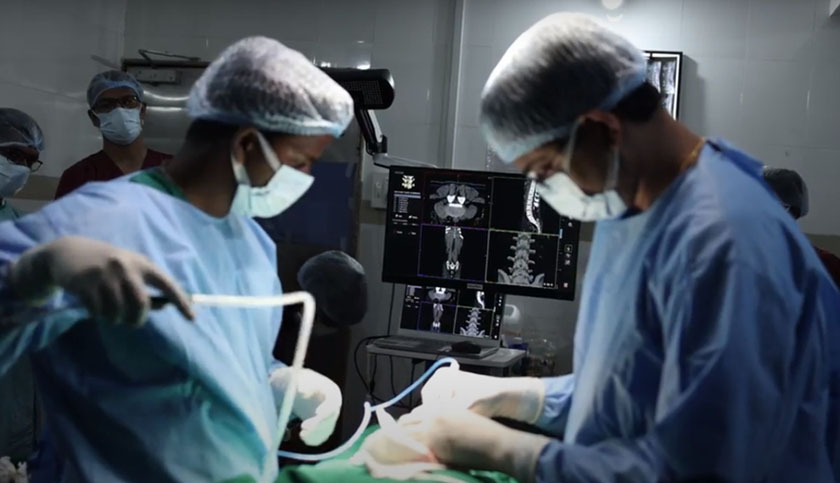
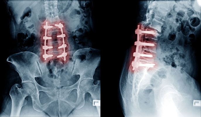
Had tumours in his spine twice: Surgical Navigation saved him

Surgical Navigation - Ravindra patil

Had tumours in his spine twice: Surgical Navigation saved him

By Dr.Ravindra Patil

Chief Neurosurgeon of Samarth Neuro and Multispeciality Hospital [Miraj, South Maharashtra] Dr Ravindra Patil writes about how he could operate a difficult case with the help of surgical navigation.

Mr Mahantesh Shellikeri is from Belgaum. He had a rare disease called Neurofibromatosis. He had two tumours pressing on the spinal cord in his back and as a result he had lost the ability to walk in the past two months.

Table of Contents

Neurofibromatosis

Neurofibromatosis (NF) is a conditions in which tumours grow in the nervous system. There are three types of neurofibromatosis, type I (NF1), type II (NF2) and Schwannomatosis. The tumours in NF are generally non-cancerous.

Non-cancerous tumours are of course good as compared to cancerous tumours, of course, because they don’t grow rapidly and spread to other parts of the body. But Mahantesh’s bad luck was that he had NF tumours in in spinal cord. He had been previously operated in the lumbar spine for the removal of an NF tumour 13 years ago. The surgeon had removed the tumour at the level of the second lumbar vertebra [L2]. But sadly for Mahantesh, the neurofibromatosis tumours recurred! This time it was at the 12th thoracic vertebra [T12] and first lumbar vertebra [L1].

Due to the regrowth of the tumour, Mahantesh developed weakness in both lower limbs. As the tumours grew, gradually he could not walk. His MRI scan showed multiple spinal tumours at the level of the T12, L1 and L2 vertebrae. So he was referred to a neurosurgeon. The neurosurgeon explained that the surgery was very risky as the tumours were small and would be difficult to locate exactly and during their removal Mahantesh was likely to suffer permanent weakness or paralysis of his lower limbs and partial or complete loss of bladder and bowel control.

And if Mahantesh was not operated soon, then too he was likely to suffer permanent weakness or paralysis of his lower limbs and partial or complete loss of bladder and bowel control!

There was another complication. Mahantesh’s wife has poliomyelitis. She is a differently abled and challenged person. She needs a stick to support herself when she walks. So if Mahantesh suffered paraplegia, she would not be able to help him survive.

Besides, the cost of surgery, if it were attempted, would be high.

Thus Mahantesh Shellikeri and his wife were indeed in a desperate situation. Their extended family helped them a lot during their helpless condition.

Many neurosurgeons were consulted for Mahantesh’s treatment. All explained to them that surgery is a risky step because there was a high chance of Mahantesh developing complete paraplegia and bladder plus bowel incontinence.

Chance referral

One of Mahantesh’s relatives is a doctor. He met Dr Somnath Kheradkar, who suggested my name for another opinion.

When Mahantesh and family they came to me, they were very apprehensive and did not have much hope. I reassured them, explained about the disease, although they knew so much.

It was clear to me that if Mahantesh was not operated soon, he would loose control over his lower limbs, bladder and bowel. The surgery was risky.

Advantage factor

But we had one advantage. At Samarth Neuro and Superspeciality Hospital where I work, we have surgical navigation system which is very useful in fine brain and spine surgeries. Mahantesh’s tumours were small and would require accurate removal. The surgical navigation equipment would help me pinpoint his tumours and remove only the tumours without damaging his surrounding spinal cord tissue. I would remove the least amount of vertebral bones and leave his spine intact.

I explained Mahantesh and family about the one almost magical and extremely tool that Samarth Hospital has, namely Surgical Navigation and how it would help me pinpoint the tumours and remove them accurately.

Mahantesh and his wife had many. I answered them all and cleared all their doubts. Then they became were hopeful. They were willing for surgery. But they had reservations about the cost.

But it was a special case. Nearly all neurosurgeons had refused to operate and almost all had advised that the risk of the surgery was far more than the benefits.

I was willing to take the risk of surgery only because of surgical navigation guidance system in Samarth Neuro and Super-speciality hospital where I work. That system would give me the almost magical advantage of doing accurate surgery without and permanent aftereffects.

Surgical navigation would help me find the tumour inside the spinal canal. Also, I could reduce the size of the incision. Besides knowing exactly where to cut and what to remove, the time of the surgery would be much reduced. This would result in less post operative pain and faster healing. The possibility of infection would also be somewhat reduced.

Without Navigation…

The surgery would have been very risky. Had I operated without navigation, I would have been forced to remove one or more levels of his vertebrae which in turn would cause complications during and after the surgery.

Day of surgery

Then we posted him for surgery at the earliest. We did the surgery at reduced charges as Mahantesh Shellikeri’s financial condition is weak. We had CT scan and MRI scan images of his neurofibromas. We started the surgical navigation system after feeding the CT and MRI images into its computer system. We fitted the trackers of the navigations system at exact locations.

Now we knew exactly where to cut and how to cut. Some of our surgical instruments have markers. Thus the markers fixed on the patients and some of our surgical instruments showed up clearly on the 3D screen of the surgical navigation system. With the help of these images and our skills we operated.

We localized the level of the tumour exactly. Then we removed the exact amount of the bone to excise the tumour. We opened the duramater [a stiff covering of the brain and the spinal cord] and I dissected that tumour carefully away from the spinal cord. Afterwards I removed both the tumours located between the T-12 and L-1 levels as also the tumour below the L-2 level.

We were able to remove both the tumours in a single surgery most important of all, the surgery was successful.

Then came the post-operative care. Apart from nursing care and doctors’ rounds came in a very important care giver, the physiotherapist. She motivated and guided Mahantesh to move his limbs as the pressure of the tumours on the nerves was relieved. Mahantesh was surprised that he could move his legs. Remember, he had lost the ability to move his lower limbs when he came to me.

From merely moving his lower limbs to being able to walk was a gradual journey for Mahantesh. But he accomplished it and was able to walk with the support of a walker. But we are moving ahead too fast.

First came Mahantesh’s discharge from Samarth Hospital. We discharge him on the seventh day after surgery.

Many months have passed since Mahantesh was discharged. His would was very small, thanks to navigation assisted surgery. So he did not require much wound dressing. His surgical wounds healed fast.

The Shellikeri couple lives in Belgaum and hence it is too far away for them to come for follow up visits. Local doctors in Belgaon take care of Mahantesh’s routine problems.

But Mahantesh’s wife Mrs Shellikeri called me once after many months to tell me that Mahantesh is progressing well.

With our efforts we were able help Mahantesh walk again. I hope to god that he does not get more episodes of Neurofibromatosis either on his spine or anywhere else.

The Shellikeri family is happy about the results.

Spine Fusion Surgery Cost in India

spinal fusion surgery cost

Spine Fusion Surgery Cost in India

By Dr.Ravindra Patil

Any type of spine surgery is thought to be a very expensive surgery. It is true to some extent as spinal fusion surgery cost is high because it needs very highly skilled surgeons, state of the art operation theatres and above all, surgical navigation for accuracy. But all these give fairly good results in patients who could not be operated at all a decade ago, because the skillset and surgical navigation were not available then. So now although back surgery cost is high, results are better.

Table of Contents

Cost of Spinal Fusion Surgery

The average Spinal Fusion Surgery cost is very high in western countries.

India is known worldwide for its advanced medical facilities and promising technology for Low Cost of Spinal Fusion Surgery. Many of the best hospitals for Spinal Fusion Surgery can be found in India.

Spinal Fusion Surgery cost in India is much less when compared to any other countries. Also the cost of Spinal Fusion Surgery in India is substantially lower than other developed countries of the world.

Private back surgery cost is higher because Private back surgery cost means treatment in a private hospital

Various deciding factors could determine the price of Spinal Fusion Surgery in India. These can be broadly classified as Hospital, Medical Team or Patient Dependent factors.

Hospital Factors

Type of the hospital (Government/Trust/Private).

Use of insurance, type of insurance or self paid.

Accreditation of the facility

Reputation and brand value of the hospital.

Medical Team Factors

Technology / Approach Used

Surgery Type

Type of Anesthesia or Sedation

Qualification / Expertise of the specialist

Extent of the surgery needed

Patient Factors

Patient’s diagnosis

Patient’s general health

Room Category selected by the patient

Other treatment required by the patient in conjunction

What is the definition of spinal fusion?

Spinal fusion is a surgical procedure wherein one or more vertebrae are united to eliminate any motion between them. The approach is similar to welding. Spinal Fusion Surgery, only does not weld the vertebrae immediately during surgery. The bone grafts are placed around the spine during surgery. The body then heals the grafts over several months.

When are the indications of spinal fusion surgery?

There are several potential reasons to consider spinal fusion. These include treatment of a fractured vertebra, rectifying deformity, elimination of pain caused during motion, treatment of instability, and treatment of cervical disc herniations.

Not all spinal fractures need surgery, some fractures, particularly those associated with spinal cord or nerve injury, generally require fusion as part of the surgical treatment.

Another condition treated is instability, i.e. the abnormal or excessive motion between two or more vertebrae. It is commonly believed that instability can be a source of back or neck pain or cause potential irritation or damage to close by nerves.

Cervical disc herniations that require surgery usually need fusion as well. In this technique, the disc is removed by an incision in the front of the neck and a small piece of bone is inserted in its place.

How is the Spinal Fusion Surgery performed?

There are many surgical methods available to fuse the spine that involve placement of a bone graft between the vertebrae. The spine may be treated from the back (posterior), from the front (anterior) or by a combination of both. The anterior approach is more common. The ultimate objective of fusion is to obtain a solid union between two or more vertebrae.

A fusion may or may not involve use of supplemental tools like plates, screws and cages. Instrumentation is sometimes used to rectify a deformity, but is usually used to hold the vertebrae together to while the bone grafts heal. Whether the hardware is used or not, it is important that bone or bone substitutes be used to get the vertebrae to fuse together.

The bone may be taken either from another bone in the patient or from a bone bank. Fusion using bone from the patient has a long history of use and results in predictable healing. Smoking, medications you are taking for other conditions and your overall health can affect the rate of healing and fusion, too.

How long does it take to recover from spinal fusion surgery?

Immediate discomfort following a spinal fusion is generally greater than with other types of surgeries. Fortunately, there are excellent methods of postoperative pain control available, including pain medications and intravenous injections.

Another option is a patient-controlled postoperative pain control pump. Here the patient presses a button that delivers a predetermined amount of narcotic pain medication through an intravenous line.

Patients generally stay in the hospital for three to four days, but a longer stay after more extensive surgery is also possible. A short stay in a rehabilitation centre after discharge from the hospital is recommended for patients who underwent extensive surgery, or for elderly or debilitated patients.

The fusion process varies in each patient. The healing process after fusion surgery is similar to that after a skeletal fracture. During this time, the patient’s activity is generally restricted. Substantial bone healing does not usually take place until three or four months after surgery.

Many a times spinal decompression surgery cost and healing time is compared with spinal fusion surgery cost. It must be understood that both are far different. Spinal decompression surgery does not need bone fusion and healing like spinal fusion surgery.

How many patients underwent Spinal Fusion Surgery in India in the last 5 years?

Private back surgery prices will vary from place to place. Scoliosis surgery cost is also of interest to many patients who have suffered kyphosis or scoliosis. Cost of spinal decompression surgery is also queried.

In recent years, India has emerged as a medical hub for patients looking for affordable, accessible and efficient low cost Spinal Fusion Surgery Treatment. Some of the best Spinal Fusion Surgery hospitals in the world are found in India. The country is known for offering advanced medical facilities at the most reasonable cost. An average increase of 15 to 20 percent annually has been observed in the number of patients in the last 5 years. The Indian Spinal Fusion Surgery hospitals deliver advanced health care and highest quality services backed by elaborate infrastructure and lower treatment cost

Here are the approximate figures of the patients underwent Spinal Fusion Surgery in the last 5 years in India

Year

2013

2014

2015

2016

2017

Surgeries

4800

6000

7500

9600

10800

What are our special services offered to international patients?

At Samarth Neuro and Super Speciality Hospital we help arranging your treatment in a way that will suit you and be affordable for you. Chief neurosurgeon at Samarth Neuro and Super Speciality Hospital Dr Ravindra Patil is vastly experienced in all types of spine and brain surgeries. Spine fusion surgeries are done there as required.

Congenital Brain and Spine Anomalies

congenital brain and spine malformations

Congenital Brain and Spine Anomalies

By Dr. Ravindra Patil

Table of Contents

What are congenital brain and spine malformations?

Congenital means present at birth.

These conditions may be mild and without symptoms or may be serious, requiring treatment. In some cases, surgery may be recommended to:

  1. Address the child’s or the patient’s symptoms.
  2. Correct the form and function of the brain and spine structures.
  3. Maximize cognitive and motor
  4. Prevent development of neurological deficits.

Congenital abnormalities, called malformations, are conditions affecting the form and function of the nervous system. There are numerous variations of congenital malformations of the bone and soft tissue of the head and spine, including neural tube defects, such as spina bifida, encephaloceles, Chiari malformations and arachnoid cysts.

Some congenital malformations are mild, and some are severe but correctable with surgery by a pediatric neurosurgeon.

Types of some of the Congenital Brain and Spine Malformations are as follows:

Chiari Malformations

This is a condition in which portions of the brain, called the cerebellar tonsils, protrude through the bottom opening of the skull into the upper spine, which can put pressure on the brain or spinal cord. Chiari malformations may block the flow of cerebrospinal fluid, leading to hydrocephalus.

Treatments often focus on removing portions of the bone and soft tissue to relieve pressure on the spinal cord and brain, as well as providing new pathways to drain cerebrospinal fluid. Surgeons use different methods for treating these malformations, including decompression, with or without cutting open a small part of the dura mater [thick membrane covering the brain].

Encephaloceles

Encephaloceles are a type of neural tube defect characterized by the brain being exposed to the outside instead of being covered by the skull and skin. It can lead to infections and hydrocephalus.

Surgical treatment of this condition involves removing bone and soft tissue, draining cerebrospinal fluid, and surgically repairing or closing the encephalocele.

Children who have developed hydrocephalus as a result of an encephalocele will require treatment for that condition, often with a cerebrospinal fluid shunt. Shunting is the placement of a tube into the open area (ventricle) of the brain that allows cerebrospinal fluid to drain to the child’s abdomen or other location where it can be safely reabsorbed into the body.

Arachnoid Cysts

Arachnoid cysts are the most common type of brain cyst. They are congenital (present at birth) lesions that occur as a result of the splitting of the arachnoid membrane. The cysts are fluid-filled sacs, not tumors, appearing in one of the three layers of tissue covering the central nervous system.

Surgical treatment of this condition involves draining the cyst by drilling a small hole or by opening the skull and making small openings in the cyst to open the natural fluid pathways in the brain. This process is called fenestrating.

What are Spinal Deformities?

A spine deformity occurs when your spine varies by more than 10 degrees from ‘healthy’ curvature. But, what does this mean exactly?

Contrary to popular belief, your spine isn’t 100% straight and vertical. The spinal shape has curvatures, but the end result is that it is vertical! Our spine consists of a series of gentle arcs. Our lumbar spine, or lower back, swoops slightly to the back, and our thoracic spine, or upper back, bends subtly forward. The backward curve of your lower back is known as lordosis and the forward stoop that runs between our shoulder blades is known as kyphosis. Lordosis and kyphosis are spinal curvature deformities. Both are abnormal curvatures of the spine.

But, when viewed head-on, our backbone should look like a straight pillar. Hence, it is also called the ‘vertebral column’.

Moreover, the curves and straight stretches of your spine make symmetry possible. Your head sits directly over your pelvis because the lordosis of your lower back and the kyphosis of your upper spine balance each other out. If one of these curves becomes greater or lesser than the other, then problems can occur. We refer to this as sagittal imbalance, because the head and pelvis no longer fall within the same, or sagittal, plane.

Too much swaying backwards can be thought of as ‘lordosis’, and too much forward stooping in the upper back is ‘kyphosis’.

Likewise, when the spine tilts away from the midline of the body, doctors refer to this problem as coronal imbalance or scoliosis. Unevenness in the ‘coronal’ plane (the view from head-on) causes asymmetry in the trunk of the body. This can include uneven hips and shoulders or one-sided bulging of the ribs.

Diagnosis of Congenital Brain and Spine Malformations

If a child is born with any of the above malformations, a thorough evaluation by a paediatrician or neurologist is needed to diagnose the problem and recommend a plan for addressing it.

After a thorough physical and detailed family and patient history, your doctor may order imaging of the brain and/or spine through an MRI scan. If the MRI scan shows any evidence of these malformations, a neurosurgical consultation is a must to plan the best treatment.

Congenital Brain and Spine Malformations Treatment

A multidisciplinary approach is often beneficial for addressing children with congenital brain and spine malformations. Neurosurgeons, craniofacial plastic specialists and geneticists, among others, may be called upon to develop your child’s treatment plan and determine what kind of surgery may be appropriate.

If a congenital brain or spine malformation is mild and not causing any signs or symptoms in your child, the neurosurgeon may recommend observation, which means regular visits and testing to monitor your child’s condition.

If your child does undergo surgery, follow-up care is extremely important in tracking the progress of your child’s recovery. Your paediatric neurosurgeon will schedule follow-up appointments to ensure your child is making the best recovery possible.

Spinal treatments are focused on both correcting the functional shortcomings as well as the structural defects of the spine.

Congenital brain and spine defects treatment in Miraj, Maharashtra

Samarth Neuro and Super Speciality hospital has facilities to surgically treat some of the above brain and spine deformities present since birth. Abnormal curvature of the spine can be corrected to some extent.

Chief Neurosurgeon at Samarth Hospital Dr Ravindra Patil has considerable experience in treating such defects. Besides, as his hospital is located in a tier two city, the cost of treatment is considerably less than in major cities of India. Patients from foreign countries may find going to Miraj for treatment of brain and spine diseases from Dr Ravindra Patil considerably cost effective.

Trigeminal Neuralgia

trigeminal neuralgia

Trigeminal Neuralgia

By Dr. Ravindra Patil

Trigeminal neuralgia (TN), is sometimes described as the most excruciating pain known to humanity. The pain typically involves the lower face and jaw, although sometimes it affects the area around the nose and above the eye. This intense, stabbing, electric shock-like pain is caused by irritation of the trigeminal nerve, a nerve in face, which sends branches to the forehead, cheek and lower jaw. It usually is limited to one side of the face. The pain can be triggered by an action as routine and minor as brushing your teeth, eating or a sudden sharp breeze. If left untreated, trigeminal neuralgia can progressively worsen. Facial neuralgia means pain on the face related to a nerve.

Trigeminal neuralgia cannot always be cured, there are treatments available to alleviate the debilitating pain. Normally, anticonvulsive medications are the first treatment choice. Surgery can be an effective option for those who become unresponsive to medications or for those who suffer serious side effects from the medications.

Table of Contents

The Trigeminal Nerve

The trigeminal nerve is one set of the cranial nerves, which means it originates from the brain. It is the nerve responsible for providing sensation to the face. There are two trigeminal nerves, one on either side of our face. The trigeminal nerve leaves the brain and travels inside the skull, where it divides into three smaller branches, controlling sensations throughout the face:

  • Ophthalmic Nerve controls sensation in our person’s eyes and forehead.
  • Maxillary Nerve controls sensation in the lower eyelid, cheek, nostril, upper lip and upper gum.
  • Mandibular Nerve controls sensations in the jaw, lower lip, lower gum and some of the muscles used for chewing.

Prevalence

It is reported that 150,000 people are diagnosed with trigeminal neuralgia every year. While the disorder can occur at any age, it is most common in people over the age of 50. TN is twice as common in women than in men.

Types of Trigeminal Neuralgia

There are two main forms of trigeminal neuralgia:

  • Typical (Type 1) trigeminal neuralgia. Patient is likely to experience painful episodes that are sharp, intense and sporadic.
  • Atypical (Type 2) trigeminal neuralgia. This will be less painful and intense but more widespread.

Causes

The exact cause of TN is still unknown, but the pain associated with it represents an irritation of the nerve. Primary trigeminal neuralgia has been linked to the compression of the nerve, typically in the base of the head where the brain meets the spinal cord. This is usually due to contact between a healthy artery or vein and the trigeminal nerve at the base of the brain. This places pressure on the nerve as it enters the brain and causes the nerve to misfire. Secondary TN is caused by pressure on the nerve from a brain lesion like a tumour, cyst, facial injury or any medical condition that damages the myelin sheaths.

Trigeminal Neuralgia Symptoms

Any one side face pain cannot be trigeminal neuralgia. Most patients report that their pain begins spontaneously. Other patients say their neuralgia pain came after a car accident, a blow to the face or dental work.

TN tends to run in cycles. Patients often suffer long stretches of frequent attacks, followed by weeks, months or even years of little or no pain. The pain typically begins with a sensation of electrical shocks that culminates in an excruciating stabbing pain within less than 20 seconds.

Pain can be focused in one spot or it can spread throughout the face. Typically, it is only one side face pain, eg… pain in right side of face and jaw and ear; however, in rare occasions and sometimes when associated with multiple sclerosis, patients may feel pain in both sides of their face. Pain areas include the cheeks, jaw, teeth, gums, lips, eyes and forehead.

Attacks of TN may be triggered by the following:

  • Touching the skin lightly
  • Washing
  • Shaving
  • Brushing teeth
  • Blowing the nose
  • Drinking hot or cold beverages
  • Encountering a light breeze
  • Applying makeup
  • Smiling
  • Talking

There are many other conditions which are similar to TN. Brain specialists use various tests to pinpoint the exact diagnosis.

Trigeminal Neuralgia Diagnosis

TN can be very difficult to diagnose, because there are no specific diagnostic tests and symptoms are very similar to other facial pain disorders. Therefore, it is important to seek medical care when feeling unusual, sharp pain around the eyes, lips, nose, jaw, forehead and scalp, especially if you have not had dental or other facial surgery recently.

TN usually is diagnosed based on the description of the symptoms provided by the patient, detailed patient history and clinical evaluation.

Testing

There are no specific diagnostic tests for TN, so physicians must rely heavily on symptoms and history, type of pain (sudden, quick and shock-like), the location of the pain and things that trigger the pain. MRI can detect tumour/s or Multiple Sclerosis and if there is compression caused by a blood vessel. Newer scanning techniques can show if a vessel is pressing on the nerve and may even show the degree of compression.

Treatment of Trigeminal Neuralgia – Non-Surgical

There are several effective ways to alleviate the pain, including a variety of medications. Medications are generally started at low doses and increased gradually based on patient’s response to the drug.

  • Carbamazepine
  • Gabapentin
  • Oxcarbazepine

Other medications include baclofen, amitriptyline, pregabalin, phenytoin, valproic acid etc.

Medicines may have side effect and may become ineffective after long usage.

Trigeminal Neuralgia Treatment: Surgery

If medications have proven ineffective in treating TN, several surgical procedures may help control the pain.

Open Surgery

Microvascular decompression surgery involves microsurgical exposure of the trigeminal nerve root, identification of a blood vessel that may be compressing the nerve and gentle movement of the blood vessel away from the point of compression. Although most effective, it is also most invasive.

Lesioning Procedures

Percutaneous radiofrequency rhizotomy treats TN through the use of electrocoagulation (heat). It can relieve nerve pain by destroying the part of the nerve that causes pain and suppressing the pain signal to the brain.

Percutaneous balloon compression utilizes a needle that is passed through the cheek to the trigeminal nerve. The balloon compresses the nerve, injuring the pain-causing fibres, and is then removed.

Percutaneous glycerol rhizotomy utilizes glycerol injected through a needle into the area where the nerve divides into three main branches. The goal is to damage the nerve selectively in order to interfere with the transmission of the pain signals to the brain.

Stereotactic radiosurgery (through such procedures as Gamma Knife, Cyberknife, Linear Accelerator (LINAC) delivers a single highly concentrated dose of ionizing radiation to a small, precise target at the trigeminal nerve root. This treatment is non-invasive and avoids many of the risks and complications of open surgery and other treatments.

Overall, the benefits of surgery or lesioning techniques should always be weighed carefully against its risks. At Samarth Neuro and Trauma Hospital in Miraj, many patients with Trigeminal neuralgia or one side face pain have recovered after medical or surgical treatment, under the specialist care of Neurosurgeon Dr Ravindra Patil.

Pain in Ears and Jaw

Pain in Ears and Jaw

By Dr. Ravindra Patil

Pain in the area near our ear, our jaw or the muscles on the side of our face, sometimes accompanied by a clicking or popping sound and/or restricted jaw movement is called Temporo-Mandibular Disorders or TMD in short. There is another abbreviation related to the joints of the jaws. It is TMJ for Temporo-Mandibular Joint. We have two TMJs on either side of our face.

Jaw pain reasons are many and may be due to TMJ disease or even mental stress!

TMD pain describes a group of conditions characterized by pain and dysfunction of the TMJ and/or the muscles surrounding it. It’s not always easy to figure out the cause of these symptoms. However, most TMD cases can be treated with conservative remedies. Only if these remedies are not useful should major procedures like dental treatment or surgery should be done.

The two TMJs that connect your lower jaw, the mandible, to the temporal bones of the skull on either side, are actually very complex joints that allow movement in three dimensions. The TMJ anatomy is that the lower jaw and temporal bone fit together as a ball and socket, with a cushioning cartilage disk in between. Large pairs of muscles in the cheeks and temples move the lower jaw. Any of these parts — the disk, the muscles or the joint itself — can become the source of a TMD problem.

For people suffering from TMD pain or for people having difficulty opening or closing their jaw, a thorough investigation workup is necessary to pinpoint the cause of the pain or difficulty in opening and closing the jaw.

Table of Contents

What causes TMD?

The temporo-mandibular joint or TMJ can suffer orthopaedic problems like any other joint. These problems include inflammation, sore muscles, strained tendons and ligaments, and cartilage problems.

Heredity also plays a role in causing TMD. Compared to men, women appear to be more prone to it. As we age, the chances of getting TMD are higher. Physical and psychological stresses can also cause TMD. In some cases, jaw pain may be related to a more widespread, pain-inducing medical condition such as fibromyalgia. Fibromyalgia is a pain of muscles and connective tissue around the TMJ [or any other joint].

Because we have two Temporo Mandibular Joints, one on each side our the face, pain in left side of head, jaw pain left side, pain on left side of head above ear, pain in right side of head, jaw pain left side near ear or jaw pain one side, all permutations and combinations of pain are possible.

Signs and Symptoms

Clicking Sounds: some people with TMD hear a clicking, popping or grating sound coming from the TMJ when opening or closing the mouth. This is usually caused by a shifting of the cartilage disk inside the joint. This clicking or popping sound can sometimes be heard by anyone standing next to the patient. Clicking by itself is actually not a significant symptom because it has been found that over one third of all the population has jaw joints which click. However, if the clicking is accompanied by pain or limited jaw movement and function, or the jaw is getting ‘stuck’ in an open or closed position often, this definitely is TMD.

TMD Muscle Pain Cycle

Every joint is operated by muscles. The jaw can be clenched by two pairs of very powerful muscles, the temporalis muscles and the masseter muscles. They are located on either side of our face. Pain in these muscles is significant and should be taken care of. Jaw pain is not so uncommon.

Pain on right side of the head or left side head pain, the side depends on which side TMJ is affected. Often both sides jaw pain may occur.

The reason for jaw pain may be muscle spasm of temporalis and masseter muscles.

The muscle pain may be felt in the cheeks (masseter muscles) and temples (temporalis muscles), where the two big pairs of jaw-closing muscles are located. If a person feels soreness and stiffness upon waking up in the morning, it’s often related to habits of clenching and/or grinding the teeth at night. If you have this type of nocturnal habit, it can be treated with a custom-made guard which is very helpful in decreasing the force applied to your teeth. This in turn will allow your muscles to relax and be painless. This will relieve pressure on your jaw joints. Some of the other self-care remedies are discussed below.

TMJ headache means headache or jaw pain caused because of TMD.

Joint Pain

Pain that originates one or both jaw joints is called arthritis of the TMJs. When we look at X-Ray images of the TM joints, we may or may not find arthritic changes. The funny part is, some people have arthritic-looking TMJs but no symptoms of pain or dysfunction; while others have significant symptoms of pain and dysfunction but their joints look normal in X-Ray images. Alas, there is no permanent cure for arthritis anywhere in the body, but medicines definitely help in relieving arthritic pain and swelling.

Pain Relief

As has been mentioned, a thorough investigative work up on a TMD patient will help in finding the cause of the pain and then treat or reduce the same. Sometimes an intervention as simple as switching to a soft diet which does not need thorough chewing can reduce stress on the muscles and joints and reduce pain.

Application of Ice and/or moist heat can help relieve swelling, soreness and inflammation. If the pain is caused because of muscle spasm, it can be relieved with gentle stretching exercises. Non-steroidal anti-inflammatory drugs [NSAIDs] and muscle relaxants can also provide relief. NSAIDs apart from reducing inflammation, also reduce pain.

Other Treatments

Severe TMD cases may require more complex forms of treatment, which might include orthodontics, dental restorations like bridgework, or minor procedures inside the joint such as cortisone injections or lavage (flushing) of the joint. It’s rare for major surgery ever to be necessary in a case of TMD. Again, it’s important to try the wide range of conservative, reversible treatments available, and give them enough time to work as they almost always prove effective.

Chewing tobacco and TMD

It is well known that chewing tobacco causes cancers of the mouth. But chewing tobacco also causes difficulty with chewing, swallowing, speaking or moving the tongue and jaw. Tobacco users find it very difficult to open their mouth and so are unable to eat properly. While the risk of cancer with tobacco chewing is great, the stiffening of the jaw and inability to open the jaw fully is also a major symptom. There may be cheek swelling too.

Many people who had been chewing tobacco and Gutkha for a few years, experience inability to open their mouth. The mouth barely opens more than 30 mm and the opening is further reduced over a period of time. The jaws become stiff. Such patient are firstly unable to eat because of small mouth opening. Besides, they are unable to eat spicy food. This condition is known as Oral Sub Mucous Fibrosis.

Stopping tobacco chewing is the first step in treating Oral Sub Mucous Fibrosis. Surgical treatment also helps open the jaw wide.

To summarise…

TMD, or Temporomandibular Disorders, is an umbrella term for various painful conditions that affect the jaw joints. There are different treatment approaches to TMD problems, but not all are based on science. It’s important to be up on the latest information and to be an educated patient. We have tried to provide useful information in this article about TMD.

Pituitary Gland Disorders

Pituitary Gland Disorders

Pituitary Gland Disorders

By Dr. Ravindra Patil

What is the pituitary gland and where is it located?

The pituitary gland is a tiny pea-sized organ located at the base of the brain. Barely a cubic centimetre in volume, it produces and stores so many hormones of our body that it is known as the ‘master gland’. These pituitary hormones control the activity of other hormones in the body.

The pituitary gland has 2 parts: the anterior (front) and the posterior (back) parts, each producing a different set of hormones. Each of these hormones acts on different parts of the body and controls their proper functioning. The pituitary hormones are:

  • Prolactin
  • Growth hormone
  • Adrenocorticotrophic hormone [ACTH]
  • Thyroid stimulating hormone [TSH]
  • Luteinizing hormone
  • Follicle stimulating hormone
  • Melanocyte stimulating hormone
  • Antidiuretic hormone [ADH]
  • Oxytocin

Function of pituitary hormones are vital for human survival. The deficiency or overproduction of each and every of the above hormones results in major medical disorders. For example, a shortage of growth hormone during childhood leads to reduced growth. The child does not grow like a normal adult but remains a dwarf. Conversely, excess of growth hormone leads to gigantism, a condition where the person keeps on growing abnormally.

Table of Contents

Hormones and Hormone Diseases

Hormones are the body’s chemical messengers, sending signals into the bloodstream and tissues. Hormones work slowly, over time, and affect many different processes, including growth and development, blood sugar, sexual functions, reproduction, urine output, managing stress and mood. Hormone diseases occur when the level of any hormone is reduced or increased because of any reason. The Pituitary Gland produces many hormones and any pituitary gland problems lead to major illnesses.

What are common pituitary disorders?

Apart from a deficiency or excess production of the above hormones, disorders of pituitary gland are as follows:

  • Tumours
  • Pituitary damage because of Head injury
  • Birth defects
  • Inherited genetic defects
  • Low blood supply to the pituitary gland
  • Previous history of pituitary disorders
  • Iron overload
  • Medication
  • Radiation therapy in the head and neck region

What are the different types of anterior pituitary disorders?

Anterior pituitary disorders occur as a result of overproduction or underproduction of hormones secreted by the anterior lobes.

     Disorders caused by over-production of anterior pituitary hormones

  • Acromegaly & gigantism
  • Prolactemia
  • Cushing’s disease


      Disorders caused by under-secretion of anterior pituitary hormones

  • Dwarfism
  • Central adrenal insufficiency
  • Gonadotropin deficiency
  • Hypothyroidism


What are the different types of posterior pituitary disorders?

Posterior pituitary disorders are caused by either under-production or overproduction of the ADH hormone. ADH helps the kidneys to prevent excess water loss through urine. Imbalance in ADH production can lead to the following disorders:

Pituitary disorders symptoms: Hypopituitarism

Hypopituitarism is a condition that results in a partial or complete loss of the anterior pituitary gland functions. Panhypopituitarism is a condition, characterized by damage to the entire pituitary gland. In such cases, the production of all pituitary hormones stops. 

Causes of hypopituitarism

  • Growth of a pituitary tumour
  • Head injuries
  • Brain surgery
  • Medication
  • Radiation therapy

Signs and symptoms of hypopituitarism

  • Tumours may cause eyesight problems.
  • The symptoms of hypopituitarism depend upon which hormones are no longer being produced.
  • Symptoms vary depending upon the age of the patients.

Diagnosis

  • Blood tests and stimulation or dynamic testing to identify low levels of pituitary hormones
  • Brain imaging using an MRI or CT scan to detect tumour or other pituitary gland problems
  • Vision tests to determine if the pituitary gland is affecting eyesight

Pituitary gland disorders treatment

  • Hormone replacement therapy: Patients need to take life-long medications to control the symptoms. The doctor monitors and adjusts the medication dose periodically based on the patient condition
  • Surgery or radiation treatment may be required in some patients suffering from pituitary tumours

What are pituitary tumours?

Pituitary tumours are abnormal growths in the pituitary glands. They may cause overproduction or low levels of pituitary hormones.  Most pituitary tumours are noncancerous growth and remain confined within the pituitary gland. These are known as an adenomas.

When the tumour size is less than one cm, it is called a Microadenoma. Most pituitary adenomas are microadenomas. When the tumour is greater than one cm in size, it is called a Macroadenoma.

Malignant Pituitary tumours (cancers) are rare and mostly affects old aged persons. Pituitary carcinomas can spread to the brain, spinal cord or the bone surrounding the pituitary glands. In some patients, the pituitary adenoma can turn cancerous and spread to other parts of the body.


What is the prevalence of pituitary tumours?

A pituitary adenoma is the third largest cause of brain tumours, accounting for 10% of all cases. The global prevalence of pituitary adenomas is approximately 17%. Studies have reported the risk of pituitary tumours to increase with age, with maximum cases being diagnosed between 30 and 60 years.

What causes pituitary tumours? Are there any risk factors?

The cause of pituitary adenoma remains unknown. Genetic alteration is thought to play an important role in adenoma development. Studies have shown that people with certain genetic factors such as multiple endocrine neoplasia, type 1 (MEN 1) are at increased risk of pituitary tumours.

What are the signs and symptoms of pituitary adenomas?

  • Tumours can put pressure on the pituitary gland and the nearby structures. This can cause headaches and loss of side-visions
  • As the tumour grows in size, it can damage the normal functioning of the gland and interfere with hormone production. Overproduction or hormonal deficiencies can cause specific signs and symptoms or sometimes a combination of them.
  • For example, ACTH tumours exhibit signs and symptoms of Cushing’s syndrome.

Are pituitary tumours life-threatening?

If diagnosed early, pituitary tumours can be managed well. However, if left undiagnosed and untreated for a long period, such tumours can become large and affect the functioning of several organs of the body, causing blindness, hypertension, diabetes, osteoporosis, heart disease and death.

Evidence suggests the 5-year survival rate of pituitary gland tumours be about 82%. 

Survival rate also depends upon factors like:

  • Type of tumour
  • Person’s age
  • How far the tumour has spread in the brain or to other parts of the body

Complications of pituitary tumours

  • Blindness
  • Permanent hormone deficiency
  • Pituitary apoplexy is a rare and serious complication

How are pituitary tumours diagnosed?

  • Blood and urine tests help to determine whether there is an overproduction of deficiency of hormones
  • Biopsy: This involves removing a small number of cells and examining them under a microscope to check for abnormal growth
  • Brain imaging: A CT scan or brain MRI scan can help the doctor to determine the size and location of the tumour
  • Vision testing to understand if the tumour has caused eyesight problem
  • Neurological exam: A set of questions and answers to check a person’s mental status and appropriate brain functioning controlling movement and coordination.

Pituitary gland diseases treatment

Not all pituitary tumours require treatment. Treatment depends upon the following:

  • Type and size of the tumour
  • If the tumour is making hormones
  • If the tumour is obstructing vision or associated with other signs or symptoms
  • If the tumour is localized or has spread to other parts of the body
  • If the tumour has occurred for the first time or has recurred
  • The patient’s overall healt

    The treatment is usually given by a brain surgeon (neurosurgeon), endocrine system specialist (endocrinologist) and a radiation oncologist. Doctors use a combination of surgery, radiation therapy and medications to normalize hormone levels.

Surgery for pituitary

Surgery is needed when the tumour presses on the optic nerve or leads to the overproduction of hormones. The Endoscopic transnasal transsphenoidal approach involves removing the tumour through the nose without making an incision. This is usually done when the tumour is small in size and no other part of the brain is affected by it. The Transcranial approach (craniotomy) is used for removing large tumours by making an incision in the scalp.

Radiation therapy

This technique uses high-energy radiation to destroy tumour cells. It can be used alone or in combination with surgery.

Medicines

They help to limit excess hormone production and shrink certain types of pituitary tumours.

Hormone replacement therapy is done in patients with reduced hormone production after surgery.

Watchful waiting: Many patients do not exhibit any symptoms and function normally without any treatment. In such cases, patients are advised only regular tests to keep a check on the tumour growth.

To summarise, a disease of pituitary gland can be very harmful. It must be diagnosed and treated as soon as possible. Such complex disease treatment is available in Samarth Neuro and Superspeciality Hospital in Miraj, Maharashtra. Dr. Ravindra Patil, the chief neurosurgeon there, is an expert in performing such operations.

Ventricles of the Human Brain

Ventricles of the Human Brain

Ventricles of the Human Brain

By Dr. Ravindra Patil

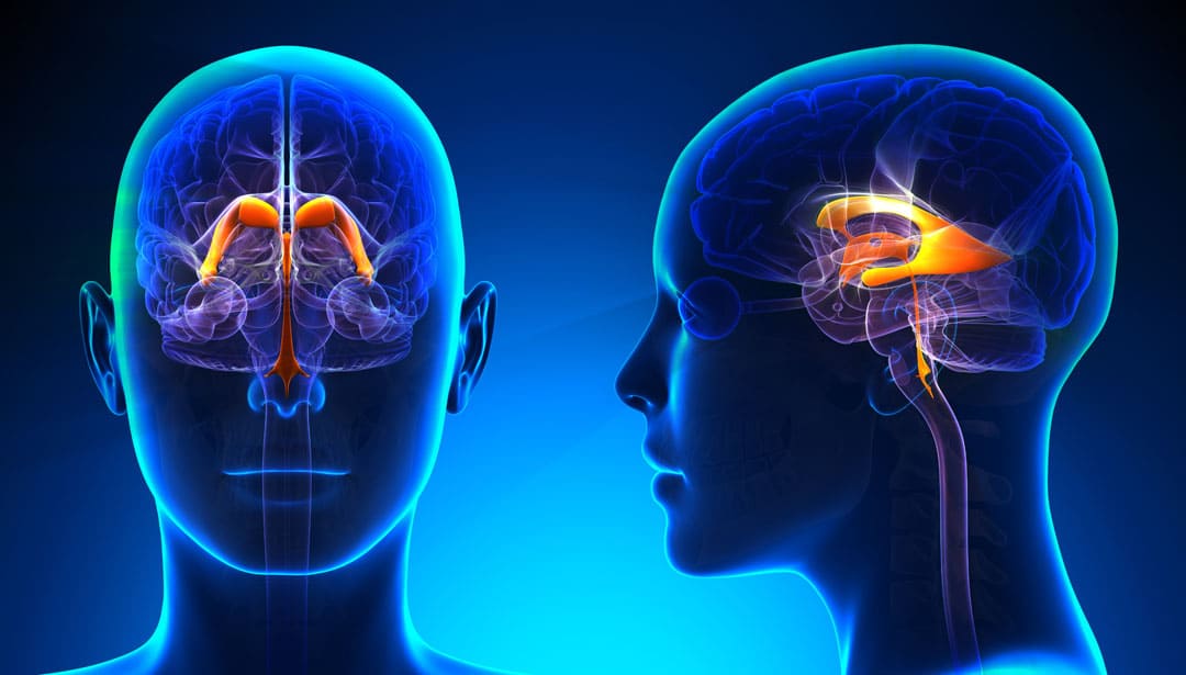
A ventricle is a hollow part or cavity in an organ. The word ventricle originates from Latin, the language which has given most of the words used in anatomy and medicine. Ventricles of brain are cavities within the brain. Ventricles of the brain have distinctive functions, which we will see in this article.

Two major organs of the major organs of the human body which have ventricles are the heart and the brain. The ventricles of the heart pump blood through our body, while the ventricles of the brain are cavities within the brain that produce and store a liquid called cerebrospinal fluid (CSF). This liquid surrounds our brain and spinal cord, cushions them and protects them from injury. The CSF is also responsible for removing waste and delivering nutrients to our brain.

Table of Contents

Where in the brain are the ventricles?

There are four ventricles in the brain. They are interconnected cavities. They extend throughout the brain. As has been mentioned, they produce, store and circulate the cerebrospinal fluid (CSF).

The two lateral ventricles of brain are C-shaped chambers found in the cerebral hemispheres, which is one in each hemisphere.

Lateral ventricles are connected to the third ventricle by an opening called the inter-ventricular foramen. The third ventricle is a very narrow cavity that runs along the midline of the diencephalon.

What structure connects the third and fourth ventricles? The third ventricle communicates with the fourth ventricle via the cerebral aqueduct. The fourth ventricle is wedged between the cerebellum on one side and the brainstem on the other; it extends to the central canal of the spinal cord. Which means in effect, the four ventricles extend from the brain to the end of the spinal cord. The CSF surrounds the brain and is also within the ventricles. The CSF is a liquid cushion in which the brain literally floats. The brain ventricles essentially supply, store and circulate the CSF around the brain.

The CSF is a thick viscous fluid which serves to cushion the brain, regulate its temperature and supply the brain with nutrients.

More about the brain ventricles

When looking at the interior of the brain, the four hollow ventricular cavities stand out in contrast to the rest of the organ, which is mostly composed of brain tissue, which in turn is made of grey matter and white matter. The main ventricle function is of course, producing CSF.

As the ventricles contained nothing except fluid, in ancient times it was thought that the ventricles contained ‘animal spirits’, a mysterious substance that allowed the immortal soul to exert control over the physical body. Later it was thought that the ventricles were credited with functions like the generation of imagination and memory. However, it was in 1764 that it was discovered that the ventricles were filled with CSF, fluid, and not animal spirits. And the CSF flowed around and inside the brain through the connections between ventricles. The ventricles gave the CSF a route to flow throughout the brain. Gradually it was discovered that it is the production, storage and circulation of CSF which is the main role of the ventricles.

How is CSF produced?

The production of CSF is done by a specialized membrane called the choroid plexus, which is made up of ependymal cells. This choroid plexus lines the ventricles in brain. Ependymal cells are glial cells tailored to produce CSF, and they secrete the fluid into the ventricles at a relatively constant rate; about a half a litre of CSF is produced by ependymal cells every day. CSF passes through the ventricular system and circulates around the brain and spinal cord in a small area between the meninges called the subarachnoid space.

Functions of the CSF

CSF is thought to play many important roles in the brain. The CSF and the brain ventricular system together keep the brain buoyant, reducing its weight and hence the physical stress it would otherwise experience from the forces of gravity and movement. The brain is a very delicate and soft organ. If the  brain would not have been suspended in the CSF, it would have been distorted with its own weight! While organs like the kidney and liver and heart are much sturdier, the brain is very soft. Without being suspended in a fluid the delicate tissue of the brain can tear.

There is about 125–150 mL of CSF at any one time. The actual mass of the human brain is about 1400–1500 grams; however, the net weight of the brain suspended in CSF is equivalent to a mass of 25-50 grams. The brain therefore exists in neutral buoyancy, which allows the brain to maintain its density without being impaired by its own weight. Had the brain been suspended in the skull without floating in the CSF, its blood supply would have been cut off and its neurons would be killed in the lower sections of the brain!

The layer of CSF surrounding the brain also acts as a buffer against potential injuries that can be caused by mechanical pressure or force (e.g. being hit hard in the head). Additionally, as the CSF circulates over the brain it carries away toxins and other waste matter and empties these into the bloodstream where they can eventually be removed by mechanisms like kidney filtration.

Ventricles and the CSF

The rate of CSF production in the ventricles is fairly constant regardless of changes in pressure within the ventricles (i.e. inter-ventricular pressure). This can be problematic if the passage of CSF is blocked somewhere within the ventricular system. In the ventricular system of brain, CSF will continue to be produced, but it will have no means of exiting the system. This will cause pressure within the ventricles to increase, and the rising pressure may eventually force the ventricles to expand. The expanding ventricles can then put pressure upon other brain structures, and cause a variety of complications depending on where exactly the blockage has occurred and which structures are most affected by the blockage.

When such a blockage of the brain ventricles occurs in children whose skull has not completely become bony [generally under age of 2 years], it results in the enlargement of the head. It is known as hydrocephalus. Hydrocephalus can be cause by blockage in the passage of CSF as well as by excess CSF production. In lay language, hydrocephalus is referred as ‘Water in the Brain’.

There can be a number of causes of a blockage that leads to hydrocephalus, such as a tumour, infection, or congenital malformation, which impedes the function of ventricles. Hydrocephalus can often be treated by surgically implanting a shunt that drains the ventricles of excess CSF and empties it into the abdominal cavity. This approach can be successful, but if the reason for the blockage isn’t resolved additional surgeries may be required in the future (e.g. to replace an outgrown shunt, treat an infected shunt, etc…).

How is blockage in ventricles diagnosed?

A contrast CT Scan can show the size and shape of the ventricles in brain. Either a radiopaque dye is injected in the patient’s veins or the CSF is replaced with air and then CT Scan of brain ventricles is done. In either case, a good contrast will be seen and the ventricles can be visualised. While the radio opaque dyes shows up as white colour, air shows up as black colour in CT scans and that helps doctors view the ventricular shape and size and reach a diagnosis about tumours, raised CSF pressure or blockage in the brain ventricular system.

Samarth Neuro and Superspeciality Hospital in Miraj has all the infrastructure and staff to treat hydrocephalus in children and other reasons of CSF blockage. The chief neurosurgeon Dr Ravindra Patil has operated upon many children suffering from hydrocephalus who needed shunts. These children are now grown up. Dr Ravindra Patil has also successfully operated on many adult patients with brain tumours.

Craniotomy surgery

Craniotomy surgery

Craniotomy surgery

By Dr. Ravindra Patil

Craniotomy is a surgery, but not to treat one specific disease. Craniotomy means opening the skull to do brain tumour surgery or epilepsy surgery. The word craniotomy has two parts: Cranium and Otomy.

The word Cranium originates from the Latin language. It means the skull. And ‘Otomy’ comes from the Greek language and it means cutting into a part of the body. Most medical terminology is from Latin and Greek. Thus, a gastrotomy would be cutting into the stomach, a pharyngotomy means cutting into the pharynx. In the same way, craniotomy means cutting into the cranium or skull.

In this surgery for the brain, a part of the skull is removed to access the brain. The bone is replaced when the surgery is done. Craniotomy is done to remove brain tumours, treat brain aneurysms, treat epilepsy, treat cerebral haemorrhage, to decrease intracranial pressure and so on. Neurosurgeons performs the procedure.

Cost of brain surgery in India

When such a major surgery is mentioned, the subject of cost must be cleared. Brain surgeries may cost between one lakh to five lakhs or more. But there are options like Ayushman Bharat Yojana, health insurance or employer given benefits to cover the costs. If you have none of the above benefits, it is highly advisable to buy a medical insurance.

Before a craniotomy procedure

Various tests are done to confirm that the patient can safely undergo the procedure. They are:

  • Physical exam
  • Blood tests
  • Neurological exam
  • Imaging of the brain (ct or mri)
  • Determination of the surgical site based on the medical condition

On the night before surgery, the patient will be NBM [nil by mouth]. Scalp will be shaven.

During the procedure the patient lies down on the operating table. The position depends on the part of the brain being operated on. Intravenous line is inserted into your arm or hand. A urinary catheter into the patient’s bladder. And then general anaesthesia is given. Surgery is done with a knife, a medical drill and a saw. Then the brain tumour operation, epilepsy surgery or brain haemorrhage is done. Then the surgeons fix the bone with wires, stitches, or plates either immediately or a few months later. Finally the skin will be stitched or stapled. A sterile dressing and a sterile bandage are applied. Craniotomy surgery may take about three to four hours.

Types of Craniotomy

There are many types of craniotomies. Each type is named for the technique or location used in the surgery.

Stereotactic craniotomy

If in a craniotomy the skull is fixated with a stereotactic frame and uses an MRI or CT scan, it’s called a stereotactic craniotomy.

Endoscopic craniotomy

Done through a tiny incision in the skull through an endoscope.

Awake craniotomy

Table of Contents

An awake craniotomy is done while the patient is awake.

Keyhole craniotomy

A keyhole craniotomy is used to remove brain tumours. It’s a minimally invasive surgery.

Supra-orbital ‘eyebrow’ craniotomy

A supra-orbital means surgery above the eye sockets, done to remove brain tumours in the front of the brain.

Pteronial (frontotemporal) craniotomy

In the skull, the pterion is where the frontal, temporal, sphenoid, and parietal bones meet. It involves removing part of the pterion.

Orbitozygomatic craniotomy

Difficult tumours and aneurysms may be treated with an orbitozygomatic craniotomy. The surgeon temporarily removes part of the bone that creates the curve of your orbit, or eye socket, and cheek.

Posterior fossa craniotomy

This is done through an incision at the base of the skull.

Translabyrinthine craniotomy

In a translabyrinthine craniotomy, the surgeon makes a cut behind a ear.

Bifrontal craniotomy

A bifrontal craniotomy, or extended bifrontal craniotomy, is done to remove difficult tumours in front of the brain.

Why is a craniotomy done?

As the cost for brain tumour surgery in India is becoming affordable through various schemes to everybody, these surgeries are done often. A craniotomy is done to treat the following conditions of the brain:

  • Tumour
  • Aneurysm
  • Infection
  • Swelling (cerebral oedema)
  • Bleeding inside the skull
  • Blood clot
  • Brain abscess
  • Skull fracture
  • Dura mater tear
  • Arteriovenous malformation
  • Arteriovenous fistula
  • Raised Intracranial pressure
  • Epilepsy
  • To implant devices for movement disorders like Parkinson’s disease.

A brain tumour after removal is usually sent for a biopsy. Brain tumour surgery cost in India is not as high as most people think.

Craniotomy risks, side effects, and complications

The risk of complications depends on many factors, including the patient’s specific brain surgery and medical condition. Possible complications include:

  • Head scarring
  • Dent where bone flap was removed
  • Injury from the head device
  • Facial nerve damage
  • Damage to the sinuses
  • Infection of the bone flap or skin
  • Seizures
  • Brain swelling
  • Leaking of cerebrospinal fluid
  • Muscle weakness
  • Stroke

Rarely, a craniotomy may lead to:

  • speech problems
  • memory problems
  • balance issues
  • paralysis
  • coma

A craniotomy can also cause general surgical side effects like:

  • Bleeding
  • Blood clots
  • Pneumonia
  • Reaction to general anesthesia
  • Unstable blood pressure

However, the surgeon is the best person who can explain about these complications.

Recovery following craniotomy

Immediately after surgery the patient is taken to the recovery unit or intensive care unit [ICU]. Nurses closely monitor the patient’s vitals as he/she gradually come out of anaesthesia. Then the patient is shifted to a room. Hospital stay will depend on type of surgery but may be as long as a week.

During this crucial recovery period:

  • Head is elevated to prevent swelling
  • oxygen is given
  • Deep-breathing exercises and spirometry are taught to prevent pneumonia
  • Special tourniquets are wound around legs into which air is pumped in and out alternately. This gives massage to the muscles of the calves and thighs. This prevents blood clots in veins.
  • The urinary catheter stays in the urinary bladder for several days.
  • Frequent neurological check-ups are done to check brain and body functions.

At home

After a craniotomy, the head wound care is critical. Medicines for pain and infection prevention are to be taken till the would heals. Recovery may take at least 6 weeks. The patient must take extreme care while attempting routine activities like:

  • Walking
  • Talking
  • Activities needing strength
  • Activities needing Balance

Physiotherapy, occupational therapy and speech therapy is usually advised. And of course, rest is necessary. The patient needs to visit his/her hospital for wound dressing and functional brain assessment. A long rest is required after brain surgery and during that time the patient cannot do any productive earning work, and this increases the brain surgery cost in India.

Life after craniotomy

After the patient has fully recovered, it’s still important to take care. This will help manage future complications and improve the long-term outlook. After recovering from such a major surgery, it is highly advisable to:

  • Exercise regularly
  • Eat a healthy diet
  • Limit or avoid alcohol
  • Quit smoking
  • Get enough sleep

Complications

Like in all surgeries, a craniotomy can cause complications like pain, haemorrhage, infection and so on. But surgeons will treat them if the occur. Patients must contact the hospital immediately if he/she has severe headaches, seizures, or signs of a wound infection.

To summarise…

Craniotomy means opening the bone, not a specific surgery. It can be done in large and medium sized cities in India. Cost of brain tumour surgery in India is affordable even to the poorest through AB-PMJAY yojana or through medical insurance to others.

Epilepsy surgery

Epilepsy surgery

Epilepsy surgery

By Dr. Ravindra Patil

Overview

Epilepsy is a brain disorder that causes seizures or unusual sensations and behaviour. Medicines can control seizures. But sometimes medicines do not work and brain surgery for seizures or brain surgery for epilepsy is done.

Epilepsy brain surgery is not the first line of treatment, because it is a super major surgery. But surgery for epilepsy is most effective when seizures always occur in a single location in the brain. So, every epileptic patient does not get relief from seizures after brain surgery.

Special tests can determine if a patient is eligible for epilepsy surgery.

In epilepsy brain surgery a part area of the brain where seizures occur is removed.

Table of Contents

Goal of Epilepsy Surgery

Is to limit seizures after brain surgery, limit their severity with or without the use of medications.

How Do Seizures Kill

Poorly controlled epilepsy can have complications like:

  • Physical injuries during a seizure – a head injury can cause permanent disability or death
  • Drowning, if the seizure occurs during a bath or swimming
  • Depression and anxiety
  • It delays growth in children
  • Sudden death, a rare complication of epilepsy
  • Memory and other thinking skills loss

Types of epilepsy surgery

The type of brain surgery for seizures depends on the location of the electrical disturbance in the brain and the age of the patient:

  • Resective surgery, the most common epilepsy surgery, is the removal of a small portion of the temporal lobes of the brain.
  • MRI guided Laser interstitial thermal therapy uses a laser to pinpoint and destroy a small portion of brain tissue.
  • Just as a pacemaker controls heart rhythms, an implantable device for the brain detects abnormal activity and corrects it – before a person with epilepsy experiences a seizure. This innovative device is called a neurostimulator or a seizure pacemaker. It is a proven treatment for adults who have disabling seizures not controlled by medication.
  • MRI guided Deep Brain Stimulation is a permanently implanted seizure pacemaker in patient’s chest, which stimulates deep inside the brain. It releases regularly timed electrical signals that disrupt abnormal seizure-inducing activity.
  • Corpus callosotomy is a surgery to completely or partially remove part of the brain that connects nerves on the right and left sides of the brain.
  • Hemispherectomy removes one side of the gray matter of the brain.
  • Functional hemispherectomy is used in children that removes the connection nerves without removing actual brain.

Risks of brain surgery for epilepsy

Different areas of the brain control different functions. Therefore, risks vary depending on the surgical site and the type of surgery. The surgical team will help patients or their relatives understand the specific risks of your procedure, as well as the strategies the team will use to reduce the risk of complications. Risks may include the following:

  • Memory and language problems
  • Visual impairment
  • Depression, mood changes
  • Headache
  • Stroke
  • Seizure after brain surgery

Before operation

Special tests will detect exactly which brain part is to be operated upon.

  • Baseline electroencephalogram (EEG).
  • Continuous EEG with video monitoring records seizures as they occur. Evaluating EEG with during a seizure helps pinpoint the area of your brain to be operated.
  • MRI imaging to identify damaged cells, tumours or other abnormalities that can cause seizures.
  • Invasive EEG monitoring.
  • Video Invasive EEG monitoring.
  • PET scan: measures brain function.
  • Single-photon emission computerized tomography (SPECT) measures blood flow in the brain during a seizure.

Evaluations to understand brain function

Depending on the surgical site, your team may recommend tests to determine the precise areas of the brain that control language, sensory functions, motor skills or other critical functions. This information helps surgeons preserve function to the greatest extent possible when removing or altering a site in your brain. They are:

  • Functional MRI identifies regions of brain activity when you’re doing a particular task, such as listening or reading. This helps the surgeon know the precise locations in your brain that control a particular function.
  • Wada test. With this test, an injected medication temporarily puts one side of brain to sleep at a time. You’re then administered a test for language and memory function. This test can help determine which side of your brain is dominant for your language usage.
  • Brain mapping. Small electrodes match tasks with measurements of brain’s electrical activity.

Neuropsychological tests

Additionally, testing is usually recommended to measure verbal and nonverbal learning skills and memory function. These tests may provide additional insight into the area of the brain affected by seizures, as well as a baseline for measuring function after surgery.

How is surgery done

Hair will be shaved over the section of your skull that before the operation. IV tubes will be placed in veins and pre-operative medicines will be given

General anaesthesia is given. In some cases, awake surgery is done, more details are given at the end. Awake surgery helps save parts brain which control language and movement. But the patient feels no pain.

The surgeon operated through a small window in the skull after surgery the window of bone is replaced and fastened to the remaining skull for healing. Pain is reduced with appropriate medicine. Antibiotics are given to prevent infection. Total hospital stay for epilepsy surgeries will be about three to seven days.

Returning to work or school may take one to three months. Normal activity must started gradually.

Results

The outcomes of epilepsy surgery vary depending on the type of surgery performed. The best expected outcome is seizure control without medication.

The most common and best-understood procedure — resection of tissue in the temporal lobe — results in seizure-free outcomes for about two-thirds of people. Studies suggest that if you do not have a seizure in the first year after temporal lobe surgery — with medication — the likelihood of being seizure-free at two years is 87% to 90%. If you have not had a seizure in two years, the likelihood of being seizure-free is 95% at five years and 82% at 10 years.

If a patient remains seizure-free for at least one year, medicines may be discontinued. If seizures occur, they are controlled by medicines.

Awake brain surgery

Awake brain surgery is a type of brain surgery for seizure performed on the brain while the patient is awake and alert. Awake brain surgery is used to treat some brain (neurological) conditions, including some brain tumours or epileptic seizures.

To summarise, surgery on the brain for epilepsy is useful for those people whose seizures originate from one part of the brain. It is a procedure for seizure which is useful when medicines don’t work.