डोक्याची मोठी दुखापत

डोक्याची मोठी दुखापत

By Dr. Ravindra Patil

डोक्याची मोठी दुखापत – मेंदूला झालेली इजा

डोक्याच्या गंभीर दुखापतींवर उपचार करण्याच्या पहिल्या तासांबद्दल (ज्याला गोल्डन अवर म्हणतात) बरेच काही लिहिले गेले आहे. जितक्या लवकर उपचार सुरू केले जातील तितके चांगले परिणाम येणार. पण वास्तविक जीवनालील परिस्थिती पूर्णपणे वेगळी असते. लोक रुग्णालयात धाव घेत नाहीत, ते प्रतीक्षा करतात आणि मते घेतात, त्यांना आशा असते की हॉस्पटलमध्ये भरती न करताच रूग्ण बरा होईल.

इथे प्रस्तुत आहे समर्थ न्यूरो हॉस्पिटलमध्ये उपचार झालेली एक खरी केस. आमच्या ट्रीटमेन्ट नंतर रुग्ण कोणत्याही न्यूरोलॉजिकल कमतरतेशिवाय जगला खरा, पण जेमतेमच!

डोक्यावर हिंसक आघात झाल्यावर काय होते ते आधी पाहू. त्याला हेड इन्जुरी म्हणतात.

Table of Contents

डोक्याला झालेल्या दुखापतीचा परिणाम

डोक्यावर कोणताही आघात म्हणजे हेड इन्जुरी. डोक्याच्या दुखापतीमुळे आपल्या मेंदूमध्ये आणि आजूबाजूला अनेक अचानक अनेक बदल होतात.

पहिले म्हणजे थेट इजा झालेली जागा. त्यामुळे मेंदूच्या कोणताही भागावर विपरीत प्रभाव होतो आणि मेंदू जखमी होऊ शकतो.

दुसरे इजा म्हणजे कॉन्ट्रापंटल [किंवा कॉन्ट्रेकू] दुखापत. ही दुखापत मेंदूच्या ज्या बाजूला डोक्यावर घाव लागला असेल त्याच्या बरोबर उलट ठिकाणी होतो. अशा मेदूच्या इजेचे स्थान मेंदूच्या विरुद्ध बाजूला असल्यामुळे अनेकदा त्याच्याकडे दुर्लक्ष केले जाते किंवा चुकीचे निदान केले जाते. जेव्हा एखाद्या आघातामुळे मेंदूला कवटीच्या बाजूने आघाताच्या बिंदूपासून विरुद्ध बाजूने आघात होतो त्या इजेला कॉन्ट्रेकू म्हणतात. हे व्हायचे कारण असे की मेंदू कवटीच्या आत व त्याच्या आवरणांच्या आत सेरेब्रोस्पाइनल फ्लुइडमध्ये तरंगत असतो. डोक्याच्या एका भागाला गंभीर इजा झाली की मेंदू त्या धक्याने दुसर्‍या बाजूवर आपटतो व कॉन्ट्रेकू इजा होते.

डोक्याला दुखापत झाल्यामुळे मेंदूच्या रक्तवाहिन्या फुटू शकतात व त्यामुळे रक्तस्त्राव होतो. कवटीच्या आत मेंदू घट्ट बंदिस्त असल्याने रक्तस्त्राव झाला कर मेंदू भोवतालचा दाब वाढतो. मेंदूत रक्ताचा संग्रह होऊ शकतो [याला हेमॅटोमा म्हणतात]. यामुळे मेंदूच्या महत्त्वाच्या भागांवर दाब पडू शकतो आणि लगेच कार्यात्मक विकार होऊ शकतात. पण जर लगेचच ऑपरेशन द्वारा रक्तसंकलनाचा किंवा हेमॅटोमाचा निचरा काढता आला तर लगेच वरील कार्यात्मक विकार बरे होतात.

कोणत्याही दुखापतीमुळे मेंदूभोवती द्रवाचा दाब वाढतो [या द्रवाला सेरेब्रोस्पाइनल फ्लुइड किंवा सी.एस.एफ. म्हणतात]. हे द्रव सहसा डोक्याला जर झटके लागले तर त्याचा दुष्परिणाम कमी करण्यास मदत करते. परंतु जर सी.एस.एफ. चा दाब वाढला तर ते अत्यंत मऊ आणि नाजूक मज्जातंतू आणि चरबीने बनलेल्या मेंदूसाठी धोकादायक असते. डोकेदुखी, मळमळ, उलट्या आणि तीव्र अस्वस्थता ही सी.एस.एफ. दाब वाढल्याची लक्षणे आहेत. डोळ्यांच्या बाहुल्या (प्युपील्स) प्रकाशावर प्रतिक्रिया देऊ शकत नाहीत. (टॉर्चचा उजेड डोळ्यात टाकला की लगेच डोळ्याच्या बाहुल्या संकुचीत होतात. रूग्ण तपासणीची ही एक अति महत्त्वाची चाचणी आहे). अगदी कमी केसेस मध्ये उच्च दाबामुळे मेंदूचा एक भाग स्पाइनल कॉलमच्या कालव्यामध्ये ढकलला जातो. याला ‘हर्निएशन’ म्हणतात.

एक वास्तविक जीवनातीक कहाणी

या खऱ्या घडलेल्या प्रसंगातील सर्वांची नावे बदलली आहेत.

10 महिन्यांचा बाळ सुजय त्याच्या घरकुलात शांतपणे खेळत होता. त्याच्या सात वर्षांच्या लाडक्या बहीणीने (सौम्याने) त्याला उचलले आणि सुजय आनंदाने खिदळला. तेवढ्यात फोन वाजला. सौम्याला फोन घ्यायचा होता आणि फोन तिच्या आईला द्यायचा होता कारण तिला आलेल्या फोनला उत्तर द्यायला आवडायचे. त्यामुळे तिने फोनकडे धाव घेतली. तिने सुजयला एका हाताने धरले आणि तीन फूट उंच पलंगावर चढून फोन उचलण्यासाठी कपाटाच्या वरती पोहोचली. त्या बालिश प्रयत्नात तिची बाळ सुजयवरची पकड सुटली आणि सुजय बिचारा तिच्या हातातून निसटून जोरात जमिनीवर कोसळला. सुजय ज्या उंचीवरून पडला ती उंची पाच फूट असल्याचा अंदाज आहे. सौम्या घाबरून ओरडली. त्यांची आई घाईघाईने आत आली आणि सौम्याला बेडवर चढल्याबद्दल रागावणार होती तेवढ्यात सौम्याने जमिनीवर पडलेल्या सुजयकडे इशारा केला.

बाळ हालचाल करत नव्हते

स्थीर पडलेल्या सुजयला पाहून सौम्या आणि सुजयची आई राशीची हीची पहिली समजूत झाली तिचे लाडके बाळ मरण पावले आहे!

त्या मानसिक धक्याने ती पण तिच्या बाळाच्या शेजारी जमिनीवर कोसळली. तिच्या मते ती तिच्या बाळाच्या मृत्यूला मूक प्रेक्षक होती. सौम्या ओरडत राहिली आणि तिच्या ओरडण्याने शेजारी त्यांच्या घरी आले.

ते कुटुंब सांगलीजवळ एका छोट्या गावात राहत होते. संध्याकाळची वेळ होती, अंधार पडत होता. जमिनीवर झोपलेले बाळ पाहण्यासाठी शेजाऱ्यांनी गर्दी केली होती. तेव्हा एका महिलेने बाळाला हळूवारपणे उचलून आपल्या मांडीत धरले. बाईने बाळाच्या नाकासमोर कापसाचे छोटे तंतू धरले. तंतू हलले. त्यामुळे बाळ श्वास घेत जिवंत होते! सर्वांनी देवाचे आभार मानले!

राशी अजूनही शॉकमध्येच होती. त्यामुळे शेजाऱ्यांनी त्यांच्या फॅमिली डॉक्टरला बोलवायला पाठवले. डॉक्टर त्यांच्या पेशंटमध्ये व्यस्त होते, तरीही ते अर्ध्या तासाने सुजयला भेटायला आले.

वास्तविक जीवनातील परिस्थितींमध्ये विलंब कसा होतो ते लक्षात घ्या. डॉक्टरांनी बाळाची तपासणी केली तेव्हा एक तास उलटून गेला होता.

डॉक्टरांनी सुजलच्या डोक्याची हळुवारपणे तपासणी केली आणि बाळाच्या डोक्याच्या डाव्या बाजूला डिप्रेशन असल्याचे दिसून आले. तसेच, फॉन्टॅनेल म्हणजे बाळाची टाळु फुगून बाहेर आली होती.

फॉन्टानेल्स (टाळु)

फॉन्टॅनेल हे लहान मुलांच्या डोक्यावरचे मऊ ठिकाणे असतात जिथे कवटी बनवणार्‍या हाडांच्या प्लेट्स अजून एकत्र जमलेल्या नाहीत. सर्व बाळांच्या डोक्यावर ही मऊ ठिकाणे असतात. ही ठिकाणे, ज्याला मराठीत टाळु म्हणतात, डोक्याच्या वरच्या बाजूला आणि मागच्या बाजूला दिसू शकतात. अलगद पणे बोट लावल्यास त्यांना आपण दाबू शकतो. (लहान बाळांच्या टाळुला कधीही हात लावू नये हे इथे नमूद करणे आवश्यक समजतो!)

सामान्य पणे दोन फॉन्टॅनेल असतात. एक डोक्याच्या मागच्या बाजूला असलेली फॉन्टॅनेल साधारणपणे 1 किंवा 2 महिन्यांच्या वयात बंद होतो. डोक्याच्या वरच्या बाजूला असलेले फॉन्टॅनेल सहसा 9 महिने ते 18 महिन्यांच्या दरम्यान बंद होते. मेंदू वाढतो आणि विकसित होतो म्हणून फॉन्टानेल्स कवटीचा आकार वाढविण्यास मदत करतात. इंट्राक्रॅनियल सी.एस.एफ. दाब वाढल्यास, फॉन्टॅनेल फुगून बाहेर येतात. सुजयच्या बाबतीत तेच झालं होतं.

प्रौढ रूग्णांमध्ये असे कोणतेही फॉन्टॅनेल नसतात आणि काहीही बाहेर पडत नाही. फक्त सी.एस.एफ.चा दाब आतल्या आत वाढत जातो.

फॅमिली डॉक्टरांचा अंदाज होता की सुजयची स्थिती गंभीर होती. त्यांनी सुजयच्या वडिलांशी फोनवर बोलून सुजयला तात्काल ब्रेन स्पेशालिस्टकडे नेण्याचा सल्ला दिला. सुजयच्या मेंदूचे सीटी स्कॅन करणे आणि कदाचित त्याच्या कवटीवर शस्त्रक्रिया करणे अत्यावश्यक होते.

डॉक्टरांनी कोणतेही औषध दिले नाही, ते म्हणाले की अशा परिस्थितीत औषध पाजणे धोकादायक ठरले असते.

आता कुटुंबाची कोंडी झाली होती. त्यांनी काय करावे हे कोणालाही सुचत नव्हते.

डॉक्टरांचे निदान बरोबर होते का? बाळाला खरोखरच तातडीने शस्त्रक्रिया करण्याची गरज होती का? डोक्यावरची सूज गेली नसती का? एवढ्या लहान बाळाची शस्त्रक्रिया कशी होणार? आणि एवढ्या संध्याकाळी ते बाळाला दवाखान्यात कसे घेऊन जाऊ शकणार? तोपर्यंत रात्रीचे ९ वाजले होते.

तोवर पूर्णपणे सावरली होती. तिने हळूच सुजयला आपल्या मांडीवर घेतलं आणि त्याच्याशी बोलली. त्याने रडतच प्रतिक्रिया दिली. तोपर्यंत तो प्रतिसाद देत नव्हता. तो फक्त श्वास घेत होता.

राशीने बाळाचे रडणे थांबवण्यासाठी दूध पाजण्याचा प्रयत्न केला, पण बाळाला दूध टिकले नाही. त्याला उलट उलट्या झाल्या. उलटीमध्ये दह्यासारखा पदार्थ बाहेर आला.

कुटुंबीयांनी पुणे आणि मुंबईतील नातेवाईकांशी संपर्क साधला. कुणीतरी तात्काळ मुंबई किंवा पुण्याला जाण्याचे सुचवले. इतरांनी सांगितले की यास खूप वेळ लागेल आणि त्या दरम्यान बाळाचा जीव गेला असता.

समर्थ हॉस्पिटल

“सांगली मिरज रोडवरच्या त्या समर्थ न्यूरो का काय, त्या हॉस्पिटलमध्ये न्यायचे काय?” कुणीतरी विचारलं.

“रात्र झालीये, ते हॉस्पिटल बंद झाले असेल…”

“पण ते २४ तास उघडे असते म्हणे…”

शेवटी राशीच्या इच्छेविरुद्ध आणि बाळ जगेल या आशेने सुजयला समर्थ रुग्णालयात आणण्यात आले.

जलद प्रतिसाद

आपत्कालीन कक्ष [इमर्जन्सी रूम] कर्मचार्‍यांनी ताबडतोब न्यूरोसर्जन आणि बालरोगतज्ञांना बोलावले. तज्ञ येईपर्यंत, परिचारिका आणि ड्युटीवर असलेल्या डॉक्टरांनी रुग्णाच्या महत्वाच्या लक्षणांची (यांना व्हायटल साईन्स म्हणतात) तपासणी केली आणि सर्जनला कळवले, त्यांनी ताबडतोब सीटी स्कॅन करण्याचे आदेश दिले.

सीटी स्कॅन दरम्यान सुजय हलू नये म्हणून त्याला थोडे गुंगीचे औषध देण्यात आले. त्यानंतर सीटी स्कॅन करण्यात आला.

सर्जन आणि बालरोगतज्ञांनी सीटी स्कॅन तपासले. सीटी स्कॅनमध्ये एक प्रचंड सबड्यूरल हेमेटोमा दिसला.

“मध्यरेखा शिफ्ट असल्याने त्वरित डीकंप्रेशन आवश्यक आहे,” सर्जन म्हणाले.

“पण मला आधी प्री-ऑपरेटिव्ह तपासणी करू द्या आणि बाळाला NBM करू द्या.” बालरोगतज्ज्ञ महणाले. डॉक्टरांच्या भाषेत NBM म्हणजे ‘Nil by mouth’. भूल देण्याआधी, सर्व रुग्णांना एनबीएम केले जाते नाहीतर भूल देतांना रूग्णांना उलट्या होऊ शकतात.

रात्रीचे ११ वाजले होते. ऍनेस्थेसिया देण्यापूर्वी बाळाला किमान सहा तास NBM राहावे लागणार होते. तेम्हा कुठे त्याचे पोट संपूर्ण पणे रिकामे झाले असते. त्या दरम्यन बाळाला सलाईन दिले जात होते.

शल्यचिकित्सकाने ऑपरेशन थिएटर [OT] कर्मचार्‍यांना पहाटे 4 वाजता तयार राहण्याचे आदेश दिले.

ओटी तयारी

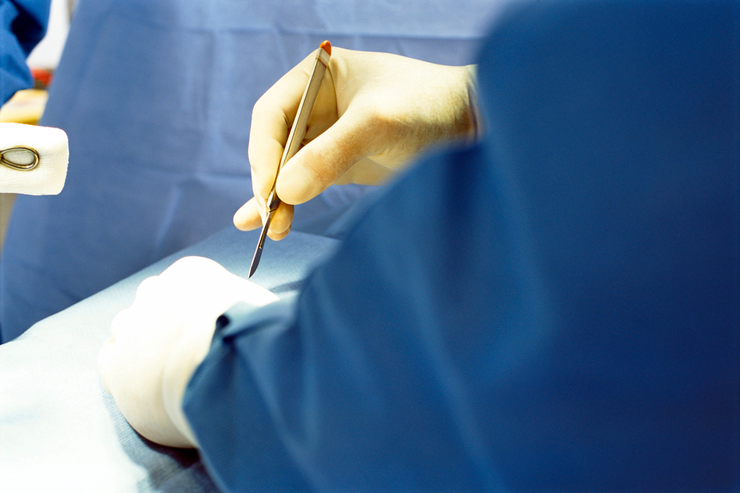
मेंदूच्या शस्त्रक्रियेसाठी ओटी तयार करणे म्हणजे फक्त दिवे लावणे आणि एअर कंडिशनर चालू करणे असे नसते. त्यासाठी शेकडो निर्जंतुकीकरण केलेली साधने हवीत, औषधे हवीत, भूल देणारी ट्रॉली (एनेस्थेशियाची ट्रॉली) हवी, तसेच कुशल शल्यचिकित्सक, स्क्रब नर्स, ओटी तंत्रज्ञ, ओटी रनर्स इत्यादी कर्मचारी तयार हवेत. प्रत्येक व्यक्तीने ओटी स्क्रब सूट घालणे आवश्यक असते. सर्जन आणि स्क्रब नर्स त्यांच्या स्क्रब सूटवर निर्जंतुकीकरण केलेले गाऊन घालतात. शेवटी ते कॅप, मास्क आणि सर्वात शेवटी निर्जंतुकीकरण केलेले हातमोजे घालतात.

ऑपरेसन करण्यास संमती

शस्त्रक्रिया धोकादायक असल्याचे सर्जनने स्पष्ट केले. सर्जरी यशस्वी झाल्यास बाळाचे प्राण वाचले असते. 

“तो पूर्णपणे नॉर्मल होईल का?” राशीने रडत विचारले. सर्जनने काय धोके आहेत हे स्पष्ट केले. पालकांनी संमती दिली आणि भूलतज्ञांनी आपले काम सुरू केले.

लहान बाळावर डीकंप्रेशन शस्त्रक्रिया करण्यात आली. साठलेल्या रक्ताचा निचरा काढण्यात आला. मेंदूवरील दाब कमी झाला. मेंदू एका बाजला ढकलला गेला होता तो त्याच्या जागी आला.

शस्त्रक्रियेनंतर सुजय बराच वेळ तंद्रीत होता. मग हळूहळू त्याने तोंड उघडले. सर्वप्रथम त्याने डोक्यावरील पट्टी काढण्याचा प्रयत्न केला. मग तो ओरडला.

पुढच्या 24 तासात सुजय स्तनपान करत होता. त्याचे इंट्राव्हेनस लिक्विड्स बंद झाले.

शस्त्रक्रियेनंतर दुसऱ्या दिवशी तो हॉस्पिटलच्या कॉटवर ताठ बसला होता आणि डॉक्टरांकडे पाहून हसत होता.

चार दिवसांत त्यांला डिस्चार्ज देण्यात आला. आठवडाभरानंतर त्याचे टाके काढण्यात आले. तो पूर्णपणे बरा होईल आणि कोणत्याही कायमस्वरूपी मेंदूच्या विकृतीशिवाय सामान्यपणे वाढेल अशी अपेक्षा आहे.

विश्लेषण

लहान मुले अनेकदा पडतात, परंतु बहुतेक वेळा ती बरे होतात. प्रौढांपेक्षा लहान मुलांवर शस्त्रक्रिया करणे अधिक कठीण असते. त्यासाठी अधिक कौशल्य आणि विशाल अनुभवाची आवश्यकता असते. बाळाच्या डोक्याला झालेल्या दुखापतीवर उपचार करणे अतिशय क्लिष्ट असते. तथापि, समर्थ न्यूरो हॉस्पिटलमधील वैद्यकीय आणि पॅरामेडिकल व्यावसायिकांच्या अनुभवी टीमने कौशल्य आणि आत्मविश्वासाने सर्जरी केली आणि त्याचे परिणाम उत्तम आले.

अपघाताच्या दुसर्‍या दिवशी जर सुजयला आमच्याकडे आणले असते तर खूप उशीर झाला असता आणि शस्त्रक्रियेचा निकाल इतका आला नसता.

Head Injury

Head Injury

By Dr. Ravindra Patil

Head Injury – Traumatic Brain Injury

Much has been written about the golden hour in treating grievous head injuries. The earlier the treatment is begun, the better the outcome. But in real life, the scenario is totally different. People do not rush to the hospital, they wait and take opinions, and they hope things will become better.

I share a real case that we treated at Samarth Neuro Hospital. The patient survived without any neurological deficit, but just!

But let us first see what happens when there is a violent impact on the head.

Table of Contents

Results of head injury

Any impact on the head is a head injury. A head injury causes many sudden changes in and around our brain.

The first cause is the direct injury and any part of the brain that might sustain the impact and be injured.

The second cause is the contrapuntal [or Contrecoup] brain injury. These injuries affect the side of the brain opposite of where the impact of the head injury occurred. They are often overlooked or misdiagnosed because of their location on the opposite side of the brain. Contrecoup occurs when a force or blow causes the brain to strike the side of the skull opposite from the point of impact. This happens because the brain is floating inside the skull and its covers membranes in the cerebrospinal fluid.

As a result of head injuries, blood vessels of the brain may be ruptured. They bleed. As the skull and the brain within are tightly enclosed, any bleeding increases the pressure around the blood. A collection of blood in the brain [called a haematoma] may press vital areas of the brain and cause functional disorders which are reversible once the blood collection or haematoma is drained.

Any injury increases the pressure of the liquid around the brain [this liquid is called the cerebrospinal fluid or CSF]. This liquid usually helps dampen shocks to the head. But if the CSF pressure increases, it is dangerous for the brain, which is made of very soft and delicate nerve fibers and fat. Symptoms of increased CSF pressure are headache, nausea, vomiting and severe discomfort. The pupils may not react to light. High pressure may rarely push a part of the brain into the canal of the spinal column. This is called ‘herniation’.

A real life scenario

All names in this real story have been changed.

10 month old baby boy Sujay was playing peacefully in his crib. His loving seven year old sister Saumya picked him up and Sujay laughed happily. Just then the phone rang. Saumya wanted to take the call and hand over the phone to her mother because she liked to answer phones. So she rushed to the phone. She held Sujay in one arm and climbed a three feet high bed to reach the top of the cupboard to pick up the phone. During that childish attempt, she lost her grip on baby Sujay and the poor fellow slipped out of her arm fell on the ground with a bang. The height from which Sujay fell was estimated to be five feet. Saumya shrieked in panic. Their mother rushed in and was about to scold Saumya for climbing on the bed when Saumya pointed to Sujay lying on the floor.

The baby was motionless.

Saumya and Sujal’s mother Rashi’s first reaction was that her lovely baby had died!

She collapsed to the ground next to her baby in shock, a silent spectator to her baby’s death. Saumya kept on screaming and her screaming brought their neighbours to their house.

They lived in a small town near Sangli. It was getting dark. The neighbours crowded to see the baby lying on the floor motionless. Then one lady gently picked up the baby and held it in her lap. The lady held small cotton fibres in front of the baby’s nose. The fibres moved. So the baby was breathing and alive! Thank God!

Rashi was still in a shock. So the neighbours sent someone to call in their family doctor. The doctor was busy with his patients, yet he came to see baby Sujay after half an hour.

Note how time delays occur in real life scenarios. When the doctor actually examined the baby, one hour had passed.

The doctor gently examined Sujal’s head and found that there was a depression on the left side of the baby’s head. Also, the fontanelle was bulging outwards and upwards.

Fontanelles

The fontanelles are the soft spots on an infant’s head where the bony plates that make up the skull have not yet fused together. It is normal for infants to have these soft spots, which can be seen and felt on the top and back of the head. There are two fontanelles. The fontanelle near the back of the head usually closes by age 1 or 2 months. While the fontanelle on the top of the head usually closes sometime between 9 months and 18 months. The fontanelles help the skull expand as the brain grows and develops. If the intracranial CSF pressure increases, the fontanelles bulge out. That is what had happened in Sujay’s case.

In adult patients there are no such fontanelles and nothing bulges out.

The doctor guessed that it was a serious condition. He talked to Sujay’s father on phone and advised that Sujay be taken to a brain specialist immediately. Sujay needed a brain CT scan and perhaps surgery.

The doctor did not administer any medicine, saying it could be dangerous.

Now the family was in a dilemma. What were they to do?

Was the doctor right? Did the baby really require immediate surgery? Would the swelling on the head go away on its own? How could such a small baby be operated? And how could they take the baby to the hospital so late in the evening? By then it was 9 PM.

Rashi had recovered her wits. She gently took Sujay in her lap and talked to him. He responded by crying. Till then he was not responding. He only breathed.

Rashi tried feeding the baby to stop its crying, but the baby did not feed. Instead, it vomited. Curd like milk came out in its vomit.

The family contacted their relatives in Pune and Mumbai. Someone suggested immediate transfer to Mumbai or Pune. Others said it would take too much time.

Samarth Hospital

“What about that hospital on Sangli Miraj road?” Someone asked.

“It must be closed…”

“But it is open for 24 hours…”

Finally against Rashi’s wishes and with not much hopes that the baby would survive, Sujay was brought to Samarth hospital.

Rapid response

The Emergency Room [ER] staff immediately called a neurosurgeon and a paediatrician. Till the specialists came, the nurses and duty doctors had examined the patient’s vital signs and reported them to the surgeon, who immediately ordered a CT scan.

Sujay was sedated so that he would lie still. Then the CT scan was done.

The surgeon and paediatrician checked the CT scan. A huge subdural haematoma was seen on the CT scan.

“Requires immediate decompression as there is a midline shift,” the surgeon said.

“But let me first do pre-operative examination and let us make the baby NBM.” The paediatrician said. NBM in doctors’ slang is ‘Nil by mouth’. Prior to anaesthesia, all patients are made NBM.

It was 11 PM. The baby had to remain NBM for at least six hours before anaesthesia could be given.

The surgeon ordered the operation theatre [OT] staff to be ready at 4 PM.

OT preparation

Preparing the OT for brain surgery does not mean just switching on the lights and the air conditioner. It requires hundreds of sterilised instruments, medicines, anaesthesia trolley, skilled surgeons, scrub nurses, OT technicians, OT runners and so on. Every person must wear OT scrub suits. The surgeons and scrub nurses wear sterilised gowns over their scrub suits. Finally they wear caps, masks and lastly the sterile gloves.

OT consent

The surgeon explained that the surgery was dangerous. However if successful the life of the baby would be saved.

“Will he be completely normal?” Rashi asked, sobbing. The surgeon explained what the risks were. The parents gave their consent and the anaesthetist began his work.

Decompression surgery was done on the little baby. The haematoma was drained. The pressure on the brain was released.

After surgery, Sujay was drowsy for a long time. Then gradually he opened his mouth. The first thing he tried was to pull off the bandage on his head. Then he cried.

In the next 24 hours Sujay was breast feeding. His intravenous liquids were stopped.

On the second day after surgery he was sitting upright on the hospital cot and smiling at the doctors.

He was discharged in a four days. His sutures were removed after a week. He is expected to heal completely and grow normally without any permanent brain deformity.

Analysis

Babies fall often, but mostly they recover. Surgeries on babies are more difficult than adults. They require more skills and experience. Treating head injuries in a babies is even more complicated. However, the team of medical and paramedical professionals at Samarth Neuro Hospital manage such cases with skill, confidence and good outcomes.

If Sujay had been brought to us the next day, it would have been too late and the outcome of surgery may not have been so good.

शस्त्रक्रियां मध्ये अचूकता

शस्त्रक्रियां मध्ये अचूकता

By Dr. Ravindra Patil

गुंतागुंतीच्या शस्त्रक्रियांमध्ये इतकी प्रगती झाली आहे की आता मानवी डोळे आणि हात यापुढे जटील ऑपरेशने करण्यास पुरेसे पडत नाहीत! अत्याधुनिक बायोमेडिकल उपकरणे सर्जनला ऑपरेशन्स करण्यास मदत करतात जे अन्यथा शक्य झाले नसते.

मानवी केसांपेक्षा पातळ धाग्याने टाके घेतल्याचे तुम्ही ऐकले असेल. इतक्या पातळ धाग्याने जखमेची शिलाई करता येण्यासाठी कौशल्य आणि सर्जिकल लूप आवश्यक असतात [दृश्य अनेक वेळा मोठे करणाऱ्या उपकरणांसारखे चष्मा, त्याला सर्जिकल लूप म्हणतात]. ऑपरेटिंग मायक्रोस्कोप सर्जनना 1 – 2 मिमी इतक्या लहान अवयवांवर किंवा ट्यूमरवर कार्य करण्यास मदत करतात. एंडोस्कोप सर्जनना नाकातून पिट्यूटरी ग्रंथीसारख्या लहान परंतु अत्यंत महत्त्वाच्या अवयवांपर्यंत पोहोचण्यास मदत करतात!

पण ते पुरेसे नाही. आता गुंतागुंतीच्या मेंदू आणि मणक्याच्या शस्त्रक्रियांमध्ये ट्यूमर किंवा रोगग्रस्त भागापर्यंत अचूकपणे पोहोचण्यासाठी आणि त्यांना काढून टाकण्यासाठी त्वचेवर अचूक चीरे आणि हाडांवर अचूक ठिकाणी ड्रिल करण्याची आवश्यकता असते.

कल्पना करा की 3 मिमीचा कर्करोग नसलेला (कॅंसर नसलेला) मेंदूत [किंवा मणक्यात] एक ट्यूमर आहे. तो ट्यूमर मेंदूचा भाषण नियंत्रित करणारा एक छोटासा भाग. त्यामुळे रुग्णाचे बोलणे हळूहळू बिघडते. ट्यूमर कॅन्सररहित असल्याने वरील गाठ दूरच्या भागात पसरण्याची भीती नसते. परंतु कर्करोग नसलेल्या ट्यूमर देखील वाढतच राहतील आणि मेंदूवर [किंवा पाठीचा कण्यावर] आणखी दबाव टाकत जाईल आणि रुग्णाच्या मेंदूची कार्यक्षमता अधिकाधिक कमी होऊ लागेल.

ट्यूमर काढून टाकणे हा नक्कीच सर्वोत्तम उपाय आहे. पण ते इतके सोपे नाही. मेंदू आणि मणक्याच्या शस्त्रक्रियांमध्ये सर्जन हाडे आणि अत्यंत कठीण अस्थिबंधन कापल्यानंतरच शस्त्रक्रियेच्या क्षेत्राकडे जाऊ शकतात. आणि लक्षात ठेवा, ऑपरेशनचा घाव शक्य तितका लहान असणे आवश्यक आहे. कवटीच्या किंवा कशेरुकामध्ये चीरा किंवा कट जास्त मोठा दिला तर सभोवतालच्या निरोगी भागांना हानी पोहोचण्याची आणि शस्त्रक्रियेच्या ठिकाणी संसर्ग होण्याची शक्यता जास्त असते.

म्हणून कमीतकमी आकाराचा चीरा देणे केवळ चांगले नाही तर अत्यंत आवश्यक असते!

नाजूक नसा आणि मेंदू किंवा पाठीच्या कण्यातील ऊतींचे संरक्षण करणारी हाडे कठीण असतात. अशी हाडे कापण्यासाठी कठोर धातुचे बनवलेले ड्रिल आणि आरी आवश्यक असते. ही उपकरणे प्रति मिनिट शेकडो किंवा हजारो वेळा फिरतात आणि त्यांचा काप किंवी त्यांनी कवटीला पाडलेले भोक.

जर ही उपकरणे मेंदूच्या ऊतींच्या किंवा मणक्याच्या ऊतींच्या संपर्कात आली तर अशा आरी किंवा ड्रिलमुळे नाजूक मऊ ऊतकांवर किती विनाश होईल याची कल्पना करा.

त्यामुळे मेंदू आणि मणक्याच्या शस्त्रक्रियेमध्ये अचूकतेला अनन्यसाधारण महत्त्व आहे.

येथे कामी येते सर्जिकल नेव्हिगेशन सिस्टम.

Table of Contents

गुगल नकाशे आणि मोटार गाडी चालवणे

अलीकडच्या काळातील सर्वात मोठे वरदान म्हणजे गुगल किंवा इतर नेव्हिगेशन सिस्टमसह कार चालवणे. ते आपल्याला ट्रॅफिक जॅमबद्दल देखील सांगतात, जेणेकरून आपण ते रस्ते टाळू शकतो.

गुगल नकाशे तुमची स्थिती ओळखण्यासाठी उपग्रह आणि भौगोलिक स्थान चिन्हकांवर अवलंबून असतात आणि त्यानूसार तुम्हाला तुमच्या गंतव्यस्थानावर पोहोचण्यास मार्गदर्शन करतात.

लोकं कारमधील गुगल नेव्हिगेशन वापरण्यावर अशी टीका करतात की त्यांना रस्ते इतके चांगले माहित असतात की त्यांना अशा फॅन्सी गॅजेट्सची जरूर नसते. असा लोकांचे म्हणणं बरोबर आहे. पण हे कबूल करायलाच पाहिजे की गुगल नकाशे हे अनोळखी मार्गांवरून चालण्यासाठी अतिशय उपयुक्त असतात. तसेच, रात्रीच्या काळोखात, गुगल नॅव्हिगेटर जवळ येणारी वळणे आणि चौक दाखवतो. दिवसाच्या प्रकाशात हे सर्व स्वच्छ दिसते, पण रात्री एवढे छान दिसत नाही आणि त्यामुळे गुगल नॅव्हिगेटरचा निश्चितच उपयोग व फायदा होतो.

मेंदू आणि मणक्याच्या शस्त्रक्रियेमध्ये देखील गुगल नेव्हिगेशन वापरण्यात होतात तसेच सर्व फायदे आणि तोटे लागू होतात.

नेव्हिगेशन असिस्टेड सर्जिकल तंत्रात देखील Google सारखेच नेव्हिगेशन मार्कर असतात. जसे गुगल मार्कर भौगोलिक स्थानांवर असताना, तसे सर्जिकल नेव्हिगेशन मार्कर शस्त्रक्रियेपूर्वी त्वचेवरील जोडणयात येतात. त्याद्वारे मेंदू आणि मणक्याचे सर्जन अचूक ठिकाणी त्वचे वर चीरा देऊ शकतात, हाडांमधून छिद्र करू शकतात हाडे किंवा कापू शकतात आणि लहान उपकरणे किंवा ड्रिल किंवा स्क्रू घालू शकतात. तसेच हाडांमध्ये अचूक बिंदूंवर के-वायर्स घुसवू शकतात. या अति प्रगत शस्त्रक्रियेच्या प्रणालीमुळे आसपासच्या निरोगी ऊतींना कमीत कमी दुखापत होते.

कुशल आणि अनुभवी मेंदू आणि मणक्याचे सर्जन सुरुवातीला सर्जिकल नेव्हिगेशन पध्धतीवर विश्वास ठेवत नव्हते. परंतु जेव्हा त्यांना सर्जिकल नेव्हिगेशनने मिळू शकणारी मिलिमीटरची अचूकता लक्षात आली तेव्हा सर्वांचे मत मेंदूच्या शस्त्रक्रियेसाठी नेव्हिगेशनच्या पक्षात होत गेले. अधिकाधिक सर्जनांचा कल सर्जिकल नेव्हिगेशन कडे आहे.

मार्कर

रुग्णाचा सीटी स्कॅन के ला जातो जो निदान आणि शस्त्रक्रिया प्रणालीचा एक भाग आहे. ट्यूमरच्या ठिकाणी मार्कर जोडलेले असतात. अत्यंत प्रगत इमेजींग द्वारे सर्जन ट्यूमरच्या संबंधात knife, ड्रिल किंवा स्क्रूचे स्थान  थ्री-डी पाहू शकतात. अशा प्रकारे सुसज्ज सर्जन अचूक ठिकाणी सर्जरी करू शकतो किंवा एखादे हाड स्थीर करण्यासाठी ड्रिल, स्क्रू, खिळे किंवा वायर घालू शकतो.

याशिवाय, सर्जिकल नेव्हिगेशन सिस्टमच्या फ्लॅट स्क्रीनवर न्यूरोसर्जनच्या आवश्यकतेनुसार ऑपरेटिंग फील्डची त्रिमितीय (थ्री-डी) दृश्ये पाहिली जाऊ शकतात.

अशा प्रकारे सर्व स्पष्ट पणे दिसणे ही सर्जरीच्या क्षेत्रात एक महाक्रांती आहे. कारण जुन्या प्रकारच्या शस्त्रक्रियेमध्ये सर्जनला दिसणारे अवयवांचे दृश्य लहान चीरांमुळे खूप संकुचीत आणि मर्यादित असते.

थ्री-डी

कागदावर चौरस किंवा वर्तुळ काढण्याचा प्रयत्न करा. खूपच सोपे असते! आता कागदाच्या उलट्या बाजूला न पाहता तेच काढण्याचा प्रयत्न करा. थोडे कठीण, पण शक्य आहे.

आता एक ग्लास घ्या आणि न पाहता त्याच्या आत तळाशी लिहिण्याचा प्रयत्न करा. तुम्हाला एक लांब पेन लागेल. तुम्ही ओरखाडे करू शकाल परंतु अचूक लिहू शकणार नाही.

मणक्याची आणि मेंदूची शस्त्रक्रिया अतिशय क्लिष्ट आणि त्रिमितीय (थ्री-डी) आहे. ऑपरेट करायच्या भागाकडे सरळ पाहता येत नाही. अशा परिस्थितीत, सर्जिकल नेव्हिगेशन सिस्टीम फक्त ‘असली तर बरे’ नसतात, त्या ‘असल्याच पाहिजेत’ अशा असतात.

जे मेंदूला लागू होते ते पाठीच्या कण्यालाही लागू होते. मेंदूभोवती कवटी आणि आत ड्युरामेटर नावाचे कठीण आवरण असते. पाठीचा कणा कशेरूक आणि ड्युरामेटरने सुरक्षितपणे झाकलेला असतो. कवटी आणि मेंदूच्या शस्त्रक्रिये इतकेच पाठीच्या कण्याच्या शस्त्रक्रिया देखील सुरक्षितपणे करणे अति कठीण असते.

नेव्हिगेशन असिस्टेड स्पाइन सर्जरी हा तंत्रज्ञानाचा एक समूह आहे, जो ऑपरेशन दरम्यान सर्जनला रीअल-टाइममध्ये (त्याच वेळी), शस्त्रक्रियेच्या साधनांच्या संबंधात मणक्याच्या त्रिमितीय आणि आभासी प्रतिमांमध्ये प्रवेश करू देते. अचूकतेमुळे शस्त्रक्रियेचे परिणाम जास्त प्रमाणत सफळ होतात.

मणक्याशी संबंधित वेदनांमध्ये ‘प्रेशर पॉइंट’ म्हणजे चिमटीत अडकलेले मज्जातंतु (नर्व) व त्यामुळे होणार्‍या तीव्र वेदना हे होण्याचे कारण असते ‘स्लिप डिस्क’.कशेरुकाच्या स्तंभाच्या संरचनेत बदल झाल्यामुळे जेव्हा मज्जातंतु पाठीच्या कण्यातून बाहेर पडतात आणि कशेरुकी स्तंभांमधील लहान छिद्रांमधून जातात तिथे त्या सरकलेल्या गादीमुळे चिमटतात.

एकंदरीत, संगणक नेव्हिगेशन रीढ़ आणि मेंदूच्या रोगग्रस्त भागापर्यंत अगदी अचूकपणे पोहोचणे आणि ट्यूमर काढून टाकणे किंवा दाब किंवा आकुंचन अगदी अचूकपणे दुरुस्त करण्यात मदत करते.नमूद केल्याप्रमाणे, केवळ काही मिलीमीटर रुंद असलेल्या गाठी सुध्धा अशा प्रकारे काढल्या जाऊ शकतात.

नॅव्हिगेशनशिवाय एक्साइज केल्यास हेच ट्यूमर एक मोठी समस्या होतात. नॅव्हिगेशनशिवाय न्यूरोसर्जनने ऑपरेशन केल्यास लहान ट्यूमरच्या सभोवतालच्या निरोगी मेंदूच्या ऊती काढून टाकल्या जाण्याची शक्यता असते. त्यामुळे शस्त्रक्रियेनंतर कार्यक्षमतेमध्ये नक्कीच नुकसान होऊ शकते. शस्त्रक्रियेपूर्वी जी कार्ये करण्यात पेशंट पूर्णपणे समर्थ होता ती दैनंदिन कार्ये करण्याची क्षमता चुकीच्या शस्त्रक्रियेमुळे कमी होऊ शकते. अशाप्रकारे रुग्णाची गाठ काढून टाकली जात असली तरी, त्याच्या निरोगी मेंदूच्या ऊतींचा एक छोटासा भाग देखील जातो! न्यूरोसर्जरी अत्यंत अचूकतेने न केल्यास हीच किंमत मोजावी लागते.

सांगली मिरजेत प्रथम

सांगली-मिरजेतील समर्थ न्यूरो हॉस्पिटल हे मेंदू आणि मणक्याच्या शस्त्रक्रियांसाठी नेव्हिगेशन सिस्टिम असलेले या भागातील पहिले हॉस्पिटल आहे. हॉस्पीटलमध्येच सीटी स्कॅन असल्यामुळे, समर्थ न्यूरो हॉस्पिटलमध्ये जवळजवळ सर्व गुंतागुंतीच्या मेंदू आणि मणक्याच्या शस्त्रक्रिया सुरक्षितपणे केल्या जाऊ शकतात.

मेंदू आणि मणक्याच्या शस्त्रक्रियांमध्ये सुरक्षिततेबद्दल खूप चिंता असायची. पण आता अशा अचूक डायग्नोस्टिक मार्किंग सिस्टीममुळे मेंदू किंवा मणक्याच्या शस्त्रक्रियेची गरज असल्यास तुम्हाला पुण्य़ाला किंवा मुंबईला जाण्याची गरज नाही, सांगली-मिरजेतील समर्थ न्यूरो हॉस्पिटलमध्ये अशा शस्त्रक्रिया होऊ शकतात!

Accuracy in Surgery

Accuracy in Surgery

By Dr. Ravindra Patil

With the advancement in complex surgeries, the human eyes and hands are no longer enough! Complex biomedical equipment helps surgeons conduct operations which would not have been possible otherwise.

You may have heard of stitches being taken with thread thinner than the human hair. To be able to knot a stitch with thread so thin requires skill and surgical loupes [spectacle like devices which magnify the view many times]. Operating Microscopes help surgeons operate on organs or tumours as small as 1 – 2 mm. Endoscopes help surgeons reach small but extremely vital organs like the pituitary gland through the nose!

But that is not enough. Now complex brain and spine surgeries require precise incisions on the skin and drills on the bones to reach the tumour or diseased portion accurately and remove it.

Imagine a 3 mm non-cancerous brain [or spine] tumour is pressing a small part of the brain which controls speech. So, the patient’s speech gradually deteriorates. As the tumour is non-cancerous there is no fear of its spreading to distant parts. But even non-cancerous tumours will keep on growing and putting further pressure on the brain [or spinal cord] and the patient will suffer more and more loss of function.

The obvious solution is to remove the tumour. But it is not so simple. In brain and spine surgeries, the surgeons can approach the area to be operated on only after cutting through bones and very tough ligaments. And remember, the opening must be as small as possible. Larger the incision or cut in the skull or vertebrae, the higher the chance of damaging surrounding healthy parts and the incidence of surgical site infection.

So minimal cutting is not only better but essential!

The bones which protect the delicate nerves and brain or spinal cord tissue within are tough. Cutting such bones requires tough wear-resistant drills and saws. These drills or saws rotate at hundreds or thousands of revolutions per minute.

Imagine how much devastation such saws or drills will cause on delicate soft tissue if these instruments come in contact with brain tissue or spine tissue.

Hence, accuracy in surgery of brain and spine is of the greatest priority.

Here is where surgical navigation systems come in.


Table of Contents

Google Maps and driving a car

The greatest boon of recent times is driving a car with Google or other navigation systems. They even tell us about a traffic jams, so that we can avoid those roads.

Google maps rely on satellites and geographical location markers to identify your position and then guide you to your destination.

People also do criticise using google navigation in cars saying that they know the roads so well that they do not need such fancy gadgets. These people are right. But whatever they say, google maps are very, very useful for driving over unexplored routes. Also, in pitch darkness at night, the navigator shows oncoming turns and crossroads which could have been seen during daylight hours and can be unexpected in the night.

All the advantages and disadvantages apply to using the navigation in brain and spine surgery.

Navigation assisted surgical techniques also have navigation markers similar to Google. While Google markers are located in geographic locations, surgical navigation markers are attached just prior to surgery to spots on the skin through which the brain and spine surgeons can incise the skin, drill through or cut open bones, and insert small instruments or drills or screws or K Wires or such to pierce the bone at the precise points and with minimal injury to surrounding healthy tissue.

Skilled and experienced brain and spine surgeons were critical of surgical navigation in the beginning. But when they all realised the millimetre sharp accuracy achievable with Surgical Navigation, the general consensus opinion is more and more in favour of using navigation for brain surgeries.

Markers

The patient undergoes a CT scan which is a part of the diagnostic and surgical system. Markers are attached at the location of the tumour. The surgical instruments are also marked and visible in the highly advanced image intensifying television system so that the surgeon can see the location of his knife, drill or screw in relation to the tumour [marked by the special markers] in three dimensions. Thus equipped, the surgeon can then make an incision and put in a drill, screw, nail or a wire to fix a vertebra or a bone.

Besides, three-dimensional views of the operating field can be seen as per the neurosurgeon’s requirements, on the flat screen viewer of the surgical navigation system.

Getting such clear views is a revolution because, in open surgery, the view that a surgeon gets is very limited and restricted because of small incisions.

3-D

Try to draw a square or a circle on paper. So easy! Now try to draw the same on the reverse side of the paper without looking at it. Slightly difficult, but possible.

Now take a glass and try to write inside it on the bottom without looking. You will need a long pen and you may be able to scratch but not write accurately.

Spine and brain surgery is far more complex and in three dimensions. There is no straightforward approach to the part to be operated. In such situations, surgical navigation systems are not just ‘GOOD TO HAVE’, they are ‘MUST HAVE’.

What applies to the brain also applies to the Spinal Cord. The brain has hard covers over it called the skull and dura mater. The spinal cord is covered and protected by the vertebrae and the extension of the dura mater. Compared to the skull and the brain, the spinal cord is perhaps equally difficult to approach safely and operate.

Navigation assisted spine surgery is a group of technologies, which allow the surgeon to access real-time, three-dimensional and virtual images of the spine in relation to the surgical instruments during the operation. Again, accuracy in approaching the ‘pressure point’ of nerves or the spinal cord leads to predictable and safer surgical outcomes.

‘Pressure point’ in spine related pain means the pain because of pinched nerves. This happens because ‘slipped discs’ or change in the structure of the vertebral column presses nerves when they emerge from the spinal cord and pass through small holes in the vertebral columns through which nerves come out.

All in all, the fundamental idea in computer navigation assisted spine and brain surgery is to reach the diseased portion very precisely and remove the tumour or correct the pressure or constriction very accurately. As has been mentioned, tumours which are only a few mm wide can be thus excised.

These same tumours would be a big problem if excised without navigation. The neurosurgeon would in all likelihood have removed healthy brain tissue surrounding the small tumours and that would have led to functional damage after surgery. Those functions which were perfectly normal before surgery might get affected because of inaccurate surgery. Thus although the patient’s tumour would be removed, a small part of his healthy brain tissue would also go! That is the price which has to be paid if neurosurgery is not done with extreme accuracy.

First in Sangli Miraj

Samarth Neuro Hospital of Sangli-Miraj is the first hospital in the area to have a navigation system for brain and spine surgeries. With their in house CT scan and cath lab, almost all complex brain and spine surgeries can be done safely in Samarth Neuro Hospital.

There used to be a lot of concern about safety in brain and spine surgeries. But now with such accurate diagnostic marking systems, you don’t have to go to Pune and Mumbai in case you need brain or spine surgery, Samarth Neuro Hospital in Sangli-Miraj has it all!