Spine Fusion Surgery Cost in India

spinal fusion surgery cost

Spine Fusion Surgery Cost in India

By Dr.Ravindra Patil

Any type of spine surgery is thought to be a very expensive surgery. It is true to some extent as spinal fusion surgery cost is high because it needs very highly skilled surgeons, state of the art operation theatres and above all, surgical navigation for accuracy. But all these give fairly good results in patients who could not be operated at all a decade ago, because the skillset and surgical navigation were not available then. So now although back surgery cost is high, results are better.

Table of Contents

Cost of Spinal Fusion Surgery

The average Spinal Fusion Surgery cost is very high in western countries.

India is known worldwide for its advanced medical facilities and promising technology for Low Cost of Spinal Fusion Surgery. Many of the best hospitals for Spinal Fusion Surgery can be found in India.

Spinal Fusion Surgery cost in India is much less when compared to any other countries. Also the cost of Spinal Fusion Surgery in India is substantially lower than other developed countries of the world.

Private back surgery cost is higher because Private back surgery cost means treatment in a private hospital

Various deciding factors could determine the price of Spinal Fusion Surgery in India. These can be broadly classified as Hospital, Medical Team or Patient Dependent factors.

Hospital Factors

Type of the hospital (Government/Trust/Private).

Use of insurance, type of insurance or self paid.

Accreditation of the facility

Reputation and brand value of the hospital.

Medical Team Factors

Technology / Approach Used

Surgery Type

Type of Anesthesia or Sedation

Qualification / Expertise of the specialist

Extent of the surgery needed

Patient Factors

Patient’s diagnosis

Patient’s general health

Room Category selected by the patient

Other treatment required by the patient in conjunction

What is the definition of spinal fusion?

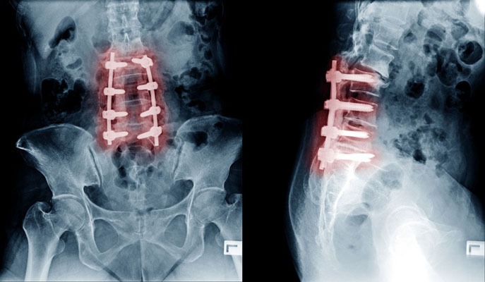
Spinal fusion is a surgical procedure wherein one or more vertebrae are united to eliminate any motion between them. The approach is similar to welding. Spinal Fusion Surgery, only does not weld the vertebrae immediately during surgery. The bone grafts are placed around the spine during surgery. The body then heals the grafts over several months.

When are the indications of spinal fusion surgery?

There are several potential reasons to consider spinal fusion. These include treatment of a fractured vertebra, rectifying deformity, elimination of pain caused during motion, treatment of instability, and treatment of cervical disc herniations.

Not all spinal fractures need surgery, some fractures, particularly those associated with spinal cord or nerve injury, generally require fusion as part of the surgical treatment.

Another condition treated is instability, i.e. the abnormal or excessive motion between two or more vertebrae. It is commonly believed that instability can be a source of back or neck pain or cause potential irritation or damage to close by nerves.

Cervical disc herniations that require surgery usually need fusion as well. In this technique, the disc is removed by an incision in the front of the neck and a small piece of bone is inserted in its place.

How is the Spinal Fusion Surgery performed?

There are many surgical methods available to fuse the spine that involve placement of a bone graft between the vertebrae. The spine may be treated from the back (posterior), from the front (anterior) or by a combination of both. The anterior approach is more common. The ultimate objective of fusion is to obtain a solid union between two or more vertebrae.

A fusion may or may not involve use of supplemental tools like plates, screws and cages. Instrumentation is sometimes used to rectify a deformity, but is usually used to hold the vertebrae together to while the bone grafts heal. Whether the hardware is used or not, it is important that bone or bone substitutes be used to get the vertebrae to fuse together.

The bone may be taken either from another bone in the patient or from a bone bank. Fusion using bone from the patient has a long history of use and results in predictable healing. Smoking, medications you are taking for other conditions and your overall health can affect the rate of healing and fusion, too.

How long does it take to recover from spinal fusion surgery?

Immediate discomfort following a spinal fusion is generally greater than with other types of surgeries. Fortunately, there are excellent methods of postoperative pain control available, including pain medications and intravenous injections.

Another option is a patient-controlled postoperative pain control pump. Here the patient presses a button that delivers a predetermined amount of narcotic pain medication through an intravenous line.

Patients generally stay in the hospital for three to four days, but a longer stay after more extensive surgery is also possible. A short stay in a rehabilitation centre after discharge from the hospital is recommended for patients who underwent extensive surgery, or for elderly or debilitated patients.

The fusion process varies in each patient. The healing process after fusion surgery is similar to that after a skeletal fracture. During this time, the patient’s activity is generally restricted. Substantial bone healing does not usually take place until three or four months after surgery.

Many a times spinal decompression surgery cost and healing time is compared with spinal fusion surgery cost. It must be understood that both are far different. Spinal decompression surgery does not need bone fusion and healing like spinal fusion surgery.

How many patients underwent Spinal Fusion Surgery in India in the last 5 years?

Private back surgery prices will vary from place to place. Scoliosis surgery cost is also of interest to many patients who have suffered kyphosis or scoliosis. Cost of spinal decompression surgery is also queried.

In recent years, India has emerged as a medical hub for patients looking for affordable, accessible and efficient low cost Spinal Fusion Surgery Treatment. Some of the best Spinal Fusion Surgery hospitals in the world are found in India. The country is known for offering advanced medical facilities at the most reasonable cost. An average increase of 15 to 20 percent annually has been observed in the number of patients in the last 5 years. The Indian Spinal Fusion Surgery hospitals deliver advanced health care and highest quality services backed by elaborate infrastructure and lower treatment cost

Here are the approximate figures of the patients underwent Spinal Fusion Surgery in the last 5 years in India

Year

2013

2014

2015

2016

2017

Surgeries

4800

6000

7500

9600

10800

What are our special services offered to international patients?

At Samarth Neuro and Super Speciality Hospital we help arranging your treatment in a way that will suit you and be affordable for you. Chief neurosurgeon at Samarth Neuro and Super Speciality Hospital Dr Ravindra Patil is vastly experienced in all types of spine and brain surgeries. Spine fusion surgeries are done there as required.

Congenital Brain and Spine Anomalies

congenital brain and spine malformations

Congenital Brain and Spine Anomalies

By Dr. Ravindra Patil

Table of Contents

What are congenital brain and spine malformations?

Congenital means present at birth.

These conditions may be mild and without symptoms or may be serious, requiring treatment. In some cases, surgery may be recommended to:

  1. Address the child’s or the patient’s symptoms.
  2. Correct the form and function of the brain and spine structures.
  3. Maximize cognitive and motor
  4. Prevent development of neurological deficits.

Congenital abnormalities, called malformations, are conditions affecting the form and function of the nervous system. There are numerous variations of congenital malformations of the bone and soft tissue of the head and spine, including neural tube defects, such as spina bifida, encephaloceles, Chiari malformations and arachnoid cysts.

Some congenital malformations are mild, and some are severe but correctable with surgery by a pediatric neurosurgeon.

Types of some of the Congenital Brain and Spine Malformations are as follows:

Chiari Malformations

This is a condition in which portions of the brain, called the cerebellar tonsils, protrude through the bottom opening of the skull into the upper spine, which can put pressure on the brain or spinal cord. Chiari malformations may block the flow of cerebrospinal fluid, leading to hydrocephalus.

Treatments often focus on removing portions of the bone and soft tissue to relieve pressure on the spinal cord and brain, as well as providing new pathways to drain cerebrospinal fluid. Surgeons use different methods for treating these malformations, including decompression, with or without cutting open a small part of the dura mater [thick membrane covering the brain].

Encephaloceles

Encephaloceles are a type of neural tube defect characterized by the brain being exposed to the outside instead of being covered by the skull and skin. It can lead to infections and hydrocephalus.

Surgical treatment of this condition involves removing bone and soft tissue, draining cerebrospinal fluid, and surgically repairing or closing the encephalocele.

Children who have developed hydrocephalus as a result of an encephalocele will require treatment for that condition, often with a cerebrospinal fluid shunt. Shunting is the placement of a tube into the open area (ventricle) of the brain that allows cerebrospinal fluid to drain to the child’s abdomen or other location where it can be safely reabsorbed into the body.

Arachnoid Cysts

Arachnoid cysts are the most common type of brain cyst. They are congenital (present at birth) lesions that occur as a result of the splitting of the arachnoid membrane. The cysts are fluid-filled sacs, not tumors, appearing in one of the three layers of tissue covering the central nervous system.

Surgical treatment of this condition involves draining the cyst by drilling a small hole or by opening the skull and making small openings in the cyst to open the natural fluid pathways in the brain. This process is called fenestrating.

What are Spinal Deformities?

A spine deformity occurs when your spine varies by more than 10 degrees from ‘healthy’ curvature. But, what does this mean exactly?

Contrary to popular belief, your spine isn’t 100% straight and vertical. The spinal shape has curvatures, but the end result is that it is vertical! Our spine consists of a series of gentle arcs. Our lumbar spine, or lower back, swoops slightly to the back, and our thoracic spine, or upper back, bends subtly forward. The backward curve of your lower back is known as lordosis and the forward stoop that runs between our shoulder blades is known as kyphosis. Lordosis and kyphosis are spinal curvature deformities. Both are abnormal curvatures of the spine.

But, when viewed head-on, our backbone should look like a straight pillar. Hence, it is also called the ‘vertebral column’.

Moreover, the curves and straight stretches of your spine make symmetry possible. Your head sits directly over your pelvis because the lordosis of your lower back and the kyphosis of your upper spine balance each other out. If one of these curves becomes greater or lesser than the other, then problems can occur. We refer to this as sagittal imbalance, because the head and pelvis no longer fall within the same, or sagittal, plane.

Too much swaying backwards can be thought of as ‘lordosis’, and too much forward stooping in the upper back is ‘kyphosis’.

Likewise, when the spine tilts away from the midline of the body, doctors refer to this problem as coronal imbalance or scoliosis. Unevenness in the ‘coronal’ plane (the view from head-on) causes asymmetry in the trunk of the body. This can include uneven hips and shoulders or one-sided bulging of the ribs.

Diagnosis of Congenital Brain and Spine Malformations

If a child is born with any of the above malformations, a thorough evaluation by a paediatrician or neurologist is needed to diagnose the problem and recommend a plan for addressing it.

After a thorough physical and detailed family and patient history, your doctor may order imaging of the brain and/or spine through an MRI scan. If the MRI scan shows any evidence of these malformations, a neurosurgical consultation is a must to plan the best treatment.

Congenital Brain and Spine Malformations Treatment

A multidisciplinary approach is often beneficial for addressing children with congenital brain and spine malformations. Neurosurgeons, craniofacial plastic specialists and geneticists, among others, may be called upon to develop your child’s treatment plan and determine what kind of surgery may be appropriate.

If a congenital brain or spine malformation is mild and not causing any signs or symptoms in your child, the neurosurgeon may recommend observation, which means regular visits and testing to monitor your child’s condition.

If your child does undergo surgery, follow-up care is extremely important in tracking the progress of your child’s recovery. Your paediatric neurosurgeon will schedule follow-up appointments to ensure your child is making the best recovery possible.

Spinal treatments are focused on both correcting the functional shortcomings as well as the structural defects of the spine.

Congenital brain and spine defects treatment in Miraj, Maharashtra

Samarth Neuro and Super Speciality hospital has facilities to surgically treat some of the above brain and spine deformities present since birth. Abnormal curvature of the spine can be corrected to some extent.

Chief Neurosurgeon at Samarth Hospital Dr Ravindra Patil has considerable experience in treating such defects. Besides, as his hospital is located in a tier two city, the cost of treatment is considerably less than in major cities of India. Patients from foreign countries may find going to Miraj for treatment of brain and spine diseases from Dr Ravindra Patil considerably cost effective.

जन्मजात मेंदू आणि मणक्याचे विसंगती

जन्मजात मेंदू आणि मणक्याचे विसंगती

जन्मजात मेंदू आणि मणक्याचे विसंगती

By Dr. Ravindra Patil

Table of Contents

जन्मजात मेंदू आणि मणक्याचे विकृती काय आहेत?

जन्मजात म्हणजे जन्मत: उपस्थित.

या विकृती सौम्य आणि लक्षणे नसलेल्या असू शकतात किंवा गंभीर असू शकतात ज्यांचे उपचार करणे आवश्यक असते. काही केस मध्ये, शस्त्रक्रियेची जरूर असू शकते. सर्जरीचे हेतु असे असतात –

  1. मुलाची किंवा रुग्णाची लक्षणे समजून घ्यावी.
  2. मेंदू आणि मणक्याच्या संरचनांचे स्वरूप आणि कार्य दुरुस्त करणे.
  3. संज्ञानात्मक (म्हणजे विचार, अनुभव आणि इंद्रियांद्वारे ज्ञान आणि समज प्राप्त करण्याची मानसिक क्रिया किंवा प्रक्रिया) आणि मोटर (म्हणजे हालचालीशी संबंधीत) कार्य वाढवा.
  4. न्यूरोलॉजिकल कमतरता वाढू नयेत त्यासाठी प्रतिबंध करा.

जन्मजात उणपा किंवा त्रुट्या, ज्याला विकृती म्हणतात, मज्जासंस्थेच्या स्वरूपावर आणि कार्यावर दुष्परिणाम करणाऱ्या स्थित्या आहेत. डोके आणि मणक्याचे हाडे आणि मऊ ऊतकांच्या जन्मजात विकृती अनेक असतात. त्यामध्ये मज्जासंस्थेतील नळीच्या दोषांचा (न्यूरल ट्यूबचा) पण समावेश आहे. याचे उदाहरण म्हणजे स्पायना बिफिडा, एन्सेफॅलोसेल्स, चियारी विकृती आणि अर्कनॉइड सिस्ट.

काही जन्मजात विकृती सौम्य असतात, आणि काही गंभीर असतात परंतु बालरोग न्यूरोसर्जनद्वारे शस्त्रक्रियेने सुधारता येतात.

काही जन्मजात मेंदू आणि मणक्याच्या विकृतींचे प्रकार खालीलप्रमाणे आहेत:

चियारी विकृती

ही अशी स्थिती आहे ज्यामध्ये मेंदूचे काही भाग, ज्याला सेरेबेलर टॉन्सिल म्हणतात, कवटीच्या खालच्या भागातून वरच्या मणक्यामध्ये पसरतात, ज्यामुळे मेंदू किंवा पाठीच्या कण्यावर दबाव येऊ शकतो. चियारी विकृती सेरेब्रोस्पाइनल फ्लुइडचा प्रवाह रोखू शकते, ज्यामुळे हायड्रोसेफलस (म्हणजे मेंदूतील द्रव वाढल्यामुळे डोके मोठे होणे) होते.

पाठीचा कणा आणि मेंदूवरील दबाव कमी करण्यासाठी हाडे आणि मऊ ऊतींचे काही भाग काढून टाकण्यावर उपचार तसेच सेरेब्रोस्पाइनल द्रवपदार्थ काढून टाकण्यासाठी नवीन मार्ग बनवण्यावर लक्ष केंद्रित केले जाते. या विकृतींवर उपचार करण्यासाठी शल्यचिकित्सक वेगवेगळ्या पद्धती वापरतात, ज्यामध्ये डीकंप्रेशनचा समावेश असतो. यात ड्युरा मॅटरचा [मेंदूला झाकणारा जाड पडदा] एक छोटासा भाग कापून किंवा न कापता सर्जरी करतात.

एन्सेफॅलोसेल्स

एन्सेफॅलोसेल्स हा एक प्रकारचा न्यूरल ट्यूबचा दोष आहे ज्यामध्ये मेंदू कवटी आणि त्वचेने झाकला जाण्याऐवजी उघडा असतो. यामुळे संक्रमण आणि हायड्रोसेफलस होऊ शकते.

या स्थितीच्या सर्जिकल उपचारांमध्ये हाडे आणि मऊ ऊतक काढून टाकणे, सेरेब्रोस्पाइनल द्रवपदार्थ काढून टाकणे आणि एन्सेफॅलोसेलची शस्त्रक्रिया करून दुरुस्ती करणे किंवा बंद करणे हे करतात.

एन्सेफॅलोसेलच्या परिणामी हायड्रोसेफलस विकसित झालेल्या मुलांना त्या स्थितीसाठी सेरेब्रोस्पाइनल फ्लुइड च्या प्रवाहा साठी शंट लावून उपचार आवश्यक असतात. शंट लावणे म्हणजे मेंदूच्या खुल्या भागात (वेंट्रिकल मध्ये) नळी बसवणे. या नळीमुळे सेरेब्रोस्पाइनल फ्लुइड मुलाच्या-रूग्णाच्या ओटीपोटात किंवा शरीरात सुरक्षितपणे पुन्हा शोषले जाऊ शकते अशा ठिकाणी वाहून जाते.

अरॅक्नॉइड सिस्ट्स

अरॅक्नॉइड सिस्ट हे ब्रेन सिस्टचे सर्वाधीक आढळणारे प्रकार आहेत. ही अशी जन्मजात (जन्माच्या वेळी उपस्थित) विकृती असते की जी जे अरक्नोइड मेम्ब्रेनच्या विभाजनामुळे उद्भवतात. या सिस्ट द्रवाने भरलेल्या पिशव्या असतात. या सिस्ट ट्यूमर नसून मध्यवर्ती मज्जासंस्थेला (सेन्ट्रल नर्व्हस सिस्टमला) झाकणाऱ्या ऊतींच्या तीन थरांपैकी एकामध्ये दिसतात.

या अवस्थेच्या सर्जिकल उपचारामध्ये मेंदूतील नैसर्गिक द्रवपदार्थाचे मार्ग उघडण्यासाठी लहान छिद्र पाडून किंवा कवटी उघडून सिस्ट काढून टाकणे आणि सिस्ट ध्ये लहान छिद्र करणे असे करतात. या प्रक्रियेला फेनेस्ट्रेटिंग म्हणतात.

स्पाइनल विकृती म्हणजे काय?

जेव्हा तुमची स्पायनल कॉर्ड म्हणजे पाठीचा कणा ‘निरोगी’ वक्रतेपेक्षा 10 अंशांपेक्षा जास्त बदलतो तेव्हा पाठीच्या कण्याची विकृती उद्भवते. पण, याचा नेमका अर्थ काय? पाठीच्या कण्याची निरोगी वक्रता म्हणजे काय?

सर्वसाधारण कल्पन प्रमाणे तुमचा पाठीचा कणा 100% सरळ आणि उभा नसते. पाठीच्या कण्यात वक्रता असते, परंतु एकूण पाठीचा कणा उभा असतो! आपल्या मणक्यामध्ये सौम्य अर्धगोलांची (आर्क्सची) मालिका असते. आपला कमरेचा पाठीचा कणा, किंवा पाठीचा खालचा भाग, किंचित पाठीमागे झुकतो आणि आपला वक्षस्थळाचा कणा किंवा पाठीचा वरचा भाग पुढे वाकतो. तुमच्या पाठीच्या खालच्या बाजूचा मागचा वक्र भाग लॉर्डोसिस म्हणून ओळखला जातो आणि तुमच्या खांद्या मधील भाग किंचित पुढे झुकलेला असतो व त्या कायफोसिस म्हणतात. लॉर्डोसिस आणि कायफोसिस हे पाठीच्या कण्याची नैसर्गिक वक्रता असेत, ती वाढली की ती विकृती होते.

पण, डोके वर पाहिले असता, आपला पाठीचा कणा सरळ खांबासारखा दिसला पाहिजे. म्हणून, त्याला ‘कशेरुकी स्तंभ’ (वर्टीब्रल कॉलम) असे म्हणतात.

शिवाय, तुमच्या मणक्याचे वक्र आणि सरळ ताणणे सममिती शक्य करतात. वरून पाहिले तर तुमचे डोके थेट तुमच्या ओटीपोटावर असते कारण तुमच्या पाठीच्या खालच्या भागाचा लॉर्डोसिस आणि तुमच्या वरच्या मणक्याचा कायफोसिस एकमेकांना संतुलित करतात. जर यापैकी एक वक्र दुसऱ्यापेक्षा मोठा किंवा कमी झाला तर समस्या उद्भवू शकतात. याला सजायटल असंतुलन म्हणूतात, कारण डोके आणि नितंबांचा भाग समतोल नसतात.

पाठीचा कणा पाठीमागे खूप डोलणे म्हणजे ‘लॉर्डोसिस’ आणि पाठीच्या वरच्या बाजूला खूप पुढे वाकणे म्हणजे ‘कायफोसिस’.

त्याचप्रमाणे, जेव्हा पाठीचा कणा शरीराच्या मध्यरेषेपासून डावीकडे किंवा उजवीकडे झुकतो, तेव्हा डॉक्टर या समस्येस कोरोनल असंतुलन किंवा स्कोलियोसिस म्हणतात. ‘कोरोनल’ पातळीतील असमानता (डोक्यावरून दिसणारे दृश्य) शरीराच्या खोडात विषमता निर्माण करते. यामध्ये असमान कूल्ले आणि खांदे किंवा बरगड्यांचा एकतर्फी फुगवटा असे असू शकते.

जन्मजात मेंदू आणि मणक्याच्या विकृतींचे निदान

जर एखाद्या मुलाचा जन्म वरीलपैकी कोणत्याही विकृतीसह झाला असेल, तर समस्येचे निदान करण्यासाठी बालरोगतज्ञ किंवा न्यूरोलॉजिस्टद्वारे सखोल मूल्यांकन करणे आवश्यक आहे आणि त्यानंतर संगळ्यात चांगला उपचार योजनेची निवड करणे आवश्यक असते.

संपूर्ण शारीरिक आणि तपशीलवार कौटुंबिक आणि रुग्णाच्या इतिहासानंतर, डॉक्टर एमआरआय स्कॅनद्वारे मेंदू आणि/किंवा मणक्याचे इमेजिंग परीक्षणे करतील. एम.आर.आय. स्कॅनमध्ये या विकृतींचे कोणतेही पुरावे आढळल्यास, सर्वोत्तम उपचारांची योजना करण्यासाठी न्यूरोसर्जनची सल्लामसलत आवश्यक आहे.

जन्मजात मेंदू आणि मणक्याच्या विकृतीचे उपचार

जन्मजात मेंदू आणि मणक्याचे विकृती असलेल्या मुलांचे उपचार करण्यासाठी अनेक वैद्यकिय तज्ञांनी मिळून काम करणे आवश्यक असते. मुलांची उपचार योजना विकसित करण्यासाठी आणि कोणत्या प्रकारची शस्त्रक्रिया योग्य असू शकते हे निर्धारित करण्यासाठी न्यूरोसर्जन, क्रॅनिओफेशियल प्लास्टिक सर्जन आणि अनुवांशिक रोगांच्या तज्ञांना बोलावणे जरूरी होऊ शकते.

जर जन्मजात मेंदू किंवा मणक्याचे विकृती सौम्य असेल आणि तुमच्या मुलामध्ये कोणतीही चिन्हे किंवा लक्षणे उद्भवत नसतील, तर न्यूरोसर्जन कडून नियमित तपासणी करणे आवश्यक असते.  मुलाच्या किंवा रूग्णाच्या स्थितीचे नियमित निरीक्षण आणि चाचण्या करून लक्षणे पाहून इलाज करता येतो. 

जर मुलावर शस्त्रक्रिया होत असल्यास, पुनर्प्राप्तीच्या प्रगतीचा मागोवा घेण्यासाठी फॉलो-अप काळजी अत्यंत महत्त्वाची आहे. बालरोग न्यूरोसर्जन त्यासाठी फॉलो-अप अपॉइंटमेंट देतील.

स्पाइनल उपचारांमध्ये कार्यात्मक उणीवा तसेच मणक्याचे संरचनात्मक दोष सुधारण्यावर लक्ष केंद्रित केले जाते.

मिरज, महाराष्ट्र येथे जन्मजात मेंदू आणि मणक्यातील दोषांवर उपचार

समर्थ न्यूरो आणि सुपर स्पेशालिटी हॉस्पिटलमध्ये वरील अनेक मेंदू आणि मणक्याच्या विकृतींवर शस्त्रक्रिया करून उपचार करण्याची सुविधा आहे. मणक्याची असामान्य वक्रता काही प्रमाणात दुरुस्त केली जाऊ शकते.

समर्थ हॉस्पिटलचे मुख्य न्यूरोसर्जन डॉ. रवींद्र पाटील यांना अशा दोषांवर उपचार करण्याचा पुरेसा अनुभव आहे. याशिवाय, त्याचे रुग्णालय लहान शहरात असल्याने, उपचारांचा खर्च भारतातील मोठ्या शहरांपेक्षा खूपच कमी आहे. डॉक्टर रवींद्र पाटील यांच्याकडून मेंदू आणि मणक्याच्या आजारांवर उपचारासाठी परदेशातील रुग्णांना मिरजला जाणे अत्यंत किफायतशीर ठरू शकते.

भारतात स्पाइन फ्यूजन सर्जरीचा खर्च

स्पाइन फ्यूजन सर्जरीचा खर्च

भारतात स्पाइन फ्यूजन सर्जरीचा खर्च

By Dr.Ravindra Patil

कोणत्याही प्रकारची मणक्याची शस्त्रक्रिया ही खूप महागडी शस्त्रक्रिया मानली जाते. हे काही प्रमाणात खरे ही आहे कारण स्पाइन ट्यूमरच्या शस्त्रक्रियेसाठी अत्यंत कुशल सर्जन, अत्याधुनिक ऑपरेशन थिएटर्स आणि सर्वात महत्त्वाचे म्हणजे अचूकतेसाठी शस्त्रक्रियेत वापरले जाणारे नेव्हिगेशन या सर्वांची आवश्यकता असते. परंतु या सर्व सोईंमुळे, काही दशकांपूर्वी ज्या शस्त्रक्रिया करता येत नव्हत्या अशाही शस्त्रक्रिया आजच्या काळात करता येतात आणि त्यांचे परिणामही चांगले येतात. याचे कारण म्हणजे पूर्वीच्या काळी सर्जरीचे कौशल्य आणि सर्जिकल नेव्हिगेशन उपलब्ध नव्हते.

Table of Contents

स्पाइनल फ्यूजन शस्त्रक्रिया

पाश्चात्य देशांमध्ये स्पाइनल फ्यूजन शस्त्रक्रियेची सरासरी किंमत खूप जास्त असते.

त्या मानाने भारतात प्रगत वैद्यकीय सुविधा आणि कमी किमतीच्या स्पाइनल फ्यूजन सर्जरीसाठी आश्वासक तंत्रज्ञानासाठी जगभरात ओळखला जातो. स्पाइनल फ्यूजन सर्जरीसाठी अनेक रुग्णालये भारतात आहेत.

इतर कोणत्याही देशांच्या तुलनेत भारतात स्पाइनल फ्यूजन सर्जरीची किंमत खूपच कमी आहे. भारतात स्पाइनल फ्यूजन सर्जरीचा खर्च जगातील इतर विकसित देशांच्या तुलनेत खूपच कमी आहे.

भारतातील स्पाइनल फ्यूजन सर्जरीची किंमत विविध निर्णायक घटक ठरवू शकतात. ते घटक म्हणजे हॉस्पिटल चे घटक, मेडिकल टीमचे घटक किंवा पेशंट वर आधारीत घटक म्हणून वर्गीकृत केले जाऊ शकतात.

हॉस्पिटलचे घटक

  • रुग्णालयाचा प्रकार (सरकारी / खाजगी)
  • विमा असलेले रूग्ण
  • स्वतः हॉस्पिटलची फी देणारे रूग्ण
  • हॉस्पिटलची एन.ए.बी.एच. अथवा जे.सी.आय. कडून मान्यता
  • रुग्णालयाची प्रतिष्ठा आणि ब्रँड मूल्य.

.

वैद्यकीय संघ (मेडिकल टीमचे) घटक

  • तंत्रज्ञान
  • शस्त्रक्रियेचा प्रकार
  • ऍनेस्थेसिया किंवा सेडेशनचा प्रकार
  • तज्ञाची पात्रता / कौशल्य
  • शस्त्रक्रियेची आवश्यक व्याप्ती

रुग्ण घटक

  • रुग्णाचे निदान
  • रुग्णाचे सामान्य आरोग्य
  • रुग्णाने निवडलेली खोली श्रेणी
  • रुग्णाला आवश्यक इतर उपचार

स्पाइनल फ्यूजन म्हणजे काय?

स्पाइनल फ्यूजन ही एक शस्त्रक्रिया प्रक्रिया आहे ज्यामध्ये एक किंवा अधिक कशेरुक (मणके) त्यांच्यातील एकमेंकाशी जोडले जातात व त्यांच्यामधील हालचाल पूर्णपणे बंद होते. हे काही अंशी वेल्डिंग सारखेच असते. पण फरक असा असतो की शस्त्रक्रियेदरम्यान मणके ताबडतोब एकमेकांशी जोडले जात नाहीत. शस्त्रक्रिये नंतर अनेक महिन्यांत मणके कायमचे जोडले जातात.

स्पाइनल फ्यूजन शस्त्रक्रिया कोणत्या रोगांत करतात?

स्पाइनल फ्यूजन शस्त्रक्रिया विचारात घेण्याची अनेक संभाव्य कारणे आहेत. यामध्ये फ्रॅक्चर झालेल्या कशेरुकाचे उपचार, विकृती सुधारणे, हालचालीदरम्यान होणार्‍या वेदना दूर करणे, अस्थिरतेवर उपचार करणे आणि मणक्यामधील डिस्क हर्निएशनवर उपचार करणे अशी कारणे असतात.

सर्व पाठीच्या फ्रॅक्चरला शस्त्रक्रियेची आवश्यकता नसते. काही फ्रॅक्चर मध्ये, विशेषत: पाठीचा कणा किंवा मज्जातंतूच्या दुखापतीशी संबंधित केसमध्ये उपचारांचा भाग म्हणून फ्यूजन शस्त्रक्रिया आवश्यक असते.

उपचार लागतो अशी दुसरी स्थिती म्हणजे पाठीच्या कण्याची अस्थिरता, म्हणजे दोन किंवा अधिक कशेरुकांमधील असामान्य किंवा जास्त हालचाल होणे. सामान्यतः असे मानले जाते की अशी मणक्यांमधील अस्थिरता पाठीच्या किंवा मानेच्या वेदनांचे स्त्रोत असू शकते किंवा त्यामुळे मज्जातंतु बिघडू शकतात किंवा मज्जातंतूंना नुकसान होऊ शकते.

दोन मणक्यांमधील चकती चे (डिस्क चे) जेव्हा हर्निएशन तेव्हा त्या रूग्णांना सहसा फ्यूजन शस्त्रक्रियेची आवश्यकता असते. या तंत्रात मानेच्या पुढच्या भागात चीर टाकून डिस्क काढून त्या जागी हाडाचा छोटा तुकडा बसवला जातो.

स्पाइनल फ्यूजन शस्त्रक्रिया कशी केली जाते?

दोन किंवा अधीक मणक्याचे फ्यूजन करण्यासाठी अनेक शस्त्रक्रिया पद्धती उपलब्ध आहेत ज्यात कशेरुकाच्या दरम्यान हाडांचे कलम (ग्राफ्ट) लावणे समाविष्ट आहे. फ्यूजन मणक्याचा मागच्या बाजूने पुढील किंवा दोन्हीच्या मिश्रणाने केले जाऊ शकते. फ्यूजनचे अंतिम उद्दिष्ट दोन किंवा अधिक कशेरुकांमध्‍ये एक ठोस मिलन मिळवणे आहे. यानंतर अर्थात आधी नमूद केल्या प्रमाण त्या दोन कशेरुकांमध्ये हालचाल होऊ शकत नाही.

फ्यूजनमध्ये प्लेट्स, स्क्रू आणि पिंजरे यांसारख्या पूरक साधनांचा वापर केला जातो. सर्जीकल साधंनांचा वापर कधीकधी विकृती सुधारण्यासाठी केला जातो, परंतु सामान्यतः हाडांच्या कलम बरे होईपर्यंत मणक्यांना एकत्र ठेवण्यासाठी केला जातो. हार्डवेअर वापरले असो किंवा नसो, हे महत्त्वाचे असते की हाड किंवा हाडांचा पर्याय मणक्यांना एकत्र जोडण्यासाठी वापरला जाणे आवश्यक आहे.

हाडाचे कलम करायला एकतर रुग्णाच्या दुसर्या हाडातून घेतले जाते किंवा हाडांच्या बँकेतून (बोन बॅंक मधून) घेतले जाऊ शकते. रुग्णाच्या हाडांचा वापर करून फ्यूजन करण्याचा इतिहास मोठा आहे आणि त्याचा परिणाम चांगले होतात. धूम्रपान, इतर परिस्थितींसाठी घेत असलेली औषधे आणि एकूण आरोग्याचाही बरे होण्याच्या दरावर आणि संलयनाच्या दरावर परिणाम होऊ शकतो.

स्पाइनल फ्यूजन शस्त्रक्रियेतून बरे होण्यासाठी किती काळ लागतो?

स्पाइनल फ्यूजननंतर बराच काळ रूग्णाला बरे वाटत नसते. सुदैवाने, वेदनाशामक औषधे आणि अंतस्नायु इंजेक्शन्ससह पोस्टऑपरेटिव्ह वेदना खूप कमी करता येतात.

दुसरा पर्याय म्हणजे रुग्ण-नियंत्रित पोस्टऑपरेटिव्ह वेदना नियंत्रण पंप. येथे रुग्णाला दुखणे सुरू झाले की रूग्ण एक बटण दाबतो / दाबते, की ज्यामुळे इंट्राव्हेनस लाइनद्वारे दुखणं कमी करणारे अमली वेदना शामक औषध पूर्व निर्धारित डोसने रक्तात वितरीत होते.

रूग्ण साधारणपणे तीन ते चार दिवस हॉस्पिटलमध्ये राहतात, परंतु अधिक व्यापक शस्त्रक्रियेनंतर जास्त काळ राहवे लागते. रूग्णालयातून डिस्चार्ज झाल्यानंतर पुनर्वसन केंद्रात थोडे दिवस राहण्याचा सल्ला दिला जाऊ शकतो. प्रत्येक रुग्णामध्ये फ्यूजन वेगळी असते. फ्यूजन शस्त्रक्रियेनंतर बरे होण्याची प्रक्रिया हाडांच्या फ्रॅक्चर नंतरच्या प्रक्रिये सारखीच असते. या काळात रुग्णाची सर्व हालचाल प्रतिबंधित असते. शस्त्रक्रियेनंतर तीन किंवा चार महिन्यांनंतर हाडांचे भरीव बरे होतात.

गेल्या 5 वर्षांत भारतात किती रुग्णांवर स्पाइनल फ्यूजन शस्त्रक्रिया झाली असेल?

गेल्या काही वर्षांत भारत म्हणजे परवडणारे, सुलभ आणि कार्यक्षम कमी खर्चात स्पाइनल फ्यूजन शस्त्रक्रिया उपचारांसाठी विख्यात झाला आहे. स्पाइनल फ्यूजन शस्त्रक्रिया करून घेण्यास हॉस्पिटल शोधत असलेल्या रुग्णांसाठी वैद्यकीय केंद्र म्हणून भारत उदयास आला आहे. जगातील काही सर्वोत्तम स्पाइनल फ्यूजन शस्त्रक्रिया रुग्णालये भारतात आहेत. अत्यंत वाजवी दरात प्रगत वैद्यकीय सुविधा उपलब्ध करून देण्यासाठी हल्ली भारत देश ओळखला जातो. गेल्या 5 वर्षांत स्पाइनल फ्यूजन शस्त्रक्रिया करून घेणार्‍या रुग्णांच्या संख्येत दरवर्षी सरासरी 15 ते 20 टक्के वाढ दिसून आली आहे. भारतीय स्पाइनल फ्यूजन सर्जरी रुग्णालये प्रगत पायाभूत सुविधा आणि कमी उपचार खर्चाद्वारे समर्थित प्रगत आरोग्य सेवा आणि उच्च दर्जाच्या सेवा देतात.

भारतात गेल्या 5 वर्षात स्पाइनल फ्यूजन शस्त्रक्रिया केलेल्या रुग्णांची अंदाजे आकडेवारी अशी आहे

वर्ष

2013

2014

2015

2016

2017

शस्त्रक्रिया

4,800

6,000

7,500

9,600

10,800

मिरजेच्या एका हॉस्पिटलमध्ये स्पाईन फ्यूजन सर्जरीची उत्तम सेवा

मिरजेतील समर्थ न्युरो एन्ड सुपरस्पेश्यालिटी हॉस्पिटल मध्ये वाजवी खर्चात स्पाईन फ्यूजन, ब्रेन सर्जरी, बॅक सर्जरी वगैरे तेथील मुख्य ब्रेन व स्पाईन सर्जन डॉ रवींद्र पाटील यांच्या देखरेखीखाली होतात. मिरज लहान शहर असल्याने तेथे ऑपरेशनचा एकूण खर्च मोठ्या शहरांच्या मानाने खूपच कमी होतो.