Pituitary Gland Disorders

Pituitary Gland Disorders

Pituitary Gland Disorders

By Dr. Ravindra Patil

What is the pituitary gland and where is it located?

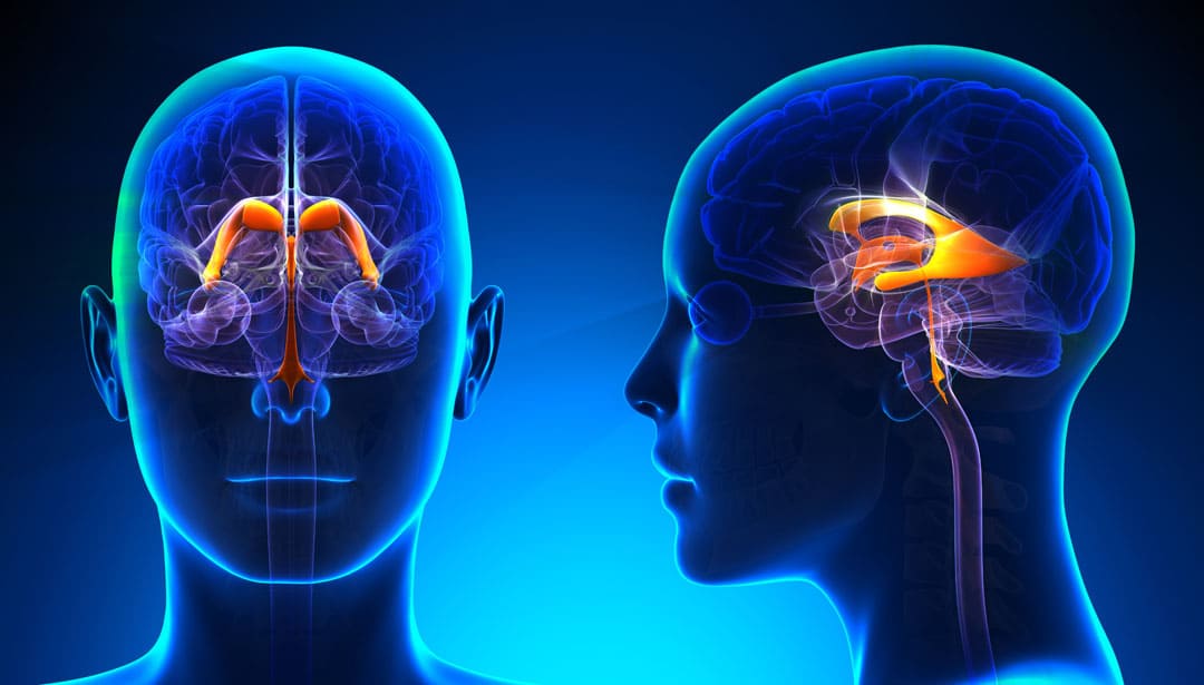
The pituitary gland is a tiny pea-sized organ located at the base of the brain. Barely a cubic centimetre in volume, it produces and stores so many hormones of our body that it is known as the ‘master gland’. These pituitary hormones control the activity of other hormones in the body.

The pituitary gland has 2 parts: the anterior (front) and the posterior (back) parts, each producing a different set of hormones. Each of these hormones acts on different parts of the body and controls their proper functioning. The pituitary hormones are:

  • Prolactin
  • Growth hormone
  • Adrenocorticotrophic hormone [ACTH]
  • Thyroid stimulating hormone [TSH]
  • Luteinizing hormone
  • Follicle stimulating hormone
  • Melanocyte stimulating hormone
  • Antidiuretic hormone [ADH]
  • Oxytocin

Function of pituitary hormones are vital for human survival. The deficiency or overproduction of each and every of the above hormones results in major medical disorders. For example, a shortage of growth hormone during childhood leads to reduced growth. The child does not grow like a normal adult but remains a dwarf. Conversely, excess of growth hormone leads to gigantism, a condition where the person keeps on growing abnormally.

Table of Contents

Hormones and Hormone Diseases

Hormones are the body’s chemical messengers, sending signals into the bloodstream and tissues. Hormones work slowly, over time, and affect many different processes, including growth and development, blood sugar, sexual functions, reproduction, urine output, managing stress and mood. Hormone diseases occur when the level of any hormone is reduced or increased because of any reason. The Pituitary Gland produces many hormones and any pituitary gland problems lead to major illnesses.

What are common pituitary disorders?

Apart from a deficiency or excess production of the above hormones, disorders of pituitary gland are as follows:

  • Tumours
  • Pituitary damage because of Head injury
  • Birth defects
  • Inherited genetic defects
  • Low blood supply to the pituitary gland
  • Previous history of pituitary disorders
  • Iron overload
  • Medication
  • Radiation therapy in the head and neck region

What are the different types of anterior pituitary disorders?

Anterior pituitary disorders occur as a result of overproduction or underproduction of hormones secreted by the anterior lobes.

     Disorders caused by over-production of anterior pituitary hormones

  • Acromegaly & gigantism
  • Prolactemia
  • Cushing’s disease


      Disorders caused by under-secretion of anterior pituitary hormones

  • Dwarfism
  • Central adrenal insufficiency
  • Gonadotropin deficiency
  • Hypothyroidism


What are the different types of posterior pituitary disorders?

Posterior pituitary disorders are caused by either under-production or overproduction of the ADH hormone. ADH helps the kidneys to prevent excess water loss through urine. Imbalance in ADH production can lead to the following disorders:

Pituitary disorders symptoms: Hypopituitarism

Hypopituitarism is a condition that results in a partial or complete loss of the anterior pituitary gland functions. Panhypopituitarism is a condition, characterized by damage to the entire pituitary gland. In such cases, the production of all pituitary hormones stops. 

Causes of hypopituitarism

  • Growth of a pituitary tumour
  • Head injuries
  • Brain surgery
  • Medication
  • Radiation therapy

Signs and symptoms of hypopituitarism

  • Tumours may cause eyesight problems.
  • The symptoms of hypopituitarism depend upon which hormones are no longer being produced.
  • Symptoms vary depending upon the age of the patients.

Diagnosis

  • Blood tests and stimulation or dynamic testing to identify low levels of pituitary hormones
  • Brain imaging using an MRI or CT scan to detect tumour or other pituitary gland problems
  • Vision tests to determine if the pituitary gland is affecting eyesight

Pituitary gland disorders treatment

  • Hormone replacement therapy: Patients need to take life-long medications to control the symptoms. The doctor monitors and adjusts the medication dose periodically based on the patient condition
  • Surgery or radiation treatment may be required in some patients suffering from pituitary tumours

What are pituitary tumours?

Pituitary tumours are abnormal growths in the pituitary glands. They may cause overproduction or low levels of pituitary hormones.  Most pituitary tumours are noncancerous growth and remain confined within the pituitary gland. These are known as an adenomas.

When the tumour size is less than one cm, it is called a Microadenoma. Most pituitary adenomas are microadenomas. When the tumour is greater than one cm in size, it is called a Macroadenoma.

Malignant Pituitary tumours (cancers) are rare and mostly affects old aged persons. Pituitary carcinomas can spread to the brain, spinal cord or the bone surrounding the pituitary glands. In some patients, the pituitary adenoma can turn cancerous and spread to other parts of the body.


What is the prevalence of pituitary tumours?

A pituitary adenoma is the third largest cause of brain tumours, accounting for 10% of all cases. The global prevalence of pituitary adenomas is approximately 17%. Studies have reported the risk of pituitary tumours to increase with age, with maximum cases being diagnosed between 30 and 60 years.

What causes pituitary tumours? Are there any risk factors?

The cause of pituitary adenoma remains unknown. Genetic alteration is thought to play an important role in adenoma development. Studies have shown that people with certain genetic factors such as multiple endocrine neoplasia, type 1 (MEN 1) are at increased risk of pituitary tumours.

What are the signs and symptoms of pituitary adenomas?

  • Tumours can put pressure on the pituitary gland and the nearby structures. This can cause headaches and loss of side-visions
  • As the tumour grows in size, it can damage the normal functioning of the gland and interfere with hormone production. Overproduction or hormonal deficiencies can cause specific signs and symptoms or sometimes a combination of them.
  • For example, ACTH tumours exhibit signs and symptoms of Cushing’s syndrome.

Are pituitary tumours life-threatening?

If diagnosed early, pituitary tumours can be managed well. However, if left undiagnosed and untreated for a long period, such tumours can become large and affect the functioning of several organs of the body, causing blindness, hypertension, diabetes, osteoporosis, heart disease and death.

Evidence suggests the 5-year survival rate of pituitary gland tumours be about 82%. 

Survival rate also depends upon factors like:

  • Type of tumour
  • Person’s age
  • How far the tumour has spread in the brain or to other parts of the body

Complications of pituitary tumours

  • Blindness
  • Permanent hormone deficiency
  • Pituitary apoplexy is a rare and serious complication

How are pituitary tumours diagnosed?

  • Blood and urine tests help to determine whether there is an overproduction of deficiency of hormones
  • Biopsy: This involves removing a small number of cells and examining them under a microscope to check for abnormal growth
  • Brain imaging: A CT scan or brain MRI scan can help the doctor to determine the size and location of the tumour
  • Vision testing to understand if the tumour has caused eyesight problem
  • Neurological exam: A set of questions and answers to check a person’s mental status and appropriate brain functioning controlling movement and coordination.

Pituitary gland diseases treatment

Not all pituitary tumours require treatment. Treatment depends upon the following:

  • Type and size of the tumour
  • If the tumour is making hormones
  • If the tumour is obstructing vision or associated with other signs or symptoms
  • If the tumour is localized or has spread to other parts of the body
  • If the tumour has occurred for the first time or has recurred
  • The patient’s overall healt

    The treatment is usually given by a brain surgeon (neurosurgeon), endocrine system specialist (endocrinologist) and a radiation oncologist. Doctors use a combination of surgery, radiation therapy and medications to normalize hormone levels.

Surgery for pituitary

Surgery is needed when the tumour presses on the optic nerve or leads to the overproduction of hormones. The Endoscopic transnasal transsphenoidal approach involves removing the tumour through the nose without making an incision. This is usually done when the tumour is small in size and no other part of the brain is affected by it. The Transcranial approach (craniotomy) is used for removing large tumours by making an incision in the scalp.

Radiation therapy

This technique uses high-energy radiation to destroy tumour cells. It can be used alone or in combination with surgery.

Medicines

They help to limit excess hormone production and shrink certain types of pituitary tumours.

Hormone replacement therapy is done in patients with reduced hormone production after surgery.

Watchful waiting: Many patients do not exhibit any symptoms and function normally without any treatment. In such cases, patients are advised only regular tests to keep a check on the tumour growth.

To summarise, a disease of pituitary gland can be very harmful. It must be diagnosed and treated as soon as possible. Such complex disease treatment is available in Samarth Neuro and Superspeciality Hospital in Miraj, Maharashtra. Dr. Ravindra Patil, the chief neurosurgeon there, is an expert in performing such operations.

Ventricles of the Human Brain

Ventricles of the Human Brain

Ventricles of the Human Brain

By Dr. Ravindra Patil

A ventricle is a hollow part or cavity in an organ. The word ventricle originates from Latin, the language which has given most of the words used in anatomy and medicine. Ventricles of brain are cavities within the brain. Ventricles of the brain have distinctive functions, which we will see in this article.

Two major organs of the major organs of the human body which have ventricles are the heart and the brain. The ventricles of the heart pump blood through our body, while the ventricles of the brain are cavities within the brain that produce and store a liquid called cerebrospinal fluid (CSF). This liquid surrounds our brain and spinal cord, cushions them and protects them from injury. The CSF is also responsible for removing waste and delivering nutrients to our brain.

Table of Contents

Where in the brain are the ventricles?

There are four ventricles in the brain. They are interconnected cavities. They extend throughout the brain. As has been mentioned, they produce, store and circulate the cerebrospinal fluid (CSF).

The two lateral ventricles of brain are C-shaped chambers found in the cerebral hemispheres, which is one in each hemisphere.

Lateral ventricles are connected to the third ventricle by an opening called the inter-ventricular foramen. The third ventricle is a very narrow cavity that runs along the midline of the diencephalon.

What structure connects the third and fourth ventricles? The third ventricle communicates with the fourth ventricle via the cerebral aqueduct. The fourth ventricle is wedged between the cerebellum on one side and the brainstem on the other; it extends to the central canal of the spinal cord. Which means in effect, the four ventricles extend from the brain to the end of the spinal cord. The CSF surrounds the brain and is also within the ventricles. The CSF is a liquid cushion in which the brain literally floats. The brain ventricles essentially supply, store and circulate the CSF around the brain.

The CSF is a thick viscous fluid which serves to cushion the brain, regulate its temperature and supply the brain with nutrients.

More about the brain ventricles

When looking at the interior of the brain, the four hollow ventricular cavities stand out in contrast to the rest of the organ, which is mostly composed of brain tissue, which in turn is made of grey matter and white matter. The main ventricle function is of course, producing CSF.

As the ventricles contained nothing except fluid, in ancient times it was thought that the ventricles contained ‘animal spirits’, a mysterious substance that allowed the immortal soul to exert control over the physical body. Later it was thought that the ventricles were credited with functions like the generation of imagination and memory. However, it was in 1764 that it was discovered that the ventricles were filled with CSF, fluid, and not animal spirits. And the CSF flowed around and inside the brain through the connections between ventricles. The ventricles gave the CSF a route to flow throughout the brain. Gradually it was discovered that it is the production, storage and circulation of CSF which is the main role of the ventricles.

How is CSF produced?

The production of CSF is done by a specialized membrane called the choroid plexus, which is made up of ependymal cells. This choroid plexus lines the ventricles in brain. Ependymal cells are glial cells tailored to produce CSF, and they secrete the fluid into the ventricles at a relatively constant rate; about a half a litre of CSF is produced by ependymal cells every day. CSF passes through the ventricular system and circulates around the brain and spinal cord in a small area between the meninges called the subarachnoid space.

Functions of the CSF

CSF is thought to play many important roles in the brain. The CSF and the brain ventricular system together keep the brain buoyant, reducing its weight and hence the physical stress it would otherwise experience from the forces of gravity and movement. The brain is a very delicate and soft organ. If the  brain would not have been suspended in the CSF, it would have been distorted with its own weight! While organs like the kidney and liver and heart are much sturdier, the brain is very soft. Without being suspended in a fluid the delicate tissue of the brain can tear.

There is about 125–150 mL of CSF at any one time. The actual mass of the human brain is about 1400–1500 grams; however, the net weight of the brain suspended in CSF is equivalent to a mass of 25-50 grams. The brain therefore exists in neutral buoyancy, which allows the brain to maintain its density without being impaired by its own weight. Had the brain been suspended in the skull without floating in the CSF, its blood supply would have been cut off and its neurons would be killed in the lower sections of the brain!

The layer of CSF surrounding the brain also acts as a buffer against potential injuries that can be caused by mechanical pressure or force (e.g. being hit hard in the head). Additionally, as the CSF circulates over the brain it carries away toxins and other waste matter and empties these into the bloodstream where they can eventually be removed by mechanisms like kidney filtration.

Ventricles and the CSF

The rate of CSF production in the ventricles is fairly constant regardless of changes in pressure within the ventricles (i.e. inter-ventricular pressure). This can be problematic if the passage of CSF is blocked somewhere within the ventricular system. In the ventricular system of brain, CSF will continue to be produced, but it will have no means of exiting the system. This will cause pressure within the ventricles to increase, and the rising pressure may eventually force the ventricles to expand. The expanding ventricles can then put pressure upon other brain structures, and cause a variety of complications depending on where exactly the blockage has occurred and which structures are most affected by the blockage.

When such a blockage of the brain ventricles occurs in children whose skull has not completely become bony [generally under age of 2 years], it results in the enlargement of the head. It is known as hydrocephalus. Hydrocephalus can be cause by blockage in the passage of CSF as well as by excess CSF production. In lay language, hydrocephalus is referred as ‘Water in the Brain’.

There can be a number of causes of a blockage that leads to hydrocephalus, such as a tumour, infection, or congenital malformation, which impedes the function of ventricles. Hydrocephalus can often be treated by surgically implanting a shunt that drains the ventricles of excess CSF and empties it into the abdominal cavity. This approach can be successful, but if the reason for the blockage isn’t resolved additional surgeries may be required in the future (e.g. to replace an outgrown shunt, treat an infected shunt, etc…).

How is blockage in ventricles diagnosed?

A contrast CT Scan can show the size and shape of the ventricles in brain. Either a radiopaque dye is injected in the patient’s veins or the CSF is replaced with air and then CT Scan of brain ventricles is done. In either case, a good contrast will be seen and the ventricles can be visualised. While the radio opaque dyes shows up as white colour, air shows up as black colour in CT scans and that helps doctors view the ventricular shape and size and reach a diagnosis about tumours, raised CSF pressure or blockage in the brain ventricular system.

Samarth Neuro and Superspeciality Hospital in Miraj has all the infrastructure and staff to treat hydrocephalus in children and other reasons of CSF blockage. The chief neurosurgeon Dr Ravindra Patil has operated upon many children suffering from hydrocephalus who needed shunts. These children are now grown up. Dr Ravindra Patil has also successfully operated on many adult patients with brain tumours.

पिट्यूटरी ग्रंथी आणि त्याचे विकार

पिट्यूटरी ग्रंथी आणि त्याचे विकार

पिट्यूटरी ग्रंथी आणि त्याचे विकार

By Dr. Ravindra Patil

पिट्यूटरी ग्रंथी म्हणजे काय आणि ती कुठे आहे?

पिट्यूटरी ग्रंथी मेंदूच्या पायथ्याशी स्थित एक लहान वाटाण्या एवढा अवयव आहे. त्याचे घनफळ जेमतेम एक क्यूबिक सेंटीमीटर असले तरी पिट्यूटरी ग्रंथी आपल्या शरीरात इतके संप्रेरक (हॉर्मोन्स) तयार करते आणि साठवते की त्याला ‘मास्टर ग्रंथी’ म्हणतात. हे पिट्यूटरी हार्मोन्स शरीरातील इतर संप्रेरकांची क्रिया नियंत्रित करतात.

पिट्यूटरी ग्रंथीमध्ये 2 भाग असतात: पुढचा भाग आणि मागचा भाग. प्रत्येक भाग संप्रेरकांचा भिन्न संच तयार करतो. यातील प्रत्येक संप्रेरक शरीराच्या वेगवेगळ्या भागांवर कार्य करतो आणि त्या भागांचे कार्य योग्य प्रकारे नियंत्रित करतो. पिट्यूटरी ग्रंथीचे खालील हॉर्मोन्स आहेत:

  • प्रोलॅक्टिन
  • ग्रोथ हॉर्मोन
  • एड्रेनोकॉर्टिकोट्रॉफिक हॉर्मोन [ACTH]
  • थायरॉईड उत्तेजक संप्रेरक [TSH]
  • ल्युटेनाइझिंग हॉर्मोन
  • फॉलिकल उत्तेजक हॉर्मोन
  • मेलानोसाइट उत्तेजक हॉर्मोन
  • अँटीडाययुरेटिक हॉर्मोन [ADH]
  • ऑक्सिटोसिन

पिट्यूटरी संप्रेरकांचे कार्य मानवी जीवनासाठी अत्यावश्यक आहेत. वरील प्रत्येक संप्रेरकांची कमतरता किंवा अतिउत्पादनामुळे मोठे विकार होतात. उदाहरणार्थ, बालपणात ग्रोथ हॉर्मोनच्या कमतरतेमुळे वाढ कमी होते. मूल सामान्य प्रौढांसारखे वाढत नाही परंतु त्यांची उंची व वाढ खुंटते व ते आकाराने लहानच राहते. याउलट, वाढीव संप्रेरकांच्या अतिरेकामुळे महाकायपणा येतो, अशी स्थिती जिथे व्यक्ती असामान्यपणे वाढत राहते.

Table of Contents

संप्रेरक आणि संप्रेरक रोग

हार्मोन्स हे शरीराचे रासायनिक संदेशवाहक आहेत, जे रक्तप्रवाहात आणि ऊतींमध्ये सिग्नल पाठवतात. हार्मोन्स हळूहळू, कालांतराने कार्य करतात आणि वाढ आणि विकास, रक्तातील साखर, लैंगिक कार्ये, पुनरुत्पादन, मूत्र उत्पादन, मानसिक तणाव आणि मूड व्यवस्थापित करणे यासह अनेक भिन्न प्रक्रियांवर परिणाम करतात. संप्रेरक रोग उद्भवतात जेव्हा कोणत्याही हॉर्मोनची पातळी कोणत्याही कारणाने कमी होते किंवा वाढते. पिट्यूटरी ग्रंथी अनेक संप्रेरकांची निर्मिती करते आणि पिट्यूटरी ग्रंथीची कोणतीही समस्या मोठ्या आजारांना कारणीभूत ठरते.

सामान्य पिट्यूटरी विकार कोणते?

वरील संप्रेरकांची कमतरता किंवा जास्त उत्पादन व्यतिरिक्त, पिट्यूटरी ग्रंथीचे विकार खालीलप्रमाणे आहेत:

  • ट्यूमर (गाठ)
  • डोक्याच्या दुखापतीमुळे पिट्यूटरी नुकसान
  • जन्मजात दोष
  • अनुवांशिक दोष
  • पिट्यूटरी ग्रंथीला कमी रक्तपुरवठा
  • पिट्यूटरी विकारांचा पूर्वीचा इतिहास
  • लोह (आयर्न) ओव्हरलोड
  • औषधोपचारांचे दुष्परिणाम
  • डोके आणि मान क्षेत्रामध्ये रेडिएशन थेरपी

पुढच्या भागातील पिट्यूटरी ग्रंथी विकार कोणते?

पुढच्या लोब्सद्वारे स्रावित हार्मोन्सच्या अतिउत्पादनामुळे किंवा कमी उत्पादनामुळे उद्भवतात.

हार्मोन्सच्या जास्त उत्पादनामुळे होणारे विकारः

  • एक्रोमेगाली आणि विशालता
  • प्रोलॅक्टेमिया
  • कुशिंग रोग

पुढच्या लोबद्वारे उत्पन्न होणार्‍या पिट्यूटरी हार्मोन्सच्या कमी स्रावामुळे होणारे विकार

  • बटु अथवा ठेंगु पणा
  • मध्य अधिवृक्क अपुरेपणा
  • गोनाडोट्रॉपिनची कमतरता
  • हायपोथायरॉईडीझम

पोस्टरियर पिट्यूटरीच्या विकारांचे प्रकार कोणते?

ए.डी.एच. संप्रेरकाच्या कमी उत्पादनामुळे किंवा जास्त उत्पादनामुळे पोस्टरियर पिट्यूटरी विकार होतात. ए.डी.एच. मूत्रपिंडांना मूत्राद्वारे अतिरिक्त पाणी कमी होण्यास मदत करते. ए.डी.एच. उत्पादनातील असंतुलन खालील विकारांना कारणीभूत ठरू शकते:

पिट्यूटरी विकारांची लक्षणे: हायपोपिट्युटारिझम

हायपो म्हणजे कमी. हायपोपिट्यूटरीझम ही अशी स्थिती आहे ज्याचा परिणाम पुढच्या पिट्यूटरी ग्रंथीच्या कार्यांचे आंशिक किंवा पूर्ण नुकसान होते. पॅन- हायपोपिट्यूटरीझम ही एक स्थिती आहे, जी संपूर्ण पिट्यूटरी ग्रंथीचे (पुढच्या व मागच्या) नुकसान होते. अशा परिस्थितीत, सर्व पिट्यूटरी हार्मोन्सचे उत्पादन थांबते

हायपोपिट्युटारिझमची कारणे

  • पिट्यूटरी ट्यूमरची वाढ
  • डोक्याला दुखापत
  • मेंदूची शस्त्रक्रिया
  • औषधोपचाराचे दुष्परिणाम
  • रेडिएशन थेरपी

हायपोपिट्युटारिझमची चिन्हे आणि लक्षणे

  • ट्यूमरमुळे दृष्टी समस्या उद्भवू शकतात.
  • हायपोपिट्युटारिझमची लक्षणे यापुढे कोणते हार्मोन्स तयार होत नाहीत यावर अवलंबून असतात.
  • रुग्णांच्या वयानुसार लक्षणे बदलतात.

निदान

  • पिट्यूटरी संप्रेरकांची कमी पातळी ओळखण्यासाठी रक्त चाचण्या आणि डायनॅमिक चाचण्या
  • ट्यूमर किंवा इतर पिट्यूटरी ग्रंथी समस्या शोधण्यासाठी एम.आर.आय. किंवा सी.टी. स्कॅन वापरून ब्रेन इमेजिंग
  • पिट्यूटरी ग्रंथीचा दृष्टीवर परिणाम होत आहे की नाही हे निर्धारित करण्यासाठी दृष्टी चाचण्या

पिट्यूटरी ग्रंथीच्या विकारांचे उपचार

  • हॉर्मोन रिप्लेसमेंट थेरपी: लक्षणे नियंत्रित करण्यासाठी रुग्णांना आयुष्यभर औषधे घ्यावी लागतात. डॉक्टर रुग्णाच्या स्थितीवर आधारित वेळोवेळी औषधांच्या डोसचे नियंत्रण करतात
  • पिट्यूटरी ट्यूमरने ग्रस्त असलेल्या काही रुग्णांमध्ये शस्त्रक्रिया किंवा रेडिएशन उपचार आवश्यक असू शकतात

पिट्यूटरी ट्यूमर म्हणजे काय?

पिट्यूटरी ट्यूमर म्हणजे पिट्यूटरी ग्रंथींमध्ये असामान्य वाढ. पिट्यूटरी ट्यूमर हॉर्मोन्सचे जास्त उत्पादन किंवा कमी उत्पादनाचे कारण होऊ शकतो. बहुतेक पिट्यूटरी ट्यूमर कर्करोगरहित असतात (कॅंसरचे नसतात) आणि पिट्यूटरी ग्रंथीमध्येच मर्यादित राहतात, पसरत नाहीत. यांना एडेनोमा म्हणतात.

जेव्हा ट्यूमरचा आकार एक सेमीपेक्षा कमी असतो तेव्हा त्याला मायक्रोएडेनोमा म्हणतात. बहुतेक पिट्यूटरी एडेनोमा मायक्रोएडेनोमा असतात. जेव्हा ट्यूमरचा आकार एक सेमीपेक्षा जास्त असतो तेव्हा त्याला मॅक्रोएडेनोमा म्हणतात.

घातक पिट्यूटरी ट्यूमर (कर्करोग किंवा कॅंसर) दुर्मिळ असतात आणि बहुतेक वृद्ध व्यक्तींना होतात. पिट्यूटरी कार्सिनोमा मेंदू, पाठीचा कणा किंवा पिट्यूटरी ग्रंथींच्या सभोवतालच्या हाडांमध्ये पसरू शकतो. काही रुग्णांमध्ये, पिट्यूटरी एडेनोमा कर्करोगात बदलू शकतो आणि शरीराच्या इतर भागांमध्ये पसरतो.

पिट्यूटरी ट्यूमरचे प्रमाण काय आहे?

पिट्यूटरी एडेनोमा हे ब्रेन ट्यूमरचे तिसरे सर्वात मोठे कारण आहे, जे सर्व प्रकरणांपैकी 10% आहे. पिट्यूटरी एडेनोमाचा जागतिक प्रसार अंदाजे 17% आहे. पिट्यूटरी ट्यूमरचा धोका वयानुसार वाढण्याची शक्यता अभ्यासांनी नोंदवली आहे, जास्तीत जास्त प्रकरणे 30 ते 60 वर्षांच्या दरम्यान आढळतात.

पिट्यूटरी ट्यूमर कशामुळे होतात? काही जोखीम घटक आहेत का?

पिट्यूटरी एडेनोमाचे कारण अज्ञात आहे. एडेनोमाच्या विकासामध्ये अनुवांशिक बदल महत्त्वपूर्ण भूमिका बजावतात असे मानले जाते. अभ्यासातून असे दिसून आले आहे की काही अनुवांशिक घटक जसे की एकाधिक अंतःस्रावी निओप्लाझिया, प्रकार 1 (मेन 1) पिट्यूटरी ट्यूमरचा धोका वाढतो.

पिट्यूटरी एडेनोमाची चिन्हे आणि लक्षणे काय आहेत?

  • ट्यूमर पिट्यूटरी ग्रंथी आणि जवळच्या संरचनेवर दबाव आणू शकतात. यामुळे डोकेदुखी आणि साइड-व्हिजन नष्ट होऊ शकतात
  • जसजसा ट्यूमरचा आकार वाढतो तसतसा तो ग्रंथीच्या सामान्य कार्यास हानी पोहोचवू शकतो आणि संप्रेरकांच्या निर्मितीमध्ये व्यत्यय आणू शकतो. अतिउत्पादन किंवा हॉर्मोनल कमतरतेमुळे विशिष्ट चिन्हे आणि लक्षणे किंवा कधीकधी त्यांचे संयोजन होऊ शकते.
  • उदाहरणार्थ, ACTH ट्यूमर कुशिंग सिंड्रोमची चिन्हे आणि लक्षणे प्रदर्शित करतात.

पिट्यूटरी ट्यूमर जीवघेणे आहेत का?

लवकर निदान झाल्यास, पिट्यूटरी ट्यूमरचे व्यवस्थापन चांगले केले जाऊ शकते. तथापि, निदान न झाल्यास आणि दीर्घकाळ उपचार न केल्यास, असे ट्यूमर मोठे होतात आणि शरीराच्या अनेक अवयवांच्या कार्यावर परिणाम करतात, ज्यामुळे अंधत्व, उच्च रक्तदाब, मधुमेह, ऑस्टियोपोरोसिस, हृदयरोग आणि मृत्यू होऊ शकतात.

पुराव्यांवरून असे सूचित होते की पिट्यूटरी ग्रंथी ट्यूमरचा 5 वर्षांचा जगण्याचा दर सुमारे 82% आहे.

जगण्याचा दर देखील यासारख्या घटकांवर अवलंबून असतो:

  • ट्यूमरचा प्रकार
  • व्यक्तीचे वय
  • मेंदूमध्ये किंवा शरीराच्या इतर भागांमध्ये ट्यूमर किती दूर पसरला आहे

पिट्यूटरी ट्यूमरची दुष्परिणाम

  • अंधत्व
  • कायमस्वरूपी हॉर्मोनची कमतरता
  • पिट्यूटरी अपोप्लेक्सी ही एक दुर्मिळ आणि गंभीर गुंतागुंत आहे

पिट्यूटरी ट्यूमरचे निदान कसे केले जाते?

  • रक्त आणि लघवीच्या चाचण्या हार्मोन्सच्या कमतरतेचे जास्त उत्पादन आहे की नाही हे निर्धारित करण्यात मदत करतात

  • बायोप्सी: यामध्ये पेशींची एक लहान भाग काढून आणि त्यांची असामान्य वाढ तपासण्यासाठी सूक्ष्मदर्शकाखाली तपासणी करतात.

  • ब्रेन इमेजिंग: सी.टी. स्कॅन किंवा ब्रेन एम.आर.आय. स्कॅन डॉक्टरांना ट्यूमरचा आकार आणि स्थान निर्धारित करण्यात मदत करू शकते.
  • ट्यूमरमुळे दृष्टी समस्या झाली आहे का हे समजून घेण्यासाठी दृष्टी चाचणी
  • न्यूरोलॉजिकल परीक्षा: एखाद्या व्यक्तीची मानसिक स्थिती तपासण्यासाठी प्रश्न आणि उत्तरांचा संच आणि मेंदूच्या हालचाली आणि समन्वय नियंत्रित करणारे योग्य कार्य.

पिट्यूटरी ग्रंथी रोग उपचार

सर्व पिट्यूटरी ट्यूमरला उपचारांची आवश्यकता नसते. उपचार खालील गोष्टींवर अवलंबून आहे:

  • ट्यूमरचा प्रकार आणि आकार
  • जर ट्यूमर हार्मोन्स बनवत असेल
  • जर ट्यूमर दृष्टीस अडथळा आणत असेल किंवा इतर चिन्हे किंवा लक्षणांशी संबंधित असेल
  • जर ट्यूमर स्थानिकीकृत असेल किंवा शरीराच्या इतर भागात पसरला असेल
  • जर ट्यूमर पहिल्यांदाच झाला असेल किंवा पुनरावृत्ती झाला असेल
  • रुग्णाचे एकूण आरोग्य

उपचार सामान्यतः मेंदू सर्जन (न्यूरोसर्जन), अंतःस्रावी प्रणाली विशेषज्ञ (एंडोक्रिनोलॉजिस्ट) आणि रेडिएशन ऑन्कोलॉजिस्टद्वारे दिले जातात. संप्रेरक पातळी सामान्य करण्यासाठी डॉक्टर शस्त्रक्रिया, रेडिएशन थेरपी आणि औषधे यांचे संयोजन वापरतात.

पिट्यूटरी साठी शस्त्रक्रिया

जेव्हा ट्यूमर ऑप्टिक नर्व्ह दाबतो किंवा हॉर्मोन्सच्या जास्त उत्पादनास कारणीभूत ठरतो तेव्हा शस्त्रक्रिया आवश्यक असते. एन्डोस्कोपिक ट्रान्सनेसझल ट्रान्सफेनॉइडल पध्दतीमध्ये चीरा न लावता नाकातून ट्यूमर काढतात. हे सहसा तेव्हा करतात जेव्हा ट्यूमर आकाराने लहान असतो आणि मेंदूच्या इतर कोणत्याही भागावर त्याचा दुष्परिणाम होत नसतो. ट्रान्सक्रॅनियल अ‍ॅप्रोच (क्रॅनिओटॉमी) टाळूमध्ये चीरा बनवून मोठ्या गाठी काढण्यासाठी वापरला जातो.

रेडिएशन थेरपी

हे तंत्र ट्यूमर पेशी नष्ट करण्यासाठी उच्च-ऊर्जा रेडिएशन वापरते. हे नुसते किंवा शस्त्रक्रियेच्या बरोबर वापरले जाऊ शकते.

औषधोपचार

ते अतिरिक्त संप्रेरक उत्पादन मर्यादित करण्यास आणि विशिष्ट प्रकारचे पिट्यूटरी ट्यूमर कमी करण्यास मदत करतात.

शस्त्रक्रियेनंतर हॉर्मोनचे उत्पादन कमी झालेल्या रुग्णांमध्ये हॉर्मोन रिप्लेसमेंट थेरपी केली जाते.

सावध प्रतीक्षा: अनेक रुग्ण कोणतीही लक्षणे दाखवत नाहीत आणि कोणत्याही उपचाराशिवाय सामान्यपणे कार्य करतात. अशा परिस्थितीत, ट्यूमरच्या वाढीवर नियंत्रण ठेवण्यासाठी रुग्णांना फक्त नियमित चाचण्यांचा सल्ला दिला जातो.

थोडक्यात, पिट्यूटरी ग्रंथीचा रोग खूप हानिकारक असू शकतो. शक्य तितक्या लवकर निदान आणि उपचार करणे आवश्यक आहे. महाराष्ट्रातील मिरज येथील समर्थ न्युरो आणि सूपरस्पेशालिटी हॉस्पिटलमध्ये अशा प्रकारच्या जटिल आजारावरील उपचार उपलब्ध आहेत. तेथील मुख्य न्युरोसर्जन डॉ रवींद्र पाटील असली ऑपरेशने करण्यात तरबेज आहेत.

मानवी मेंदूची वेंट्रिकल्स

मेंदूची वेंट्रिकल्स

मानवी मेंदूची वेंट्रिकल्स

By Dr. Ravindra Patil

वेंट्रिकल म्हणजे एखाद्या अवयवातील पोकळी. व्हेंट्रिकल हा शब्द लॅटिनमधून आला आहे. शरीरशास्त्रात वापरले जाणारे अनेक शब्द लॅटिन भाषेतून आले आहेत. मेंदूची वेंट्रिकल्स म्हणजे मेंदूतील पोकळ्या. त्या एकूण चार असतात. मेंदूच्या वेंट्रिकल्सची विशिष्ट कार्ये असतात, जी आपण या लेखात पाहू.

मानवी शरीराच्या दोन प्रमुख अवयवांमध्ये वेंट्रिकल्स असतात. ते दोन अवयव म्हणजे हृदय आणि मेंदू. हृदयाची वेंट्रिकल्स आपल्या शरीरातील रक्त पंप करतात, तर मेंदूचे वेंट्रिकल्स मेंदूतील पोकळ्यांमधून सेरेब्रोस्पाइनल फ्लुइड (सी.एस.एफ.) नावाचा द्रव तयार करतात, तो साठवतात आणि त्याचे अभिसरण करतात. सी.एस.एफ. मानवी मेंदू भोवती असते आणि या द्रव पदार्थात आपला मेंदू तरंगत असतो असे म्हंटले तरी योग्य ठरेल. सी.एस.एफ. पाठीच्या कण्याभोवती (स्पायनल कॉर्ड भोवती) पण असते. सी.एस.एफ. मेंदूचे धक्के आणि दुखापातीं यापासून संरक्षण करते. सी.एस.एफ. मेंदूला पोषक द्रव्ये पोहोचवण्याचे काम करते आणि मेंदूतील कचरा पण काढून टाकते.

Table of Contents

मेंदूमधील वेंट्रिकल्स कुठे असतात ?

 मेंदूमध्ये एकूण चार वेंट्रिकल्स असतात. या चारी पोकळ्या एकमेकांशी जोडलेल्या असतात. वर नमूद केल्याप्रमाणे, वेंट्रिकल्स सेरेब्रोस्पाइनल फ्लुइड (CSF) तयार करतात, साठवतात आणि त्याचे अभिसरण करतात.

मेंदूची दोन लेटरल वेंट्रिकल्स सेरेब्रल गोलार्धांमध्ये आढळणाऱ्या सी-आकाराचे पोकळ्या असतात.

लेटरल वेंट्रिकल्स तिसऱ्या वेंट्रिकलशी इंटर-व्हेंट्रिक्युलर फोरामेन नावाच्या छीद्राद्वारे जोडलेले असतात. तिसरे वेंट्रिकल म्हणजे एक अतिशय अरुंद पोकळी असते. ती डायएनकेफेलॉनच्या (मेंदूच्या एक भागाच्या) मध्यरेषेने चालते.

तिसऱ्या आणि चौथ्या वेंट्रिकल्सला कोणती रचना जोडते? तिसरे वेंट्रिकल चौथ्या वेंट्रिकलशी सेरेब्रल एक्वाडक्टद्वारे जोडलेले असते. चौथे वेंट्रिकल एका बाजूला सेरेबेलम आणि दुसऱ्या बाजूला ब्रेनस्टेममध्ये या दोन्ही मध्ये असते. ते पाठीच्या कण्याच्या (स्पायनल कॉर्डच्या) मध्यवर्ती कालव्यापर्यंत पसरते. याचा अर्थ, चार वेंट्रिकल्स मेंदूपासून स्पायनल कॉर्डच्या शेवटपर्यंत पसरलेली असतात. सी.एस.एफ. मेंदूभोवती असतो आणि वेंट्रिकल्समध्ये देखील असते. सी.एस.एफ. म्हणजे एका द्रव भरलेल्या उशी सारखे असते. त्यामध्ये मेंदू अक्षरशः तरंगतो. मेंदूचे वेंट्रिकल्स मूलत: मेंदूभोवती सी.एस.एफ. पुरवतात, त्याचा साठा करतात आणि त्याचे मेंदू व स्पायनल कॉर्ड भोवतालची अभिसरण करतात.

सी.एस.एफ. एक जाड व चिकट द्रव असतो. तो मेंदूला उशी सारखा टेका आणि रक्षण देतो. सी.एस.एफ. मेंदूचे तापमान नियंत्रित करते आणि मेंदूला पोषक तत्वांचा पुरवठा करते.

मेंदूच्या वेंट्रिकल्सची अधिक माहिती

ही चार पोकळ वेंट्रिकल्स बाकी मेंदू पेक्षा अगदी वेगळी असतात. मेंदू मेंदूच्या ऊतींनी बनलेला असतो. तो ग्रे मॅटर व व्हाईट मॅटरने बनलेला असतो. ग्रे (राखाडी रंग) व व्हाईट (पांढरा रंग) मॅटर ही नावे मेंदूच्या रंगामुळे दिली गेली आहेत. आणि वेंट्रिकलचे मुख्य कार्य म्हणजे अर्थातच सी.एस.एफ. तयार करते व ते मेंदूत सगळीकडे फिरवणे.

वेंट्रिकल्समध्ये द्रवपदार्थाशिवाय काहीही नसल्यामुळे, प्राचीन काळी असे मानले जात होते की व्हेंट्रिकल्समध्ये ‘प्राणी आत्मे’ असतात. या रहस्यमय पदार्थ ज्यामुळे अमर आत्म्याला भौतिक शरीरावर नियंत्रण ठेवता येते असे मानले जायचे. नंतर असे मानले गेले की वेंट्रिकल्सला कल्पनाशक्ती आणि स्मरणशक्तीच्या निर्मितीसारखी कार्ये करतात. तथापि, १७६४ मध्ये असे आढळून आले की वेंट्रिकल्स सी.एस.एफ. द्रवपदार्थाने भरलेले असतात, प्राण्यांच्या आत्म्याने भरलेले नसतात. आणि सी.एस.एफ. चारी वेंट्रिकल्समधील कनेक्शनद्वारे मेंदूच्या आजूबाजूला आणि आत वाहते. वेंट्रिकल्सने सी.एस.एफ.ला संपूर्ण मेंदूमध्ये प्रवाहित होण्याचा मार्ग दिला असतो. हळूहळू असे आढळून आले की वेंट्रिकल्सची मुख्य भूमिका आहे सी.एस.एफ.चे उत्पादन, साठवण आणि परिसंचरण.

सी.एस.एफ.कसे तयार केले जाते?

सी.एस.एफ.चे उत्पादन कोरॉइड प्लेक्सस नावाच्या विशेष पडद्याद्वारे केले जाते, जे एपेन्डिमल पेशींनी बनलेले असते. कोरॉइड प्लेक्सस मेंदूतील वेंट्रिकल्सच्या आतील बाजुवर असते. एपेन्डिमल पेशी सी.एस.एफ. तयार करण्यासाठी तयार केलेल्या ग्लियाल पेशी आहेत आणि ते स्थिर दराने वेंट्रिकल्समध्ये सी.एस.एफ.द्रव स्राव करतात. एपेन्डिमल पेशींद्वारे दररोज सुमारे अर्धा लिटर सी.एस.एफ. तयार होते. सी.एस.एफ. वेंट्रिक्युलर सिस्टिममधून जाते आणि मेंदू आणि पाठीच्या कण्याभोवती मेनिन्जेस (मेंदू भोवतीची आवरणे) च्या दरम्यानच्या एका लहान भागात फिरते ज्याला सबएरेक्नॉइड स्पेस म्हणतात.

सी.एस.एफ. ची कार्ये

सी.एस.एफ.मेंदूमध्ये अनेक महत्त्वाच्या भूमिका बजावते. आधी नमूद केल्याप्रमाणे सी.एस.एफ. मध्ये मेंदू तरंगत असतो. त्यामुळे मेंदूचे वजन कमी होते आणि त्यामुळे मेंदूवर गुरुत्वाकर्षणाचा पूर्ण प्रभाव पडत नाही. मेंदू हा अतिशय नाजूक आणि मऊ अवयव आहे. मेंदू सी.एस.एफ. मध्ये तरंगत नसता तर तो स्वतःच्या वजनाने त्याचा आकार बदलला असता व तो विकृत झाला असता! मूत्रपिंड, यकृत आणि हृदय यासारखे अवयव अधिक मजबूत असले तरी मेंदू अतिशय मऊ असतो. सी.एस.एफ.च्या द्रवपदार्थात मेंदू नसला तर मेंदूची नाजूक ऊतक फाटू शकतात.

कोणत्याही वेळी सुमारे १२५ ते १५० मिली लिटर सी.एस.एफ.अस्तित्वात असते. मानवी मेंदूचे वास्तविक वजन सुमारे १४०० ते १५०० ग्रॅम असते. पण मेंदू सी.एस.एफ.मध्ये तरंगत असल्याने मेंदूचे वजन निव्वळ  btrx २५ ते ३० ग्रॅम इतकेच असते. त्यामुळे मेंदूचे जणूकाही वजनच नसते व त्यामुळे मेंदूला त्याची घनता व कार्य राखता येते. सी.एस.एफ. मध्ये न तरंगता मेंदू कवटीत लटकला असता तर त्याचा रक्तपुरवठा खंडित झाला असता आणि मेंदूच्या खालच्या भागात त्याचे न्यूरॉन्स मेंदूच्या स्वतःच्या वजनानेच मारले गेले असते!

मेंदूच्या सभोवतालचा CSF चा थर यांत्रिक दबाव किंवा प्रहारामुळे (उदा. डोक्याला जोरदार मार लागल्याने) संभाव्य जखमांविरुद्ध बफर म्हणून काम करतो. याव्यतिरिक्त, सी.एस.एफ. मेंदूवर फिरत असताना ते निकास करण्यायोग्य आणि इतर टाकाऊ पदार्थ वाहून नेते आणि ते रक्तप्रवाहात रिकामे करते जिथून ते पदार्थ शेवटी किडनी फिल्टरेशनच्या यंत्रणेद्वारे शरीरातून बाहेर काढले जातात.

वेंट्रिकल्स आणि सी.एस.एफ.

वेंट्रिकल्समधील सी.एस.एफ. उत्पादनाचा दर वेंट्रिकल्समधील दाब (म्हणजे इंटर-व्हेंट्रिक्युलर प्रेशर) मध्ये बदल लक्षात न घेता बऱ्यापैकी स्थिर असतो. जर सी.एस.एफ.चा रस्ता वेंट्रिक्युलर सिस्टीममध्ये कुठेतरी अवरोधित असेल तर मोठी समस्या होते. मेंदूच्या वेंट्रिक्युलर सिस्टीममध्ये सी.एस.एफ. तयार होत राहते परंतु त्यास सिस्टममधून बाहेर पडण्याचे कोणतेही साधन नसेल तर वेंट्रिकल्समध्ये दबाव वाढेल आणि वाढत्या दाबामुळे वेंट्रिकल्सचा विस्तार होऊ शकतो. विस्तारणारे वेंट्रिकल्स नंतर मेंदूच्या इतर संरचनेवर दबाव आणू शकतात आणि अडथळा नेमका कुठे झाला आहे आणि कोणत्या संरचनेवर ब्लॉकेजचा सर्वाधिक परिणाम होतो यावर अवलंबून विविध गंभीर रोग निर्माण करू शकतात.

जेव्हा लहान बाळांमध्ये मेंदूच्या वेंट्रिकल्समध्ये अडथळा येतो (अशी मुले ज्यांची कवटी पूर्णपणे हाड बनलेली नसते व त्यांना टाळू असते, सामान्यत: 2 वर्षांखालील बाळे ) तेव्हा त्याचा परिणाम होतो डोक्याच्या आकाराची वाढ होण्यात. याला हायड्रोसेफलस असे म्हणतात. हायड्रोसेफलस हे सी.एस.एफ. च्या मार्गात अडथळे तसेच जास्त सी.एस.एफ. उत्पादनामुळे होऊ शकते. सामान्य भाषेत हायड्रोसेफलसला ‘मेंदूतील पाणी’ असे संबोधले जाते.

सी.एस.एफ. प्रावाहात अडथळा निर्माण होऊन हायड्रोसेफलस होते त्याची अनेक कारणे असू शकतात, जसे की ट्यूमर, संसर्ग किंवा जन्मजात विकृती, ज्यामुळे वेंट्रिकल्सच्या कार्यात अडथळा येतो. हायड्रोसेफलसवर अनेकदा शस्त्रक्रियेने शंट रोपण करून उपचार केले जाऊ शकतात. ही शंट (म्हणजे एक प्लास्टिकची नळी असते) अतिरिक्त सी.एस.एफ. पोटातील पोकळीत रिकामे करते. हे ऑपरेशन यशस्वी होते, परंतु जर अडथळ्याचे मुख्य कारण सोडवले गेले नाही तर भविष्यात अजून शस्त्रक्रिया आवश्यक असू शकतात, उदा. वय व उंची वाढल्याने शंट हलणे किंवा शंट संक्रमित होणे व त्यावरचे उपचार.

वेंट्रिकल्समधील अडथळ्याचे निदान कसे केले जाते?

कॉन्ट्रास्ट सीटी स्कॅन मेंदूतील वेंट्रिकल्सचा आकार दर्शवू शकतो. एकतर रुग्णाच्या शिरामध्ये रेडिओपेक डाय इंजेक्ट केला जातो किंवा सी.एस.एफ. एवजी वेंट्रिकल्समधे हवा भरली जाते आणि नंतर मेंदूच्या वेंट्रिकल्सचे सीटी स्कॅन केले जाते. दोन्ही प्रकारे चांगला कॉन्ट्रास्ट दिसतो आणि व्हेंट्रिकल्सच्या आकाराची कल्पना केली जाऊ शकते. सी.टी. स्कॅन मध्ये रेडिओ ओपेक डाय पांढरा दिसतो तर हवा असलेली जागा काळी दिसते. त्यामुळे डॉक्टरांना वेंट्रिकल्सचा आकार पाहण्यास आणि ट्यूमर, वाढलेला सी.एस.एफ. दाब किंवा मेंदूच्या वेंट्रिक्युलर सिस्टीममध्ये अडथळा याविषयी निदान करण्यात मदत होते.

मिरजेतील समर्थ न्यूरो आणि सुपरस्पेशालिटी हॉस्पिटलमध्ये मुलांमधील हायड्रोसेफलस आणि सी.एस.एफ. अवरोधाच्या ब्लॉकेजच्या इतर कारणांवर उपचार करण्यासाठी सर्व पायाभूत सुविधा आणि कर्मचारी आहेत. मुख्य न्यूरो सर्जन डॉ. रवींद्र पाटील यांनी अनेक हायड्रोसिफॅलस असलेल्या अनेक मुलांवर शस्त्रक्रिया केल्या आहेत व ती मुले आज मोठी झाली आहे. या मुलांना शंट शस्त्रक्रियेची गरज होती. तसेच डॉ रवींद्र पाटील यांनी मेंदूतील गाठी असलेल्या प्रौढ रुग्णांवर देखील अनेक यशस्वी शस्त्रक्रिया केल्या आहेत.