थ्रोम्बोलिसिस

थ्रोम्बोलिसिस

By Dr.Ravindra Patil

थ्रोम्बोलिसिस रक्तकणविघाताचीउपचारपद्धती

रक्त हा आपल्या शरीराचा एक जादुई अवयव आहे जो आपल्या शरीरात धमन्यांमधून आणि शिरांमधून आयुष्यभर वाहत राहतो. रक्त फुफ्फुसातून आवश्यक असलेल्या सर्व ऊतींमध्ये ऑक्सिजन वाहून नेते आणि सर्व उतींमधून कार्बन डायऑक्साईड परत फुफ्फुसात वाहून नेते. हे शरीराच्या प्रत्येक पेशीचे पोषण करते आणि सर्व उती आणि अवयवांमधून टाकाऊ पदार्थ बाहेर टाकण्यासाठी मूत्रपिंडात वाहून नेले जाते. हे आपण हायस्कूलच्या विज्ञान वर्गात शिकतो.

शाळेत असताना आपण सगळेच स्वतःला इजा करून घेतो. आपली त्वचा फाटू शकते आणि आपल्याला रक्तस्त्राव होऊ शकतो. पण आपल्या जादुई रक्ताच्या विशेष गुणांमुळे रक्त गोठते व पुढील रक्तस्त्राव थांबतो. हे आपण सर्वांनी अनुभवले आहे.

निरोगी लोकांच्या रक्ताची जादू अशी आहे की, दुखापत झाल्यावरच ते गोठते व आपला रक्तस्त्राव थांबतो. रक्त गोठण्यामुळे शरीरातून अधीक रक्त वाया जात नाही. निरोगी लोकांमध्ये, रक्त कधीही धमन्या, नसा किंवा हृदयात गोठत नाही किंवा त्याच्या गाठी होत नाहीत. ते नेहमी द्रव अवस्थेत राहते. आणि नमूद केल्याप्रमाणे आपला रक्त पुरवठा प्राणवायु आणि पोषक तत्वे पुरवण्याचे आणि टाकाऊ पदार्थ आणि कार्बन डायऑक्साइड बाहेर काढण्याचे सतत काम करत असतो. जर रक्ताच्या गुठळ्यामुळे अचानक रक्त वाहणे थांबले, तर प्रभावित अवयवांना प्रथम ऑक्सिजनची उपासमार होईल आणि थोड्याच वेळात त्या अवयवांचा मृत्यू होऊ शकतो.

मेंदू किंवा हृदय या अवयवांवर परिणाम झाला तर तो माणूसच मरेल!

त्यामुळे रक्त कोणत्याही किंमतीत द्रव अवस्थेत राहिले पाहिजे व वाहत राहिले पाहिजे.

Table of Contents

थ्रोम्बोसिस आणि एम्बोलिझम

तथापि, काही प्रसंगी वेगवेगळ्या रोगापूर्वीच्या घटकांमुळे, रक्तवाहिन्यामध्ये रक्ताच्या गुठळ्या होतात. याला थ्रोम्बोसिस असे म्हणतात.

या गुठळ्या काहीवेळा तुटतात, बाहेर पडतात आणि रक्ताभिसरण प्रणालीच्या दूरच्या भागात जातात, तिथे त्या अडकतात आणि त्या पुढील रक्तपुरवठा रोखतात. याला एम्बोलिझम म्हणतात.

कधीकधी थ्रोम्बोसिस आणि एम्बोलिझम दोन्ही एकत्र होतात आणि नंतर त्याला थ्रोम्बो-एम्बोलिक घटना म्हणून ओळखले जाते.

जर रक्तवाहिनी शीरे मध्ये गुठळी तयार झाली आणि ती सैल झाली व तिचा तुकड पडला, तर तो फुफ्फुसांच्या धमन्यांपर्यंत नेला जाईल आणि त्यांना फुफ्फुसांच्या धमनीला ब्लॉक करेल. याला पल्मोनरी एम्बोलिझम म्हणतात आणि ही एक अतिशय गंभीर व जीवघेणी स्थिती आहे.

वरील सर्व प्रकरणांमध्ये, मानवी शरीराच्या धमन्यांमध्ये रक्ताची गुठळी झाली आहे, ज्यामुळे त्या धमनीद्वारे पुरवल्या जाणार्‍या अवयवांची उपासमार होते व तो अवयव मरू शकतो. पण वरी रक्ताच्या गुठळ्या असलेल्या धमन्या जर हृदयाला किंवा मेदूला रक्तपुरवठा करत असल्या तर ती व्यक्ती गंभीर आजारी पडू शकते किंवा मृत्यू देखील पाऊ शकते.

इतर सर्व अवयवांचा रक्तपुरवठा ठप्प झाला तर परिणाम वाईटच होतो, परंतु हृदय किंवा मेंदूच्या रक्तपुरवठ्यात अडथळा आल्यास त्वरित मृत्यू होऊ शकतो.

पक्षघाताच्या (लकवा किंवा इंग्रजीत ज्याला स्ट्रोक म्हणतात त्या रूग्णांना) रूग्णांना थ्रोम्बोलिसिस हा उपचार सर्वोत्तम उपचारांपैकी एक आहे.

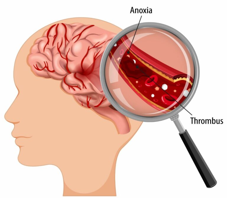
मेंदूच्या धमनीत गुठळ्या झाल्यामुळे सेरेब्रोव्हस्कुलर स्ट्रोक किंवा ब्रेन स्ट्रोक नावाचा तीव्र आजार होतो.

हृदयाच्या धमनीत गुठळ्या झाल्यामुळे मायोकार्डियल इन्फार्क्शन किंवा हृदयविकाराचा झटका म्हणून ओळखला जाणारा एक तीव्र रोग होतो.

फुफ्फुसाच्या धमन्यांमधील गुठळ्याला पल्मोनरी एम्बोलिझम असे म्हणतात.

थ्रोम्बोलिसिस

सोप्या शब्दात, थ्रोम्बोलिसिस [थ्रॉम्बोलाइटिक थेरपी म्हणूनही ओळखले जाते] रक्तवाहिन्यांमधील धोकादायक गुठळ्या विरघळवणे, रक्त प्रवाह सुधारणे आणि ऊती आणि अवयवांना होणारे नुकसान टाळण्यासाठी हा उपचार आहे. थ्रोम्बोलिसिस म्हणजे गुठळ्या विरघळवणाऱ्या औषधे इंट्राव्हेनस इंजेक्शनद्वारे किंवा लांब कॅथेटरद्वारे शीरेत इंजेक्शने दिली जातात व जी थेट ब्लॉकेजच्या ठिकाणी औषधे पोहोचवतात.

हे एका लांब कॅथेटरच्या टोकाला जोडलेल्या यांत्रिक उपकरणाने कॅथ लॅब मध्ये देखील केले जाऊ शकते. या प्रकारात एकतर गठ्ठा काढून टाकतात किंवा तो हळुहळु तोडतात.

थ्रोम्बोलिसिस प्रक्रियेचा उपयोग हृदय व मेंदूला रक्तप्रवाह देणाऱ्या धमन्यांमध्ये रक्ताच्या गुठळ्या विरघळवण्यासाठी आणीबाणीच्या उपचार म्हणून केला जातो – हृदयाच्या धमन्यांमध्ये क्लॉट झाला तर हृदयविकाराचा झटका येतो आणि मेंदूच्या धमनीत क्लॉट झाला इस्केमिक स्ट्रोकचे होतो. त्या शिवाय फुफ्फुसांच्या धमन्यांमध्ये क्लॉट झाला तर त्याला पल्मोनरी एम्बोलिझम म्हणतात व तेथे पण थ्रोम्बोलिसिस प्रक्रिया वापरून उपचार करतात.

खालील भागात देखील रक्ताच्या गुठळ्यांवर उपचार करण्यासाठी थ्रोम्बोलिसिस प्रक्रिया वापरली जाते:

  • शिरांच्या (व्हेन्सच्या आत) आत: याला डीप व्हेन थ्रोम्बोसिस (डी.व्ही.टी.) असे म्हणतात. हे पायांच्या नसा, ओटीपोटाचा भाग आणि वरच्या बाजूला असलेल्या नसांमधील गुठळ्या असतात; उपचार न केल्यास, गुठळ्याचे तुकडे फुटू शकतात आणि फुफ्फुसातील धमनीमध्ये जाऊ शकतात, परिणामी तीव्र पल्मोनरी एम्बोलिझम होतो.
  • बायपास ग्राफ्ट्सच्या आत: या म्हणजे हृदयाच्या बायपास शस्त्रक्रियेमध्ये वापरल्या जाणार्‍या नसांमध्ये तयार झालेल्या गुठळ्या आहेत, हृदयविकाराच्या उपचारासाठी केल्या जातात. या शिरा रुग्णाच्या पायातून काढल्या जातात. या नसांमध्ये रक्ताची गुठळी झाल्यास हृदयविकाराचा दुसरा झटका येऊ शकतो.
  • डायलिसिस कॅथेटर्सच्या आत: ज्या रुग्णांना वारंवार डायलिसिसची गरज असते त्यांच्या मानेच्या शिरामध्ये या जीवनरक्षक नळ्या घातल्या जातात. अशा नळीच्या आत गुठळी असणे म्हणजे रुग्णाला डायलिसिस करता येत नाही आणि अत्यंत गंभीर स्थिती उद्भवते.

जर रक्ताची गुठळी जीवघेणी ठरली असेल आणि हृदयविकाराचा झटका, स्ट्रोक किंवा पल्मोनरी एम्बोलिझमचे निदान झाले असले तर – शक्य तितक्या लवकर उपचार करता आला– आदर्श रीते एक ते दोन तासांच्या आत – तर थ्रोम्बोलिसिस हा उपचाराचा उत्तम पर्याय असतो. सुरू केल्यास एक पर्याय असू शकतो

थ्रोम्बोलिसिसची विविध औषधे

सर्वात सामान्यपणे वापरल्या जाणारी क्लोट-बस्टिंग औषधे — ज्यांना थ्रोम्बोलाइटिक एजंट्स किंवा थ्रोम्बोलिसिस इंजेक्शन्स देखील म्हणतात – ती खालील प्रमाणे आहेत:

  • एमिनेस (एनिस्ट्रेप्लेस)
  • रेटावेस (रेटीप्लेस)
  • स्ट्रेप्टेज (स्ट्रेप्टोकायनेज, काबिकिकायनेज)
  • टी-पीए (औषधांचा वर्ग — ज्यामध्ये अ‍ॅक्टिव्हेस समाविष्ट असते)
  • टी एन केस (टेनेक्टेप्लेस)
  • अॅबोकिनेज, किनलायटिक (रोकिनेज)

परिस्थितीनुसार डॉक्टर कॅथेटरद्वारे ऍक्सेस साइटमध्ये क्लॉट-बस्टिंग औषधे इंजेक्ट करू शकतात. तथापि, बहुतेकदा, डॉक्टर रक्तवाहिनीमध्ये एक लांब कॅथेटर घालतात आणि रक्ताच्या गुठळ्यांजवळ औषधे थेट गुठळ्यांपर्यंत पोहोचवतात.

दोन्ही प्रकारच्या थ्रोम्बोलिसिस दरम्यान, रक्ताची गुठळी विरघळत आहे की नाही हे पाहण्यासाठी डॉक्टर रेडिओलॉजिक इमेजिंग वापरतात. जर गठ्ठा तुलनेने लहान असेल तर प्रक्रियेस अनेक तास लागू शकतात. परंतु गंभीर अडथळ्यासाठी अनेक दिवस उपचार आवश्यक असू शकतात.

थ्रोम्बोलिसिस केव्हा करू नये

जरी थ्रोम्बोलिसिस सुरक्षितपणे करता येते आणि त्यामुळे प्रभावीपणे रक्त प्रवाह सुधारू शकतो आणि अधिक आक्रमक शस्त्रक्रियेशिवाय अनेक रुग्णांमध्ये लक्षणे दूर करू शकतो तरी खालील प्रकरणांमध्ये थ्रोम्बोलिसिस केले जाऊ शकत नाही:

  • उच्च रक्तदाब
  • सक्रिय रक्तस्त्राव किंवा रक्त कमी होणे
  • मेंदूमध्ये रक्तस्त्राव झाल्यामुळे पक्षघात
  • मूत्रपिंडाचा गंभीर आजार
  • अलीकडे झालेली शस्त्रक्रिया
  • गरोदरपण
  • वृध्ध रुग्णांमध्ये

गंभीर अंतर्गत रक्तस्त्राव होण्याच्या जोखमीव्यतिरिक्त, थ्रोम्बोलिसिसच्या इतर संभाव्य जोखमींमध्ये खालील धोके समाविष्ट आहेत:

  • जेथून थ्रोम्बोलिसि साठी सुई घालतात त्या ठिकाणी जखम होणे किंवा रक्तस्त्राव होणे
  • रक्तवाहिनीला नुकसान
  • रक्ताच्या गुठळ्याचे रक्तवहिन्या मार्गे दुसर्‍या भागात स्थलांतर
  • मूत्रपिंडाचे नुकसान व आजार

सर्वात गंभीर संभाव्य गुंतागुंत म्हणजे इंट्राक्रॅनियल रक्तस्त्राव, परंतु हे होणे दुर्मिळ असते.

थ्रोम्बोलिसिस केल्या नंतर रूग्णाचे भविष्य

थ्रोम्बोलिसिस सहसा जरी यशस्वी होत असले तरी, 25% रुग्णांमध्ये रक्ताची गुठळी विरघळण्यास उपचार सक्षम होत नाही. आणखी 12% रुग्णांच्या रक्तवाहिनीत गुठळ्या किंवा अडथळे पुन्हा विकसित होतात.

याव्यतिरिक्त, केवळ थ्रोम्बोलायसिस – यशस्वी असतानाही – आधीच रक्त प्रवाह बंद झाल्यामुळे आधीच खराब झालेल्या ऊतींवर उपचार करता येत नाहीत. त्यासाठी अधीक उपचारांची आवश्यकता असू शकते.

स्ट्रोकच्या रूग्णांना थ्रोम्बोलिसिस उपचार

मिरजेच्या समर्थ न्यूरो आणि सुपरस्पेशालिटी हॉस्पिटल सारख्या प्रगत मेंदू उपचार रुग्णालयांमध्ये, स्ट्रोकच्या रूग्णांमध्ये थ्रोम्बोलायसीस अतिशय विशिष्ट पद्धतीने केले जाते. स्ट्रोकचा रूग्ण हॉस्पिटलमध्ये येत असल्याची माहिती रुग्णालयाला मिळताच आपत्कालीन कक्ष आणि सीटी स्कॅन कक्ष सज्ज ठेवण्यात येतात. रुग्णाला सीटी स्कॅन रूममध्ये पोहोचवल्यानंतर मेंदूची सीटी स्कॅन तपासणी केली जाते. मेंदूमध्ये रक्ताची गुठळी आढळल्यास रिकॉम्बिनंट टिश्यू प्लास्मिनोजेन अॅक्टिव्हेटर [rtPA] या औषधाने त्वरित उपचार सुरू केले जातात आणि सतत बारीक निरीक्षण करण्यासाठी रुग्णाला आय.सी.यु. मध्ये स्थानांतरित केले जाते.

जर रुग्ण “गोल्डन अवर” मध्ये आला असेल किंवा स्ट्रोकनंतर पहिल्या ६० मिनिटांत आला असेल तर, रुग्ण स्ट्रोकमधून अगदी कमी वेळात पूर्णपणे बरा होण्याची शक्यता असते.

लॅकुनर इन्फार्क्ट्स

लॅकुनर इन्फार्क्ट्स

By Dr.Ravindra Patil

लॅकुनर इन्फार्क्ट्स मराठीतील मुख्य कीवर्ड असू शकतात. डॉ. रवींद्र आर. पाटील सुपरस्पेशालिटी

परिचय

पक्षाघात आणि संवेदना कमी होण्यास कारणीभूत असणारा सर्वात सामान्य आजारांपैकी एक म्हणजे स्ट्रोक. मेंदूचा एक मोठा भाग रक्ताच्या गुठळ्या होऊन रक्त प्रवाह बंद होऊन किंवा सेरेब्रल हॅमरेजमुळे रक्तापासून वंचित राहिल्यास अनेकदा शरीराच्या एक बाजूत अर्धांगवायू होतो.

तथापि, स्ट्रोकचा आणखी एक प्रकार आहे, पण तो खूपच कमी प्रमाणात होतो. याला लॅकुनर स्ट्रोक म्हणतात, हा एक अशा प्रकारचा स्ट्रोक आहे जो मेंदूच्या मुख्य धमन्यांच्या लहान व खोल जाणार्या फांद्यात रक्ताच्या लहान गाठी अडकल्यामुळे होतो. लॅकुनर स्ट्रोक मध्ये सर्व सेरेब्रल धमन्यांच्या शाखांचा समावेश होतो.

बहुतेक लॅकुनर स्ट्रोकमध्ये नॉन-कॉर्टिकल भागांचा समावेश होतो आणि त्या विशेष लक्षणे दिसत नाहीत. कारण यात मेंदूचे फक्त लहान भाग प्रभावित होतात. तथापि, जर मेंदूच्या अनेक लहान भागांना इस्केमिया होतो [अशी स्थिती जिथे शरीराच्या एखाद्या भागाला रक्तपुरवठा होत नाही आणि त्याचा मृत्यू होतो, ज्याला इन्फार्क्ट म्हणून ओळखले जाते], तर अनेक लहान लॅक्युनर इन्फार्क्ट्समुळे बरेच मोठे हालचाल, संवेदनाक्षमता आणि कार्यात्मक अपंगत्व येऊ शकते.

Table of Contents

कारणे

लॅकुनर इन्फार्कशनमध्ये, सबकोर्टिकल भागांना पुरवठा करणार्‍या लहान भेदक सेरेब्रल वाहिन्या, विविध धमन्यांच्या रोगांमुळे बंद होतात.

काही प्रकरणांमध्ये, रक्ताच्या गुठळ्यांचे लहान तुकडे, ज्याला एम्बोलिक तुकडे म्हणतात, ते मेंदूच्या लहान रक्तवाहिन्यांमधून वाहू शकतात आणि पण त्यांच्या शाखा, ज्या खोल भेदक धमन्या असतात, तिथे अडतात, रक्तप्रवाह थांबतो, व इस्केमिया आणि इन्फार्कशन होऊ शकते.

उच्च रक्तदाब, मधुमेह, धूम्रपान, रक्तात एल.डी.एल कोलेस्टेरॉलटी उच्च पातळी, कॅरोटीड धमनी एथेरोस्क्लेरोसिस, पेरिफेरल धमनी रोग, टी.आय.ए. आणि हायपरहोमोसिस्टीनेमिया यामुळे लॅकुनर इन्फार्क्ट्स आणि स्ट्रोक होतात.

काही अनुवांशिक घटकांमुळे लहान वाहिन्यांचे आजार होण्याचा धोकाही वाढतो.

पॅथोफिजियोलॉजी

लहान भेदक धमन्यांमधील अडथळ्यामुळे लहान लॅक्युनर स्ट्रोक होतात. त्यांचा आकार 3 मिमी ते 20 मिमी पर्यंत असु शकतो. तथापि, केवळ 17% लॅकुनर स्ट्रोक 10 मिमी पेक्षा कमी आकाराचे असतात.

लहान धमन्यांचे अडथळे लिपहायलिनोसिस आणि मायक्रो-एथेरोमा निर्मितीमुळे होतात.

रोगाचा इतिहास आणि शारीरिक परीक्षा

इस्केमिक स्ट्रोक प्रमाणे, लॅकुनर इन्फार्क्ट्स सहसा अचानक न्यूरोलॉजिकल डेफिसिटच्या प्रारंभासह उपस्थित होतात. तथापि, काही लॅक्युनर इन्फार्क्ट्स पायरी पायरीने वाढू शकतात आणि थोड्याच वेळात खूब खराब होतात.

थॅलेमस, बेसल गॅंग्लिया, पोन्स आणि अंतर्गत कॅप्सूलच्या पांढर्या पदार्थामध्ये लॅकुनर इन्फार्क्ट्स सामान्यपणे होतात.

लॅकुनर इन्फार्क्ट्स असून अजिबत लक्षणे नसू शकतात.

लॅकुनर इन्फार्क्ट्सची लक्षणे मेंदूच्या कुठल्य क्षेत्रात इन्फार्क्ट्स झाले आहेत त्यावर अवलंबून असतात.

काही लॅकुनर इन्फार्क्ट्स, जसे की अंतर्गत कॅप्सूल किंवा पोन्सच्या मागील अंगात, गंभीर हेमिप्लेजिया म्हणजे पक्षघात होऊ शकतो.

लॅकुनर स्ट्रोक, स्मृती, भाषा आणि निर्णय घेण्याच्या कलेवर क्वचितच दुष्परिणाम करतात.

लॅकुनर इन्फार्क्ट्सचे प्रकार

शुद्ध मोटर हेमिपेरेसिस – चेहरा, हात आणि पाय यांचे कॉन्ट्रालेटरल (विरुध्ध बाजूला) हेमिपेरेसिस. [४५%].

10% ते 18% प्रकरणांमध्ये अॅटॅक्सिक हेमिपेरेसिसचा समावेश होतो. यामुळे कॉन्ट्रालेटरल चेहरा आणि पाय आणि कॉन्ट्रालेटरल हातापायचा हेमिपेरेसिस (कमी प्रमाणात लकवा) होते. समन्वयाचा अभाव हे या स्ट्रोकचे प्रमुख वैशिष्ट्य आहे.

शुद्ध संवेदी स्ट्रोकमध्ये थॅलेमस किंवा कोरोना रेडिएटा समाविष्ट असतात; हे चेहरा, हात आणि पाय यांच्या विरुद्ध बाजूच्या अनुपस्थित किंवा असामान्य संवेदनासह सादर करते. लॅकुनर स्ट्रोकच्या 7% प्रकरणांमध्ये हे होते.

डिसआर्थ्रिया: क्लमझी हँड सिंड्रोम आणि व्हॉईस बॉक्स स्नायूंच्या कमकुवतपणामुळे शब्द उच्चारण्यात समस्या येते.

सेन्सरी मोटर स्ट्रोक हा लॅकुनर स्ट्रोकचा दुसरा सर्वात सामान्य प्रकार आहे. हे 20% प्रकरणांणध्ये होते.

मूल्यमापन

न्यूरोलॉजिकल डेफिसिट अचानक सुरू झाल्यास डोके आणि मानेचा साधा सी.टी. स्कॅन आणि सी.टी. अँजिओग्राम करवणे आवश्यक आहे. एम.आर.आय. ही एक उत्कृष्ट इमेजिंग पद्धत आहे. कॅरोटीड अल्ट्रासाऊंड एक्स्ट्रॅक्रॅनियल कॅरोटीड धमनीच्या एथेरोस्क्लेरोटिक संकुचिततेचे निदान करण्यास मदत करते.

उपचार

तीव्र-टप्प्यावरील उपचारांचे प्रारंभिक लक्ष्य वैद्यकीय स्थिरता सुनिश्चित करणे आणि रुग्ण थ्रोम्बोलिसिससाठी योग्य आहे की नाही हे निर्धारित करणे आहे. ‘टिश्यू प्लास्मिनोजेन अॅक्टिव्हेटर’ (टी.पी.ए.) वापरून उपचार केल्यास इस्केमिक स्ट्रोकच्या रुग्णांचे बरे होण्याचे परिणाम सुधारतात. पण या साठी टी.पी.ए. पहिली लक्षणे आल्यावर 4.5 तासांच्या आत दिले गेले पाहिजे. परंतु टी.पी.ए. उपचारापूर्वी सेरेब्रल हॅमरेज झालेले नाही, याची खात्री केली पाहिजे. तीव्र लॅकुनर इन्फार्क्टवर टी.पी.ए. या औषधाने प्रभावीपणे उपचार होऊ शकतात.

4.5 तासांनंतर, लक्षणे दिसू लागल्यानंतर 24 तासांच्या आत ऍस्पिरिन आणि क्लोपीडोग्रेल अशा दुहेरी अँटीप्लेटलेट उपचार सुरू करतात आणि २१ दिवस उपचार चालू ठेवतात. तीव्र अवस्थेतील लक्षणे सुरू झाल्यापासून दुहेरी अँटीप्लेटलेट उपचार 90 दिवसांपर्यंत दिल्या इस्केमिक स्ट्रोक होण्याची शक्यता प्रभावीपणे कमी करते.

जर रक्तदाब असेल तर रक्तदाबाचा उपचार. पण यात रक्तदाब सामान्य पातळीवर कमी झाला तर मेंदूला ऑक्सिजनची उपासमार होऊ शकते. म्हणून १८५/११० मीलीमीटर मर्क्युरी हून खाली रक्तदाब ठेवणे योग्य असते. रक्तातील साखरेचे व्यवस्थापन ६०-१८० मीलीग्राम प्रति डेसीलीटर इतकी ग्लुकोज पातळी राखण्यात येते. आयसोटोनिक सलाईन देऊन शरीरातील रक्ताचे घन-प्रमाण योग्य केले जाते आणि ऑक्सिजन संपृक्तता (ऑक्सिजन सॅचुरेशन) 90% पेक्षा जास्त ठेवणे आवश्यक असते. जर एल.डी.एल. कॉलेस्टेरॉल जास्त असेल तर एल.डी.एल. कमी करण्यासाठी स्टॅटिन थेरपी सुरू केली जाते.

स्ट्रोक प्रतिबंध

लॅकुनर इन्फार्क्ट रुग्णाला स्ट्रोक होण्याची शक्यता असते. त्यामुळे प्राथमिक आणि दुय्यम स्ट्रोक प्रतिबंध हा उपचार योजनेचा एक आवश्यक भाग आहे. प्राथमिक प्रतिबंधामध्ये स्ट्रोकच्या पहिल्या भागाचा प्रतिबंध समाविष्ट आहे आणि दुय्यम प्रतिबंधामध्ये पुनरावृत्ती रोखणे समाविष्ट आहे.

प्राथमिक प्रतिबंधासाठी एन्टी-हायपरटेन्सिव्ह औषधे, मधुमेह नियंत्रण, कोलेस्ट्रॉल-कमी करणारी औषधे, धूम्रपान बंद करणे, आहारात योग्य हस्तक्षेप, वजन कमी करणे आणि योग्य तो व्यायाम करणे याचा समावेश होतो.

स्ट्रोकची पुनरावृत्ती टाळण्यासाठी दुय्यम प्रतिबंध केला जातो. त्यात ऍस्पिरिन, क्लोपीडोग्रेल, हळु-हळु रिलीज होणारे डिपायरिडामोल, आणि टिक्लोपीडाइन आणि अंतर्निहित जोखीम घटकांचे व्यवस्थापन करणे यासारख्या अँटीथ्रोम्बोटिक औषधांचा समावेश असतो. अँटीप्लेटलेट एजंट्स लॅकुनर स्ट्रोक असलेल्या रुग्णांमध्ये पुनरावृत्ती होण्याचा धोका कमी करतात.

विभेदक निदान (डिफरन्शियल डायगनोसीस, इतर रोग नाहीत ते पहाणे)

लॅकुनर इन्फार्क्ट्सच्या विभेदक निदानांमध्ये खालील गोष्टींचा समावेश होतो:

  • मध्य सेरेब्रल धमनी प्रदेशात इस्केमिक स्ट्रोक
  • इंट्राक्रॅनियल रक्तस्त्राव
  • फेफरांचे दौरे
  • गुंतागुंतीचे मायग्रेन
  • मेंदूतील गाठी
  • मल्टीपल स्क्लेरोसिस (याला एम.एस. म्हणतात)

लॅकुनर स्ट्रोकच्या रूग्णांचे भविष्य

इतर स्ट्रोकच्या तुलनेत लॅकुनर स्ट्रोकच्या रोग्यांचे भविष्य अधिक चांगले असते असे भूतकाळात केलेल्या शास्त्रीय अभ्यासांनी सुचवले आहे. यात उच्च जगण्याचा दर, कमी पुनरावृत्ती दर आणि तुलनेने चांगली कार्यात्मक पुनर्प्राप्ती असते. परंतु मुख्यतः हृदय व रक्तवाहिन्यासंबंधी कारणांमुळे मृत्यूचा धोका वाढतो.

गुंतागुंत (कॉम्पलिकेशन्स)

लॅकुनर स्ट्रोक हे रक्तवहिन्यांच्या रोगांमुळे होणारा स्मृतिभ्रंश आणि संज्ञानात्मक कमजोरीचे प्रमुख कारण मानले जाते. अनेक जुने-नवे लॅकुनर इन्फार्क्ट्स मिळून शारीरिक अपंगत्वामुळे इतर रोग-संबंधित गुंतागुंत करवू शकतात, ज्यामध्ये एस्पिरेशन न्यूमोनिया, डीप व्हेन थ्रोम्बोसिस, पल्मोनरी एम्बोलिझम, मूत्रमार्गात संसर्ग, नैराश्य आणि डेक्यूबिटस अल्सर यांचा समावेश आहे, परंतु इतके रोगच होतील याची खात्री नाही, याहून जास्तही गुंतागुंती (कॉम्पलिकेशन्स) होऊ शकतात.

पुनर्वसन

शारीरिक अपंगत्व असलेल्या लॅकुनर स्ट्रोक रुग्णांसाठी फिझियोथेरपी आणि पुनर्वसन ही आवश्यक पावले आहेत. स्पीच थेरपी, ऑक्युपेशनल थेरपीची, शक्ती आणि कार्ये पुन्हा मिळविण्यासाठी हॉस्पिटलमधून रजा मिळाल्यावर आवश्यक असू शकतात. पुनर्वसनाचे उद्दिष्ट असते कार्यात्मक पुनर्प्राप्ती मिळवणे.

स्ट्रोक कसे टाळावेत

लोकांनी स्ट्रोकच्या जोखीम घटकांबद्दल जागरूक असणे आवश्यक आहे. मधुमेह, बीपी, अँटीथ्रोम्बोटिक एजंट्स वगैरें सारखी औषधे आवश्यक असल्यास नियमितपणे घ्या. आहार निरोगी ठेवा, नियमित व्यायाम करा, धूम्रपान टाळा आणि जास्त मद्यपान टाळा. लिपिडचा विकार असल्यास औषधांनी दुरुस्त करा.

लॅकुनर स्ट्रोकमधून बरे होण्याची टाइमलाइन प्रत्येकासाठी वेगळी असते. घरात रूग्णाची सुरक्षा आवश्यक आहे. शारीरिक अपंगत्वामुळे पडण्याचा धोका असतो. ज्यांना पक्षाघाताचा झटका आला आहे अशा लोकांमध्ये नैराश्य सामान्य रीते येते आहे आणि ते उपस्थित असल्यास त्याकडे लक्ष दिले पाहिजे. अनेक सबकॉर्टिकल स्ट्रोकमुळे होणारी संज्ञानात्मक कमजोरी व्हॅस्कुलर डिमेंशियामध्ये प्रगती करू शकते आणि त्याचे परीक्षण केले पाहिजे.

आरोग्य सेवा कार्यसंघ परिणाम वाढवणे

लॅकुनर इन्फ्रक्शन असलेल्या रुग्णांना सर्वसमावेशक काळजी घेण्यासाठी न्यूरोलॉजिस्ट, कौटुंबिक चिकित्सक, नर्सिंग स्टाफ आणि शारीरिक, व्यावसायिक आणि सामाजिक थेरपिस्ट, सर्व एक आंतरव्यावसायिक संघ म्हणून कार्य करणार्‍या व्यक्तींनी व्यवस्थापित केले पाहिजे. सांगली जिल्ह्यातील मिरज शहरातील समर्थ न्यूरो आणि सुपर स्पेशालिटी हॉस्पिटलमध्ये तीव्र लॅकुनर स्ट्रोकवर उपचार करण्यासाठी सर्व सुविधा आणि तज्ञ डॉक्टर आहेत.

इन्फार्कशन होण्यापूर्वीची रुग्णाची परिस्थिती परत आणण्यासाठी न्यूरोलॉजिकल फंक्शनला जास्तीत जास्त वाढवण्यासाठी पुनर्वसन थेरपी चालू ठेवली पाहिजे.

दीर्घकालीन काळजी ही फॅमिली डॉक्टरची जबाबदारी असते असे म्हणता येईल. स्ट्रोकच्या जोखीम घटकांचे व्यवस्थापन करण्यावर भर देण्यामध्ये तीव्र अँटीहाइपरटेन्सिव्ह थेरपी, लिपिड व्यवस्थापन आणि लॅकुनर इस्केमिक इव्हेंटनंतर रक्तातील साखरेचे कडक नियंत्रण आवश्यक आहे.

Thrombolysis

Thrombolysis

Thrombolysis

By Dr.Ravindra Patil

Blood is a magical organ of our body which keeps flowing throughout our body through arteries and veins all our life. It carries oxygen from the lungs to all the tissues needing it and carries carbon dioxide back to the lungs. It carries nutrition to every cell of the body and carries waste products from all tissues and organs to the kidney to be excreted. This is what we learn in high school science class.

While in school, we all injure ourselves. Our skin may be torn and we bleed. But our magical blood clots and stops further bleeding. We all have experienced this.

The magic of healthy people’s blood is, it clots only when there is an injury and we bleed and further blood loss is prevented by clotting. In healthy people, the blood never clots inside arteries, veins or the heart. It always stays in a liquid state. And as has been mentioned our blood does the supply chain work of providing O2 and nutrients and flushing out waste products and CO2. If blood suddenly stops flowing because of a clot, the organs affected will first be starved of oxygen, and may die in a very short while.

If the organs so affected are the brain or the heart, the person will die!

So, the blood must keep flowing at any cost.

Table of Contents

Thrombosis and Embolism

However, on some occasions because of different pre disease factors, blood clots inside a blood vessel. This is known as thrombosis.
These clots sometimes break, dislodge, and are carried to distant portions of the circulatory system, where they get stuck, and again block the blood supply beyond. This is known as embolism.
Sometimes both thrombosis and embolism occur together, and then it is known as a thrombo-embolic event.
If a clot forms in a vein and breaks loose, it will be carried to the arteries of the lungs and block them. This is known as pulmonary embolism and this is also a very serious life threatening condition.
In all the above cases, a blood clot has occurred inside the arteries of the human body, which is starving the organs being supplied by that artery, and the person is very seriously ill if the arteries with the blood clot supply blood to the heart or the brain.
All other organs are equally affected if their blood supply is blocked, but a block in the blood supply of the heart or the brain may cause immediate death.
Thrombolysis in stroke is one of the best treatments for brain stroke.
Clot in a brain artery results in an acute disease known as Cerebrovascular Stroke or Brain Stroke.
Clot in a heart artery results in an acute disease known as Myocardial Infarction or Heart Attack.
Clot in the pulmonary arteries is known as Pulmonary Embolism.

Thrombolysis

In simple words, thrombolysis [also known as thrombolytic therapy] is a treatment to dissolve dangerous clots in blood vessels, improve blood flow, and prevent damage to tissues and organs. Thrombolysis is done by the injection of clot-dissolving drugs through an intravenous (IV) line or through a long catheter that delivers drugs directly to the site of the blockage. It also may also be done by using a long catheter with a mechanical device attached to the tip that either removes the clot or physically breaks it up.

Thrombolysis procedure is often used as an emergency treatment to dissolve blood clots that form in arteries feeding the heart and brain — the main cause of heart attacks and ischemic strokes — and in the arteries of the lungs (acute pulmonary embolism).

Thrombolysis procedure is also used to treat blood clots in the following areas:

  • Inside veins: it is known as deep vein thrombosis (DVT). These are clots in the veins of legs, pelvic area, and upper extremities; if left untreated, pieces of the clot can break off and travel to an artery in the lungs, resulting in an acute pulmonary embolism.
  • Inside Bypass Grafts: these are clots formed in veins used in heart bypass surgeries, done for the treatment of a heart attack. These veins are harvested from the patient’s legs. This may lead to a second heart attack.
  • Inside Dialysis Catheters: these are lifesaving tubes inserted inside the neck veins of patients who need dialysis very often. A clot inside such a tube means the patient cannot be dialysed and this is a very serious condition.

If a blood clot is determined to be life threatening, thrombolysis may be an option if initiated as soon as possible — ideally within one to two hours — after the onset of symptoms of a heart attack, stroke, or pulmonary embolism (once a diagnosis has been made).

Different Thrombolysis Medications

The most commonly used clot-busting drugs — also known as thrombolytic agents or thrombolysis injections — include:

  • Eminase (anistreplase)
  • Retavase (reteplase)
  • Streptase (streptokinase, kabikinase)
  • t-PA (class of drugs that includes Activase)
  • TNKase (tenecteplase)
  • Abbokinase, Kinlytic (rokinase)

Depending on the circumstances, a doctor may choose to inject clot-busting drugs into the access site through a catheter. More often, however, doctors insert a longer catheter into the blood vessel and guide it near the blood clot to deliver medications directly to the clot.

During both types of thrombolysis, doctors use radiologic imaging to see if the blood clot is dissolving. If the clot is relatively small, the process may take several hours. But treatment for a severe blockage may be necessary for several days.

Thrombolysis Contraindications

Although thrombolysis can safely and effectively improve blood flow and relieve or eliminate symptoms in many patients without the need for more invasive surgery, it can’t be done in the following cases:

  • High blood pressure
  • Active bleeding or severe blood loss
  • Haemorrhagic stroke from bleeding in the brain
  • Severe kidney disease
  • Recent surgery
  • In pregnancy
  • In advanced age patients

Besides risk of serious internal bleeding, other possible risks of thrombolysis include:

  • Bruising or bleeding at the access site
  • Damage to the blood vessel
  • Migration of the blood clot to another part of vascular system
  • Kidney damage

The most serious possible complication is intracranial bleeding, but it is rare.

Prognosis after Thrombolysis

Although thrombolysis is usually successful, the treatment is not able to dissolve the blood clot in up to 25% of patients. Another 12% of patients subsequently redevelop the clot or blockage in the blood vessel.

In addition, thrombolysis alone — even when successful — cannot treat tissue that has already been damaged by compromised blood circulation. So, further treatment may be needed.

Thrombolysis in stroke

In advanced brain treatment hospitals like Samarth Neuro and Superspeciality Hospital of Miraj, thrombolysis is done in stroke patients in a very special manner. As soon as the hospital receives information about an incoming stroke patient, the Emergency Room and the CT Scan room are kept ready. The patient is transferred on arrival to the CT scan room and brain CT is done. If a blood clot in brain is detected, immediate treatment with Recombinant Tissue Plasminogen Activator [rtPA] is immediately begun and the patient is transferred to the ICU for continuous close monitoring.

If the patient has arrived in the “GOLDEN HOUR” or the first 60 minutes after a stroke, it is likely that the patient will recover fully from the stroke in a very short time.

Lacunar Infarcts

Diagnosis and Treatment of Lacunar Infarcts

Diagnosis and Treatment of Lacunar Infarcts

By Dr.Ravindra Patil

Introduction

Stroke is one of the most common illnesses causing paralysis and loss of sensation. Often one side of the body is paralysed when a major part of the brain is deprived of blood either by a blood clot or cerebral haemorrhage.
However there is another type of stroke on a much smaller scale. It is called Lacunar strokes, a type of stroke caused by the occlusion of small deep penetrating branches of the main brain arteries. They include branches from all the cerebral arteries.
Most lacunar strokes involve the non-cortical areas and remain asymptomatic because only small parts of the brain are affected. However, if many small parts of the brain suffer ischaemia [a condition where a body part suffers lack of blood supply and dies, which is known as an infarct], the multiple small lacunar infarcts can lead to significant movement, sensory and functional disabilities.

Table of Contents

Causes of Lacunar Infarcts

In lacunar infarction, the small penetrating cerebral vessels supplying the subcortical areas are occluded due to various vascular disease conditions.

In some cases, small pieces of blood clots called embolic fragments from a distant part can travel the small arteries of the brain and cause deep penetrating artery occlusion, ischemia and infarction.

Lacunar infarcts and strokes are in turn caused by hypertension, diabetes, smoking, high LDL levels, carotid artery atherosclerosis, peripheral artery disease, TIA, and hyperhomocysteinemia.


Certain genetic factors also increase the risk of developing small vessel disease. 

Pathophysiology

Occlusion or blockage of the small penetrating arteries causes small lacunar strokes. Their size varies from 3 mm to 20 mm in dimension. However, only 17% of lacunar strokes are less than 10 mm in size.

Small arterial occlusions are caused by lipohyalinosis and micro-atheroma formation.

History and Physical Examination

As with ischemic strokes, lacunar infarcts usually present with sudden onset of neurological deficits. However, some lacunar infarctions may present in a stepwise pattern and are known to worsen in a short time.

Lacunar infarcts are common in the thalamus, basal ganglia, pons, and white matter of the internal capsule.

Lacunar infarcts can be asymptomatic.

Clinical presentation depends on the area of brain involvement.

Certain lacunar infarcts, like in the posterior limb of the internal capsule or the pons, can present with severe hemiplegia.

Lacunar strokes seldom affect memory, language, and judgment.

Types of lacunar Infarcts

Pure motor hemiparesis – contralateral hemiparesis of the face, arm, and leg. [45%].

Ataxic hemiparesis comprises 10 to 18% of cases. It causes hemiparesis of the contralateral face and leg and ataxia of the contralateral limb. Lack of coordination is the prominent feature of this stroke.

Pure sensory stroke involves the thalamus or corona radiata; it presents with the absent or abnormal sensation of the contralateral of the face, arm, and leg. It accounts for 7% of cases of lacunar strokes.

Dysarthria: clumsy hand syndrome and problems pronouncing words due to voice box muscle weakness

A sensory: motor stroke is the second most common type of lacunar strokes. They account for 20% of cases.

Evaluation

Sudden onset of neurological deficit requires a plain head CT scan and CT angiogram of the head and neck. MRI is a superior imaging method. Carotid ultrasound also helps diagnose an atherosclerotic narrowing of the extracranial carotid artery.

Treatment

The initial goal of acute-stage treatment is ensuring medical stability and determining if the patient is fit for thrombolysis. Treatment with ‘tissue plasminogen activator’ (TPA) improves outcomes for ischemic stroke patients if TPA is administered within 4.5 hours of the first symptoms. But cerebral haemorrhage must be ruled out before TPA treatment. An acute lacunar infarct is efficiently treated with TPA.

After 4.5 hours, dual antiplatelet therapy [DAPT] with aspirin and clopidogrel within 24 hours of symptom onset is begun and continued for 21 days. DAPT in the acute phase effectively lowers recurrent ischemic stroke for 90 days from symptom onset.

Management of blood pressure if present, is done allowing for permissive hypertension. This means, if the BP is lowered to normal levels, the brain may be starved of oxygen. Lowering the blood pressure below185/110 mmHg is ideal. Blood sugar management is done to maintain normal blood glucose levels at 60–180 mg/dL. Blood volume is corrected by giving isotonic saline and oxygen saturation must be above 90%. Statin therapy to reduce LDL is begun if LDL is high.

Stroke Prevention

A lacunar infarct patient is likely to suffer stroke. Hence primary and secondary stroke prevention remains an essential part of the treatment plan. Primary prevention includes the prevention of the first episode of stroke, and secondary prevention includes the prevention of recurrence.

Antihypertensive medications, diabetes control, cholesterol-lowering agents, smoking cessation, dietary intervention, weight loss, and exercise as appropriate are done for primary prevention.

Secondary prevention is done to prevent recurrence of stroke. It includes antithrombotic agents like aspirin, clopidogrel, extended-release dipyridamole, and ticlopidine and managing underlying risk factors. Antiplatelet agents reduce the risk of recurrence in patients with lacunar strokes.

Differential Diagnosis

Differential diagnoses of lacunar infarcts include the following:

  • Ischemic stroke in the middle cerebral artery territory
  • Intracranial haemorrhages
  • Seizures
  • Complicated migraine
  • Brain tumours
  • Multiple sclerosis (MS)

Prognosis

Earlier studies suggested that lacunar stroke has a better prognosis compared to other strokes. It has a high survival rate, a low recurrence rate, and a relatively good functional recovery. But there is an increased risk of death, mainly from cardiovascular causes.

Complications

Lacunar strokes are thought to be the leading cause of vascular dementia and cognitive impairment. Accumulated lacunar infarcts can lead to other disease-related complications due to physical disability, including, but not limited to, aspiration pneumonia, deep vein thrombosis, pulmonary embolism, urinary tract infection, depression, and decubitus ulcers.

Rehabilitation

Physical therapy and rehabilitation are essential steps for lacunar stroke patients with physical disabilities. Speech therapy, occupational therapy may be needed after hospital care to regain strength and functions. The goal of rehabilitation is to optimize functional recovery.

How to prevent Stroke

People need to be aware of risk factors of stroke. Take medicines regularly for diabetes, BP, antithrombotic agents etc… Maintain a healthy diet, exercise regularly, avoid smoking, and avoid excessive alcohol use. Correct lipid disorders with medicines.

The timeline of recovery from a lacunar stroke is different for everyone. Home safety is essential. Fall risk due to physical disability is common. Depression is common in people who have experienced a stroke and should be addressed if present. Cognitive impairment due to multiple subcortical strokes can progress to vascular dementia and should be monitored.

Enhancing Healthcare Team Outcomes

Patients with lacunar infarctions must be managed by a neurologist, family clinicians, nursing staff, and physical, occupational, and social therapists, all operating as an interprofessional team, to receive comprehensive care. Samarth Neuro and Super Speciality Hospital has all the facilities and specialist doctors to mange acute Lacunar Stroke.

Rehabilitation therapy must be continued to maximize the patient’s neurologic function to bring them close to their baseline before the infarction.

Long-term care coordination is the responsibility of the Family Physicians. Emphasis on managing stroke risk factors includes intense antihypertensive therapy, lipid management, and strict control of blood sugars are essential after the lacunar ischemic event.