Spine Disc Prolapse

Spine Disc Prolapse

Spine Disc Prolapse

Spinal disc prolapse, commonly known as a slipped disc or herniated disc, is a prevalent spinal condition that affects millions of individuals worldwide. It occurs when the soft, gel-like material within the intervertebral discs protrudes through a tear or weakness in the outer layer, causing pain, discomfort, and sometimes nerve compression. This comprehensive blog aims to explore the details of spinal disc prolapse, understand its causes, symptoms, diagnosis, treatment options, and some details about how to preventive spine disc prolapse.

By Dr.Ravindra Patil

Table of Contents

Anatomy of the Spine:

To understand spinal disc prolapse, understanding the anatomy of the spine is a must. The spine is composed of 32 vertebrae stacked upon each other, with intervertebral discs acting as a cushion between them. These discs consist of a tough outer layer called the annulus fibrosus and a soft inner core known as the nucleus pulposus. Their primary function is to absorb shock and facilitate spinal movement.
Of the 32 vertebrae, five vertebrae at the lower end are fused together to form one bone called the sacrum. From above, the spinal vertebrae are: seven cervical vertebrae, 12 thoracic or dorsal vertebrae and five lumbar vertebrae. The coccyx, or tailbone, is located below the five sacral fused vertebrae and is made up of three fused vertebrae. The coccyx is the lowermost bone in the vertebral column.

Causes of Spinal Disc Prolapse:

Several factors can contribute to the development of spinal disc prolapse. Common causes include age-related degeneration, repetitive strain injuries, improper lifting techniques, obesity, and sudden trauma. Degenerative changes, often related to age or injury, weaken the discs over time, making them more susceptible to herniation. Activities that involve repetitive bending, lifting, or twisting can also strain the discs and increase the risk of prolapse.

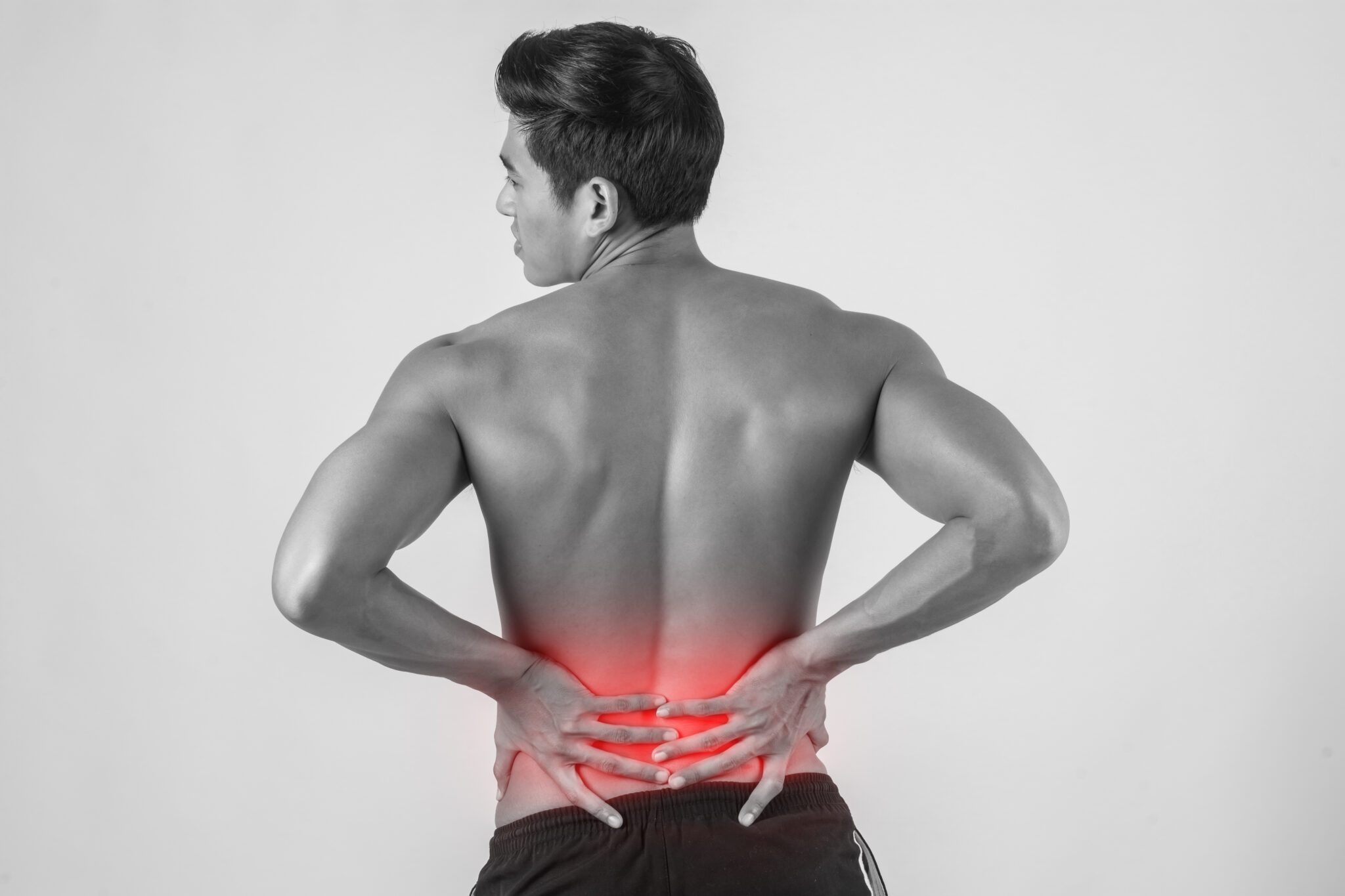
Symptoms of Spinal Disc Prolapse:

The symptoms of spinal disc prolapse can vary depending on the location and severity of the herniation. Common symptoms include localized back pain, radiating pain into the buttocks, legs, or arms, numbness, tingling, muscle weakness, and difficulty performing daily activities. In severe cases where the herniated disc compresses spinal nerves, individuals may experience bowel or bladder dysfunction, known as cauda equina syndrome, requiring immediate medical attention.

Where can a spinal disc prolapse occur?

It can occur in any of the 24 vertebrae in the spine which lie above the sacrum. However, is commonest in the lower back (lumbar spine) and neck (cervical spine).

The most common sites for a lumbar disc prolapse are between the fourth and fifth lumbar vertebrae and between the fifth lumbar vertebra and the sacrum. In the cervical spine, the most vulnerable levels are C4 to C7. The spine in the mid-back region is rarely affected. Symptoms of disc prolapse will depend on the location of the prolapse.


Diagnosis of Spinal Disc Prolapse:

Diagnosing spinal disc prolapse is done thorough medical history review, physical examination, and imaging studies. During the physical examination, surgeons will assess range of motion, reflexes, muscle strength, and sensation. Imaging tests such as X-rays, MRI scans, or CT scans to help understand the spinal structure injury and the level and extent of the prolapse or herniation of the spinal disc.

Treatment Options:

The treatment approach for spinal disc prolapse depends on the severity of symptoms and individual patient factors. Initial management often involves conservative measures such as rest, pain medication, physical therapy, and activity modification. Nonsteroidal anti-inflammatory drugs (NSAIDs) may help reduce pain and inflammation. Physical therapy aims to strengthen the surrounding muscles, improve flexibility, and promote proper posture to reduce strain on the spine.

In cases where conservative treatments fail to provide relief or symptoms worsen, more invasive interventions may be considered. Epidural steroid injections can deliver anti-inflammatory medication directly to the affected area, providing temporary pain relief. Surgical options, such as discectomy or microdiscectomy, involve removing the herniated portion of the disc to alleviate pressure on the nerves and alleviate symptoms. However, surgery is typically reserved for severe cases or when neurological deficits are present.

Surgical treatment for Spine Disc Prolapse

Surgical treatment for spinal disc prolapses is often used when conservative treatments fail or symptoms become debilitating. Surgery aims to reduce pain, restore function, and prevent neurological complications. We will see the various surgical techniques employed for the treatment of spinal disc prolapse in this blog.

Different Surgical Techniques:

Surgical techniques to cure spinal disc prolapse are tailored to the specific needs of the patient. Some surgical approaches are:

1. Discectomy:

Discectomy involves the partial or complete removal of the herniated portion of the disc that is compressing spinal nerves. This procedure can be performed using open surgery or minimally invasive techniques such as microdiscectomy. It has smaller incisions and specialized instruments, resulting in reduced tissue damage, shorter recovery times, and better surgical outcomes.

2. Laminectomy:

In cases where the herniated disc is causing spinal canal stenosis or compression of the spinal cord, laminectomy may be performed. This procedure involves the removal of a portion of the lamina (the bony arch of the vertebra) to create more space within the spinal canal, relieving pressure on the nerves.

3. Spinal Fusion:

Spinal fusion may be recommended for patients with severe instability or recurrent disc herniation. This procedure involves fusing two or more vertebrae together using bone grafts, metal screws, or rods to stabilize the spine and prevent further movement at the affected segment. Fusion is often combined with discectomy or laminectomy to address both instability and nerve compression.

Outcomes and Complications:

Overall, surgical treatment for spinal disc prolapse is associated with favourable outcomes, with the majority of patients experiencing significant pain relief and functional improvement following surgery. However, as with any surgical procedure, there are potential risks and complications to be aware of, including infection, bleeding, nerve injury, and recurrence of disc herniation. Minimally invasive techniques have been developed to mitigate these risks and optimize surgical outcomes, offering reduced postoperative pain, shorter hospital stays, and faster recovery times compared to traditional open surgery.

Surgical treatment plays a crucial role in the management of spinal disc prolapse, providing effective relief for patients with persistent symptoms or neurological deficits. By employing various surgical techniques and considering individual patient factors, healthcare providers can tailor treatment plans to meet the unique needs and preferences of each patient, ultimately optimizing outcomes and restoring spinal function. Close collaboration between patients and healthcare providers is key to ensuring informed decision-making and achieving successful surgical outcomes.

Preventive Measures:

While certain risk factors for spinal disc prolapse, such as age and genetics, cannot be modified, adopting healthy lifestyle habits and proper body mechanics can help prevent its occurrence. Maintaining a healthy weight, engaging in regular exercise to strengthen the core muscles, and practicing proper lifting techniques can reduce the risk of disc herniation. Avoiding activities that place excessive strain on the spine and using ergonomic furniture and tools can also mitigate the risk of injury.

To summarise

Spinal disc prolapse is a common spinal condition. It causes pain, discomfort and reduces quality of life. Medical and surgical treatments usually take care of spinal disc problems. Samarth Neuro & Trauma Hospital in Miraj, Sangli [Maharashtra] has the infrastructure and surgical team under the guidance of Neurosurgeon Dr Ravindra Patil to treat spinal disc prolapses.

स्पाइन डिस्क प्रोलॅप्स

स्पाइन डिस्क प्रोलॅप्स

स्पाइन डिस्क प्रोलॅप्स

स्पाइनल डिस्क प्रोलॅप्स, सामान्यतः स्लिप डिस्क किंवा हर्निएटेड डिस्क म्हणून ओळखली जाते. हा एक अनेकांना होणारा रोग आहे. याने जगभरातील लाखो व्यक्तीं प्रभावित होतात. जेव्हा इंटरव्हर्टेब्रल डिस्क्समधील मऊ, जेल सारखा पदार्थ बाहेरील कणखर थर फायल्यामुळे किंवा बाहेरील स्तराच्या कमकुवतपणाद्वारे बाहेर पडते तेव्हा वेदना होतात, अस्वस्थता वाटते आणि कधीकधी मज्जातंतूं वर दबाव येतो व सतत पाठीत किंवा मानेत दुखते. या ब्लॉगचे उद्दिष्ट स्पाइनल डिस्क प्रोलॅप्सचा तपशील प्रदान करणे हे आहे. स्पाइनल डिस्क प्रोलॅप्सची कारणे, लक्षणे, निदान, उपचार पर्याय आणि स्पाइन डिस्क प्रोलॅप्सचा प्रतिबंध कसा करावा याबद्दल काही माहिती या ब्लॉग मध्ये आहे.

By Dr. Ravindra Patil

Table of Contents

मणक्याचे शरीरशास्त्र:

स्पाइनल डिस्क प्रोलॅप्स समजून घेण्यासाठी, मणक्याचे शरीरशास्त्र समजून घेणे आवश्यक आहे. पाठीचा कणा एकमेकांवर रचलेल्या ३२ मणक्यांनी बनलेला असतो. दर दोन मणक्यांच्या मधे इंटरव्हर्टेब्रल डिस्क्स असतात व त्या दोन मणक्यामधीक उशी सारखे काम करतात. या डिस्क अथवा चकतींच्या बाहेरील भागात ॲन्युलस फायब्रोसस नावाचा कठीण स्तर असतो व त्याच्या आतील भागात न्यूक्लियस पल्पोसस नावाचा मऊ भाग असतो. डिस्कचे कार्य असते शॉक (धक्के) शोषून घेणे आणि पाठीच्या हालचाली सुलभ करणे.

32 कशेरुकांपैकी (म्हणजेच मणक्यांपैकी) खालच्या टोकाला असलेले पाच कशेरुक एकत्र मिळून सेक्रम नावाचे एक हाड तयार करतात. वरून सुरू केले तर कशेरुकांची माळ या प्रकारे असते: सात मानेच्या मणक्यांची, 12 थोरॅसिक किंवा पृष्ठीय मणक्यांची आणि पाच लंबर मणक्यांची माळ असते. कोक्सीक्स, किंवा शेपटीहाड, सॅक्रमच्या (कायमचे जोडलेले पाच मणके) खाली स्थित असते व ते तीन कायच्या जोडलेल्या कशेरुकांपासून बनते. कोक्सीक्स हे कशेरुकाच्या माळेतील किंवा स्तंभातील सर्वात खालचे हाड आहे.

स्पाइनल डिस्क प्रोलॅप्सची म्हणजे काय:

स्पाइनल डिस्क प्रोलॅप्सच्या विकासामध्ये अनेक घटक कारणीभूत शकतात. सामान्य कारणांमध्ये वय-संबंधित ऱ्हास, पुन्हा-पुन्हा होणाऱ्या दुखापती, वजन उचलण्याची अयोग्य पद्धत, लठ्ठपणा आणि अचानक झालेले पाठीवरचे  आघात यांचा समावेश होतो. डीजनरेटिव्ह बदल (ऱ्हास) बहुतेकदा वय किंवा दुखापतीशी संबंधित असतात व ते कालांतराने डिस्कला कमकुवत करतात, ज्यामुळे त्यांना डिस्कमध्ये हर्नियेशन होण्याची वाढते. पुन्हा-पुन्हा वाकणे, सारखे वजन उचलणे किंवा जास्त प्रमाणात वळणे यांचा समावेश असलेले क्रियाकलाप देखील डिस्कवर ताण आणू शकतात आणि प्रोलॅप्सचा धोका वाढवू शकतात.

स्पाइनल डिस्क प्रोलॅप्सची लक्षणे:

स्पाइनल डिस्क प्रोलॅप्सची लक्षणे हर्नियेशनच्या स्थानावर आणि तीव्रतेनुसार बदलू शकतात. (डिस्क हर्नियेशन व डिस्क प्रोलॅप्स समानार्थी आहेत). सामान्य लक्षणांमध्ये प्रोलॅप्स झालेल्या ठिकाणी पाठदुखी, नितंब, पाय किंवा हातांमध्ये पसरणारी वेदना, सुन्नपणा, मुंग्या येणे, स्नायू कमकुवत होणे आणि दैनंदिन क्रियाकलाप करण्यात अडचण येणे यांचा समावेश होतो. गंभीर प्रकरणांमध्ये जेथे हर्निएटेड डिस्क असते तिथे ती पाठीच्या नसा संकुचित करते. रूग्णाला मल-मूत्रावर नियंत्रण ठेवता येत नाही. याला कौडा इक्विना सिंड्रोम म्हणून ओळखले जाते. या परिस्थितीत ज्याला त्वरित वैद्यकीय मदतीची आवश्यकता असते.

स्पाइनल डिस्क प्रोलॅप्स कुठे होऊ शकते?

स्पाइनल डिस्क प्रोलॅप्स सॅक्रमवरील 24 मणक्यांपैकी कोणत्याही दोन मणक्यामध्ये होऊ शकते. तथापि, पाठीच्या खालच्या भागात (लंबर स्पाइन) आणि मान (मानेच्या मणक्यामध्ये) स्पाइनल डिस्क प्रोलॅप्स सर्वात अधीक प्रमाणात होते. लंबर डिस्क प्रोलॅप्ससाठी सर्वात सामान्य ठिकाणे चौथ्या आणि पाचव्या लंबर मणक्यांच्या मध्ये आणि पाचव्या लंबर मणक्यांच्या आणि सॅक्रमच्या दरम्यान आहेत. मानेच्या मणक्यामध्ये, सर्वात असुरक्षित पातळी C4 ते C7 आहेत. पाठीच्या मध्यभागी असलेल्या पाठीचा कणा क्वचितच प्रभावित होतो. डिस्क प्रोलॅप्सची लक्षणे प्रोलॅप्सच्या स्थानावर अवलंबून असतात.

स्पाइनल डिस्क प्रोलॅप्सचे निदान:

स्पाइनल डिस्क प्रोलॅप्सचे निदान वैद्यकीय इतिहासाचे संपूर्ण आवलोकन, शारीरिक तपासणी आणि इमेजिंग अभ्यास याने केले जाते. शारीरिक तपासणी दरम्यान शल्यचिकित्सक गती, प्रतिक्षेप, स्नायूंची ताकद आणि संवेदना यांच्या श्रेणीचे मूल्यांकन करतात. इमेजिंग चाचण्या जसे की एक्स-रे, एम.आर.आय. स्कॅन किंवा सी.टी. स्कॅन स्पाइनल मणक्यांच्या स्तंभाला दुखापत आणि स्पाइनल डिस्कच्या प्रोलॅप्स किंवा हर्निएशनची पातळी आणि व्याप्ती समजून घेण्यास मदत करतात.

उपचार पर्याय:

स्पाइनल डिस्क प्रोलॅप्ससाठी उपचार पद्धती लक्षणांच्या तीव्रतेवर आणि रुग्णाच्या वैयक्तिक परिस्थितीवर अवलंबून असते. प्रारंभिक व्यवस्थापनामध्ये सहसा विश्रांती, वेदना औषधोपचार, फिझियोथेरपी व्यायाम उपचार आणि क्रियाकलाप बदल यासारख्या सर्जरी शिवायच्या उपायांचा समावेश असतो. नॉनस्टेरॉइडल अँटी-इंफ्लेमेटरी ड्रग्स (NSAIDs) वेदना आणि जळजळ कमी करण्यात मदत करू शकतात. फिझियोथेरपीचा उद्देश आसपासच्या स्नायूंना बळकट करणे, लवचिकता सुधारणे आणि मणक्यावरील ताण कमी करण्यासाठी योग्य पवित्रा वाढवणे हे असतात.

ज्या प्रकरणांमध्ये सर्जरी शिवयचे उपचार आराम देण्यास अयशस्वी ठरतात किंवा लक्षणे खराब होतात, तेव्हा अधिक आक्रमक उपचारांचा विचार केला जाऊ शकतो. एपिड्यूरल स्टिरॉइड इंजेक्शन्स थेट प्रभावित भागात दाहक-विरोधी औषधे वितरीत देऊ शकतात, ज्यामुळे तात्पुरता वेदनांमध्ये आराम मिळतो. सर्जिकल पर्याय, जसे की डिसकेक्टॉमी किंवा मायक्रोडिसकेक्टोमी, मज्जातंतूंवरील दबाव कमी करण्यासाठी आणि लक्षणे कमी करण्यासाठी डिस्कचा हर्नियेटेड भाग काढून टाकतात. तथापि, शस्त्रक्रिया केवळ गंभीर प्रकरणांसाठी राखीव असते. जेव्हा न्यूरोलॉजिकल लक्षणे येतात तेव्हा देखील ऑपरेशनच करवावे लागते.

स्पाइन डिस्क प्रोलॅप्ससाठी सर्जिकल उपचार

जेव्हा औषधोपचार अयशस्वी होतात किंवा लक्षणे कमकुवत होतात तेव्हा स्पाइनल डिस्क प्रोलॅप्ससाठी सर्जिकल उपचार वापरले जातात. सर्जरीची उद्दिष्टे असतात वेदना कमी करणे, कार्य पुनर्संचयित करणे आणि न्यूरोलॉजिकल गुंतागुंत टाळणे. स्पाइनल डिस्क प्रोलॅप्सच्या उपचारासाठी वापरलेली विविध शस्त्रक्रिया तंत्रे आपण या ब्लॉगमध्ये पाहू.

विविध सर्जिकल तंत्रे:

स्पाइनल डिस्क प्रोलॅप्स बरा करण्यासाठी सर्जिकल तंत्र रुग्णाच्या विशिष्ट परिस्थितीनुसार तयार केली जातात. काही सर्जिकल पद्धती खालील प्रमाणे आहेत:

1. डिस्केक्टॉमी:

डिस्केक्टॉमी म्हणजे पाठीच्या नसा संकुचित करणाऱ्या डिस्कचा हर्निएटेड भाग आंशिक किंवा पूर्णपणे काढून टाकणे. ही प्रक्रिया खुली शस्त्रक्रिया किंवा मायक्रोडिस्केक्टोमी सारख्या कमीत कमी आक्रमक तंत्रांचा वापर करून केली जाऊ शकते. मायक्रोडिस्केक्टोमी मध्ये लहान चीरे आणि विशेष उपकरणे वापरतात, ज्यामुळे आजूबाजूच्या ऊतींचे नुकसान कमी होते, बरे होण्यास कमी वेळा लागतो आणि शस्त्रक्रिया चांगले परिणाम देते.

2. लॅमिनेक्टॉमी:

ज्या प्रकरणांमध्ये हर्निएटेड डिस्कमुळे स्पाइनल कॅनाल स्टेनोसिस होतो म्हणजे स्पाइनल कॅनाल संकुचीत होतो व स्पाईनल कॉर्ड वर दाब येतो तेव्हा लॅमिनेक्टॉमी केली जाऊ शकते. या प्रक्रियेमध्ये मेरुदंडाच्या कालव्यामध्ये (स्पाइनल कॅनाल मध्ये) अधिक जागा तयार करण्यासाठी लॅमिना, म्हणजे कशेरुकाच्या हाडांची कमान, त्यातील एक भाग काढून टाकतात. यामुळे मज्जातंतूंवरील दबाव कमी होतो.

3. स्पाइनल फ्यूजन:

पाठीची गंभीर अस्थिरता किंवा वारंवार डिस्क हर्नियेशन होणे, हे ज्यांना होते तसल्या रुग्णांसाठी स्पाइनल फ्यूजनचा सल्ला दिला जाऊ शकतो. या प्रक्रियेमध्ये हाडांच्यात कलम (बोन ग्राफ्टींग), धातूचे स्क्रू किंवा रॉड्स वापरून दोन किंवा अधिक मणक्यांना एकत्र जोडून पाठीचा कणा स्थिर करणे आणि प्रभावित भागात पुढील हालचाल रोखणे एवढे करतात. अस्थिरता आणि मज्जातंतूंवर येणारा दाब व त्यांची संकुचितता या दोहोंचा उपचार करण्यासाठी स्पाइनल फ्यूजन अनेकदा डिस्केक्टोमी किंवा लॅमिनेक्टोमीसह एकत्र केले जाते.

परिणाम आणि गुंतागुंत:

एकूण स्पाइनल डिस्क प्रोलॅप्ससाठी शस्त्रक्रियांचे परिणाम चांगले असतात. बहुतेक रुग्णांना शस्त्रक्रियेनंतर वेदनांपासून आराम आणि त्यांच्या दैनंदीन कार्यात सुधारणा झाल्याचे अनुभवत येतात. तथापि, कोणत्याही शल्यक्रिये प्रमाणेच या शल्यक्रियेतही संसर्ग, रक्तस्त्राव, मज्जातंतूला दुखापत आणि डिस्क हर्नियेशनची पुनरावृत्ती यासह संभाव्य धोके आणि गुंतागुंतीची जाणीव असणे आवश्यक आहे. हे धोके कमी करण्यासाठी आणि शस्त्रक्रियेच्या परिणामांना अनुकूल करण्यासाठी कमीत कमी आक्रमक तंत्रे विकसित केली गेली आहेत. यामुळे शस्त्रक्रियेनंतरच्या वेदना कमी होतात, रूग्णांना रूग्णालयात कमी दिवसा राहावे लागते आणि पारंपारिक खुल्या शस्त्रक्रियेच्या तुलनेत रूग्ण लवकर बरे होतात.

स्पाइनल डिस्क प्रोलॅप्सच्या व्यवस्थापनात सर्जिकल उपचार महत्त्वपूर्ण भूमिका बजावतात, ज्यामुळे सतत लक्षणे किंवा न्यूरोलॉजिकल लक्षणे असलेल्या रुग्णांना बर्यापैकी आराम मिळतो. विविध शल्यचिकित्सा तंत्रांचा वापर करून आणि रुग्णाच्या वैयक्तिक परिस्थितीचा विचार करून स्पाइन सर्जन प्रत्येक रुग्णाच्या गरजा आणि प्रकृती यांचा संपूर्ण विचार करून उपचार योजना तयार करतात. याने परिणाम सर्वात चांगले येऊ शकतात आणि पाठीचा कणा त्याचे कार्य पूर्वी सारखे करू शकतो. रुग्ण आणि स्पाईन सर्जन यांच्यात विश्वास व सहकार्य हे सर्व गोष्टी समजून निर्णय घेण्यास आणि यशस्वी शस्त्रक्रिया परिणाम प्राप्त करण्यास महत्त्वाचे आहे.

प्रतिबंधात्मक उपाय:

स्पाइनल डिस्क प्रोलॅप्ससाठी काही जोखीम घटक, जसे की वय आणि आनुवंशिकता, बदलता येत नाहीत. परंतु निरोगी जीवनशैलीच्या सवयी आणि योग्य शरीर यांत्रिकी अंगीकारणे स्पायनल डिस्क प्रोलॅप्स टाळण्यासाठी मदत करू शकते. योग्य वजन राखणे, पाठीच्या मुख्य स्नायूंना बळकट करण्यासाठी नियमित व्यायाम करणे आणि कोणतीही वस्तू उचलण्यासाठी योग्य तंत्रांचा सराव केला तर स्पयनल डिस्क हर्नियेशनचा धोका कमी होऊ शकतो. मणक्यावर जास्त ताण पडणाऱ्या क्रियाकलाप जरूर टाळावेत व अर्गोनॉमिक फर्निचर आणि साधने वापरणे हे देखील इजा होण्याचा धोका कमी करू शकतात.

थोडक्यात सांगायचे म्हणजे…

स्पाइनल डिस्क प्रोलॅप्स हा अनेकांना होणारा रोग आहे. यामुळे वेदना होतात, अस्वस्थता येते आणि जीवनाची गुणवत्ता कमी होते. वैद्यकीय आणि शस्त्रक्रिया उपचार सहसा पाठीच्या डिस्कच्या समस्या कमी करू शकतात. मिरज, सांगली [महाराष्ट्र] येथील समर्थ न्यूरो आणि ट्रॉमा हॉस्पिटलमध्ये स्पायनल डिस्क प्रोलॅप्सवर उपचार करण्यासाठी न्यूरो सर्जन डॉ. रवींद्र पाटील यांच्या मार्गदर्शनाखाली पायाभूत सुविधा आणि सर्जिकल टीम आहे.

Brain Disorders

Brain disorders

Brain Disorders

Brain disorders encompass a vast array of conditions that affect the structures and function of the brain, leading to alterations in cognition, behaviour, emotion, and physical abilities. From neurodevelopmental disorders like autism spectrum disorder to neurodegenerative diseases such as Alzheimer’s, these conditions pose significant challenges to affected individuals and their families. Understanding the complexity and diversity of brain disorders is crucial for advancing research, developing effective treatments, and improving the quality of life for those affected.

By Dr.Ravindra Patil

Table of Contents

Alzheimer’s Disease

One of the most well-known and prevalent brain disorders is Alzheimer’s disease, a progressive neurodegenerative disorder characterized by cognitive decline, memory loss, and changes in behaviour. It primarily affects older adults and is the leading cause of dementia worldwide. Alzheimer’s is associated with the accumulation of abnormal proteins in the brain, including beta-amyloid plaques and tau tangles, which interfere with neuronal function and communication. While there is currently no cure for Alzheimer’s, ongoing research aims to identify potential therapeutic targets to slow or halt disease progression.

Parkinson’s disease

Parkinson’s disease is another common neurodegenerative disorder, characterized by motor symptoms such as tremors, bradykinesia (slowed movement), rigidity, and postural instability. It results from the loss of dopamine-producing neurons in the substantia nigra region of the brain. In addition to motor symptoms, Parkinson’s can also cause non-motor symptoms, including cognitive impairment, mood changes, and sleep disturbances. Treatment typically involves medications to manage symptoms, as well as physical therapy and lifestyle modifications.

Neurodevelopmental Disorders

Neurodevelopmental disorders encompass a group of conditions that typically emerge in early childhood and affect brain development, leading to difficulties in social interaction, communication, and behaviour. Autism spectrum disorder (ASD) is one such disorder, characterized by challenges with social communication and interaction, restricted interests, and repetitive behaviours. The exact cause of ASD remains unclear, but both genetic and environmental factors are thought to play a role. Early intervention and behavioural therapy can help individuals with ASD improve communication skills and adaptive behaviours.

Attention-Deficit/Hyperactivity Disorder (ADHD)

Attention-deficit/hyperactivity disorder (ADHD) is another neurodevelopmental disorder characterized by inattention, hyperactivity, and impulsivity. It often manifests in childhood and can persist into adulthood. ADHD can significantly impact academic performance, social relationships, and daily functioning. While the exact cause of ADHD is unknown, it is believed to involve a combination of genetic, neurological, and environmental factors. Treatment typically involves a combination of medication, behavioral therapy, and support services.

Mood Disorders

Mood disorders, such as depression and bipolar disorder, involve disruptions in mood regulation and emotional stability. Depression is characterized by persistent feelings of sadness, hopelessness, and loss of interest or pleasure in activities. It can also manifest physical symptoms such as changes in appetite, sleep disturbances, and fatigue. Bipolar disorder involves episodes of depression alternating with periods of mania or hypomania, characterized by elevated mood, increased energy, and impulsivity. Both depression and bipolar disorder are associated with alterations in brain chemistry and function, as well as genetic predisposition. Treatment may include medication, psychotherapy, and lifestyle modifications.

Schizophrenia

Schizophrenia is a chronic and severe mental disorder characterized by distorted thinking, hallucinations, delusions, and impairments in social and occupational functioning. It typically emerges in late adolescence or early adulthood and can have a profound impact on an individual’s life. The exact cause of schizophrenia is not fully understood but is believed to involve a combination of genetic, environmental, and neurobiological factors. Treatment often involves antipsychotic medications, psychotherapy, and supportive services to help individuals manage symptoms and improve quality of life.

Traumatic brain injury (TBI)

Traumatic brain injury (TBI) refers to damage to the brain caused by an external force, such as a blow to the head or a penetrating injury. TBIs can range from mild concussions to severe injuries resulting in long-term cognitive, physical, and emotional impairments. Symptoms of TBI can vary widely depending on the severity and location of the injury but may include headaches, dizziness, memory problems, and changes in mood or behaviour. Treatment typically involves a multidisciplinary approach, including medical intervention, rehabilitation, and cognitive therapy.

Epilepsy

Epilepsy is a neurological disorder characterized by recurrent seizures, which result from abnormal electrical activity in the brain. Seizures can vary in severity and may involve convulsions, loss of consciousness, or subtle changes in awareness. Epilepsy can have various causes, including genetic factors, brain injury, infection, or structural abnormalities in the brain. While epilepsy cannot be cured, many individuals with the condition can effectively manage seizures with antiepileptic medications, lifestyle modifications, and, in some cases, surgery.

Brain tumours

Brain tumours are abnormal growths of cells in the brain that can be benign or malignant. Symptoms vary depending on the tumour’s size, location, and rate of growth, often including headaches, seizures, cognitive impairment, and changes in behaviour. Treatment may involve surgery, radiation therapy, chemotherapy, or a combination, depending on the tumour type and stage. Early diagnosis and intervention are crucial for improving outcomes and quality of life for those affected by brain tumours. Ongoing research aims to improve understanding, develop targeted therapies, and ultimately find a cure for these complex and challenging conditions.

Cerebrovascular stroke

Cerebrovascular stroke occurs when blood flow to the brain is disrupted, either due to a blockage (ischemic stroke) or a rupture (haemorrhagic stroke) in a blood vessel. Ischemic strokes, the most common type, typically result from a blood clot blocking an artery supplying blood to the brain. Haemorrhagic strokes occur when a weakened blood vessel ruptures and bleeds into the brain. Strokes can lead to brain damage and permanent neurological deficits if not promptly treated.

Symptoms of stroke can vary but often include sudden weakness or numbness on one side of the body, difficulty speaking or understanding speech, severe headache, and trouble walking. Immediate medical attention is critical to minimize brain damage and improve outcomes.

Treatment for ischemic stroke may involve thrombolytic therapy to dissolve the clot, mechanical thrombectomy to remove it, or anticoagulant medications to prevent further clots. Haemorrhagic strokes may require surgical intervention to repair the blood vessel and reduce bleeding.

Rehabilitation, including physical therapy, speech therapy, and occupational therapy, is essential for stroke survivors to regain function and independence. Lifestyle modifications, such as maintaining a healthy diet, exercising regularly, and managing risk factors like high blood pressure and diabetes, can also help prevent future strokes. Public education campaigns emphasize the importance of recognizing stroke symptoms and seeking immediate medical attention. Early intervention significantly improves outcomes and reduces disability.

Birth injuries to the brain

Birth injury to the brain, often known as birth asphyxia or neonatal hypoxic-ischemic encephalopathy (HIE), occurs when an infant’s brain is deprived of oxygen during childbirth. This can result from complications such as umbilical cord compression, placental abruption, or prolonged labour. Birth injuries may lead to neurological deficits, developmental delays, and lifelong disabilities. Prompt medical intervention, including resuscitation and therapeutic hypothermia, can mitigate brain damage and improve outcomes. However, some infants may require long-term medical care and rehabilitation to address cognitive, motor, and sensory impairments resulting from birth-related brain injury.

These are just a few examples of the diverse range of brain disorders that affect millions of people worldwide. Significant progress has been made in understanding the underlying mechanisms and developing treatments. Yet, much remains to be learned. Continuing research is the key to progress in brain disorders.