Homepage Marathi-समर्थ हॉस्पिटल
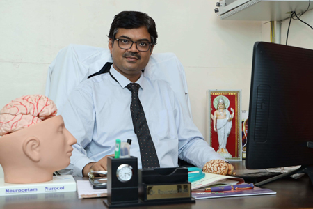
नमस्कार! मी
डॉ. रवींद्र आर. पाटील
MS, MCh(Neuro Surgery)
डायरेक्टर
समर्थ न्यूरो आणि सुपरस्पेशालिटी हॉस्पिटल, मिरज.
पश्चिम महाराष्ट्रातील सुवर्ण पदक विजेता न्यूरोसर्जन
पश्चिम महाराष्ट्रातील उभरते
उत्कृष्ट हॉस्पिटल
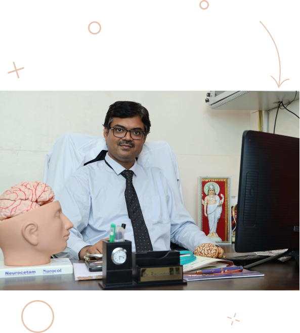
नमस्कार! मी
डॉ. रवींद्र आर. पाटील
MS, MCh(Neuro Surgery)
डायरेक्टर समर्थ न्यूरो आणी ट्रॉमा हॉस्पीटल
दक्षिण महाराष्ट्रातील सुवर्ण पदक विजेता न्यूरोसर्जन
पश्चिम महाराष्ट्रातील उभरते उत्कृष्ट हॉस्पिटल

रोज आरोग्य संबंधित टिप्स मिळवा…तुम्हाला निरोगी रहायचे आहे ना ? मग एक पाऊल पुढे टाका… क्लिक करा आणि पहा आरोग्य संबंधित रोज एक नवी टीप…न्यूरोसर्जनडॉ. रवींद्र पाटील यांनी मोठमोठ्या शहरातील आकर्षक व्यावसायिक संधी सोडून दक्षिण महाराष्ट्रात मिरज मध्ये न्यूरोसर्जरी होस्पिटल सुरु केले.परवडणारी न्यूरो आणि ट्रॉमा केअर – मिरज आणि सांगलीमध्ये!जरी न्यूरोसर्जरीला हॉस्पिटलच्या उच्च तंत्रज्ञान पायाभूत सुविधांची आवश्यकता असली, तरीदेखील समर्थ हॉस्पिटल मध्ये वाजवी किंमतीत उच्च दर्जाचे उपचार उपलब्ध आहेत.एव्हिडन्स बेस्ड मेडिसिन [EBM]समर्थ हॉस्पिटल मध्ये प्रत्येक रुग्णाचे उपचार पर्याय एव्हिडन्स बेस्ड मेडिसिन प्रमाणेच ठरवले जातात. ही उपचार पद्धत मेडिकल शास्त्रात सर्वोत्तम मानली जाते. इमर्जन्सी आणि ट्रॉमा केअर साठी तज्ञांची टीमफास्ट आणि बेस्ट – हे आहे आमच्या इमर्जन्सी रुमचे [ER] बोधवाक्य ! आमची डॉक्टर, नर्सीस व पॅरामेडिकल स्टाफची टीम तत्काल आपात कालीन सेवा देण्यास नेहमीच तत्पर असते.माझी पत्नी डॉ. शिल्पा पाटील, बालरोग तज्ञआम्ही दोघं ही डॉक्टर आहोत व समर्थ हॉस्पिटलच्या वरती राहतो. आमचे व्यवसायिक व घरगुती जीवनं एकमेकात मिसळली असतात.
थोडेसे आमच्या विषयी
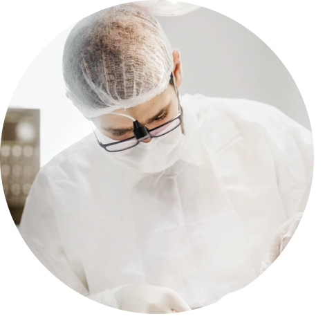
न्यूरोसर्जरी आणि संबंधित शाखांमध्ये सर्वोत्तम पेशंट मॅनेजमेंट
पश्चिम महाराष्ट्रातील, न्यूरोसर्जरी आणि संबंधित शाखांसाठी सर्वोत्कृष्ट हॉस्पिटल बनणे, हे आमचे ध्येय आहे आणि आम्ही आमचे कार्य पूर्ण करण्यासाठी अहोरात्र प्रयत्न करत आहोत. आम्ही न्यूरोसर्जरी, न्यूरोलॉजी, स्पाइन सर्जरी, ट्रॉमा सर्जरी आणि पेडियाट्रिक्स या शाखांमध्ये उपचार करतो. उत्तम, कुशल कर्मचारी नियुक्त करणे, त्यांना सतत ट्रेनिंग देणे, जागतिक दर्जाची बायोमेडिकल उपकरणे वापरणे, आणि बिल्डिंगचे इन्फ्रास्ट्रक्चर उत्कृष्ट राखणे, यासाठी प्रयत्न करतो. इमर्जन्सी केसेससाठी समर्थ हॉस्पिटल 24 तास कार्यरत असते.
न्यूरोसर्जरी
हा मज्जातंतूं (नर्व्हस सिस्टीम) च्या रोगांचे निदान, मेडिकल / सर्जिकल ट्रीटमेंट, रिहॅबिलिटेशन आणि प्रिव्हेन्शन (प्रतिबंध) विभाग आहे.
पेडियाट्रिक्स
नवजात बाळे, मुले आणि पौगंडावस्थेतील मुलांची वैद्यकीय काळजी घेणारी वैद्यकीय शाखा.
न्यूरॉलॉजी
न्यूरोलॉजी मेंदू, पाठीचा कणा आणि नर्व्ह यांच्या आजाराचे निदान आणि त्यावर उपचार करणारी शाखा आहे. यामध्ये स्ट्रोक, सिजर्स, पार्किन्सन, डिमेंशिया इत्यादी प्रमुख आजारांवर उपचार केले जातात.
निदान
आधुनिक निदानामध्ये पॅथॉलॉजी लॅबोरेटरी टेस्टिंग आणि सर्व प्रकाच्या मेडिकल डायग्नोस्टिक इमेजिंगचा म्हणजे एक्स-रे, USG, CT स्कॅन, MRI स्कॅन इत्यादींचा समावेश आहे.

![]()
![]()
![]()
थोडेसे आमच्या विषयी
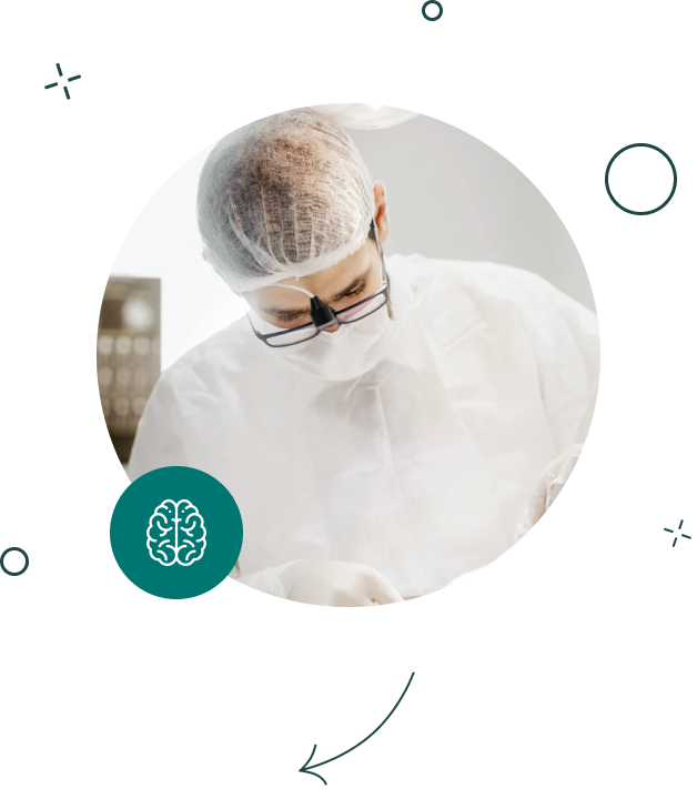
न्यूरोसर्जरी आणि संबंधित शाखांमध्ये सर्वोत्तम पेशंट मॅनेजमेंट
पश्चिम महाराष्ट्रातील, न्यूरोसर्जरी आणि संबंधित शाखांसाठी सर्वोत्कृष्ट हॉस्पिटल बनणे, हे आमचे ध्येय आहे आणि आम्ही आमचे कार्य पूर्ण करण्यासाठी अहोरात्र प्रयत्न करत आहोत. आम्ही न्यूरोसर्जरी, न्यूरोलॉजी, स्पाइन सर्जरी, ट्रॉमा सर्जरी आणि पेडियाट्रिक्स या शाखांमध्ये उपचार करतो. उत्तम, कुशल कर्मचारी नियुक्त करणे, त्यांना सतत ट्रेनिंग देणे, जागतिक दर्जाची बायोमेडिकल उपकरणे वापरणे, आणि बिल्डिंगचे इन्फ्रास्ट्रक्चर उत्कृष्ट राखणे, यासाठी प्रयत्न करतो. इमर्जन्सी केसेससाठी समर्थ हॉस्पिटल 24 तास कार्यरत असते.
न्यूरोसर्जरी
हा मज्जातंतूं (नर्व्हस सिस्टीम) च्या रोगांचे निदान, मेडिकल / सर्जिकल ट्रीटमेंट, रिहॅबिलिटेशन आणि प्रिव्हेन्शन विभाग आहे.
पेडियाट्रिक्स
नवजात बाळे, मुले आणि पौगंडावस्थेतील मुलांची वैद्यकीय काळजी घेणारी वैद्यकीय शाखा.
न्यूरॉलॉजी
न्यूरोलॉजी मेंदू, पाठीचा कणा आणि नर्व्ह यांच्या आजाराचे निदान आणि त्यावर उपचार करणारी शाखा आहे. यामध्ये स्ट्रोक, सिजर्स, पार्किन्सन, डिमेंशिया इत्यादी प्रमुख आजारांवर उपचार केले जातात.
निदान
आधुनिक निदानामध्ये पॅथॉलॉजी लॅबोरेटरी टेस्टिंग आणि सर्व प्रकाच्या मेडिकल डायग्नोस्टिक इमेजिंगचा म्हणजे एक्स-रे, USG, CT स्कॅन, MRI स्कॅन इत्यादींचा समावेश आहे.
सेवा
व्यापक स्तरावर / कॉम्प्रिहेंसिव न्यूरो केअर
हेड इंज्युरी (डोक्याला दुखापत)
टाळू, कवटी किंवा मेंदूला झालेला कोणताही आघात म्हणजे हेड इंज्युरी होय. ह्या मेंदूच्या किरकोळ किंवा गंभीर दुखापती असू शकतात. डोक्याच्या सर्व दुखापतींची वैद्यकीय तपासणी करणे आवश्यक आहे.
Know more
ब्रेन ट्यूमर
ही मेंदूमधील असामान्य पेशींची वाढ आहे, ही वाढ कॅन्सर संबंधित असू शकते किंवा नसू शकते. कॅन्सर नसलेले ब्रेन ट्यूमर देखील गंभीर असतात आणि त्यांची तपासणी करून, त्यावर उपचार करणे आवश्यक आहे.
Know more
स्पाईन ट्यूमर
पाठीचा कणा आणि / किंवा पाठीच्या कणास्तंभामध्ये किंवा त्याच्या सभोवतालच्या भागात टिश्यूचा असामान्य दबाव म्हणजे स्पाईन ट्यूमर होय. हा सौम्य [कॅन्सर नसलेला] किंवा घातक [कॅन्सर] असू शकतो.
Know more
डोकेदुखी
डोकेदुखी बहुतेकवेळा सर्दी किंवा सायनस सारख्या साध्या समस्यांमुळे होते, तरीदेखील सतत होणाऱ्या डोकेदुखीची वैद्यकीय तपासणी करणे आवश्यक आहे. कारण ती एखाद्या गंभीर आजाराचे लक्षण असू शकते.
Know more
CV स्ट्रोक
ही एक मेडिकल इमर्जन्सी आहे. लक्षणे: चालणे, बोलणे, समजून घेण्यात अडचण, अशक्तपणा/ सुन्नपणा / पॅरालिसिस/ चेहरा, हात, पाय किंवा संपूर्ण शरीराच्या एका बाजूला किंवा दोन्ही बाजूला. यासाठी त्वरित उपचारांची आवश्यकता असते.
Know more
एपिलेप्सी (फेफरे)
हा एक मेंदूचा विकार आहे. यामध्ये मेंदूचे कार्य अस्वाभाविक होते, यामुळे वागणूक, संवेदना असामान्य होते, कधीकधी जागरूकता कमी होते आणि सिजर्स देखील होते. यासाठी अचूक निदान आणि आजीवन उपचारांची आवश्यक असते.
Know more
ट्रायजेमिनल न्यूराल्जिया
ट्रायजेमिनल नर्व्ह आपल्या चेहऱ्यापासून मेंदूपर्यंत सेन्सेशन (जाणिवा) नेते. ट्रायजेमिनल न्यूराल्जिया ही चेहरा किंवा दातांमध्ये होणारी एक तीव्र वेदना आहे आणि अगदी सौम्य उत्तेजनामुळे देखील ती तीव्र होऊ शकते.
Know more
पार्किन्सन रोग
हा एक मेंदूचा विकार आहे ज्यामध्ये थरथरणे, कडकपणा येणे आणि चालणे, संतुलन करणे आणि को-ऑर्डिनेशन यामध्ये अडचण होते. लक्षणे सहसा हळूहळू सुरू होतात आणि जसा रोग वाढत जातो, तशी ती अधिक तीव्र व गंभीर होतात.
Know more
फेशियल पाल्सी (चेहऱ्याचा पक्षघात)
चेहऱ्यावरील स्नायूंचा अशक्तपणा किंवा पॅरालिसिस, यामुळे मुख्यतः चेहऱ्याच्या नर्व्हला तात्पुरते किंवा कायमचे नुकसान होते. यामुळे डोळे आणि तोंडाच्या हालचालीवर परिणाम होतो.
Know more
हेड इंज्युरी (डोक्याला दुखापत)
टाळू, कवटी किंवा मेंदूला झालेला कोणताही आघात म्हणजे हेड इंज्युरी होय. ह्या मेंदूच्या किरकोळ किंवा गंभीर दुखापती असू शकतात. डोक्याच्या सर्व दुखापतींची वैद्यकीय तपासणी करणे आवश्यक आहे.
ब्रेन ट्यूमर
ही मेंदूमधील असामान्य पेशींची वाढ आहे, ही वाढ कॅन्सर संबंधित असू शकते किंवा नसू शकते. कॅन्सर नसलेले ब्रेन ट्यूमर देखील गंभीर असतात आणि त्यांची तपासणी करून, त्यावर उपचार करणे आवश्यक आहे.
Know more
स्पाईन ट्यूमर
पाठीचा कणा आणि / किंवा पाठीच्या कणास्तंभामध्ये किंवा त्याच्या सभोवतालच्या भागात टिश्यूचा असामान्य दबाव म्हणजे स्पाईन ट्यूमर होय. हा सौम्य [कॅन्सर नसलेला] किंवा घातक [कॅन्सर] असू शकतो.
Know more
डोकेदुखी
डोकेदुखी बहुतेकवेळा सर्दी किंवा सायनस सारख्या साध्या समस्यांमुळे होते, तरीदेखील सतत होणाऱ्या डोकेदुखीची वैद्यकीय तपासणी करणे आवश्यक आहे. कारण ती एखाद्या गंभीर आजाराचे लक्षण असू शकते.
Know more
CV स्ट्रोक
ही एक मेडिकल इमर्जन्सी आहे. लक्षणे: चालणे, बोलणे, समजून घेण्यात अडचण, अशक्तपणा/ सुन्नपणा / पॅरालिसिस/ चेहरा, हात, पाय किंवा संपूर्ण शरीराच्या एका बाजूला किंवा दोन्ही बाजूला. यासाठी त्वरित उपचारांची आवश्यकता असते.
Know more
एपिलेप्सी (फेफरे)
हा एक मेंदूचा विकार आहे. यामध्ये मेंदूचे कार्य अस्वाभाविक होते, यामुळे वागणूक, संवेदना असामान्य होते, कधीकधी जागरूकता कमी होते आणि सिजर्स देखील होते. यासाठी अचूक निदान आणि आजीवन उपचारांची आवश्यक असते.
Know more
ट्रायजेमिनल न्यूराल्जिया
ट्रायजेमिनल नर्व्ह आपल्या चेहऱ्यापासून मेंदूपर्यंत सेन्सेशन (जाणिवा) नेते. ट्रायजेमिनल न्यूराल्जिया ही चेहरा किंवा दातांमध्ये होणारी एक तीव्र वेदना आहे आणि अगदी सौम्य उत्तेजनामुळे देखील ती तीव्र होऊ शकते.
Know more
पार्किन्सन रोग
हा एक मेंदूचा विकार आहे ज्यामध्ये थरथरणे, कडकपणा येणे आणि चालणे, संतुलन करणे आणि को-ऑर्डिनेशन यामध्ये अडचण होते. लक्षणे सहसा हळूहळू सुरू होतात आणि जसा रोग वाढत जातो, तशी ती अधिक तीव्र व गंभीर होतात.
Know more
फेशियल पाल्सी (चेहऱ्याचा पक्षघात)
चेहऱ्यावरील स्नायूंचा अशक्तपणा किंवा पॅरालिसिस, यामुळे मुख्यतः चेहऱ्याच्या नर्व्हला तात्पुरते किंवा कायमचे नुकसान होते. यामुळे डोळे आणि तोंडाच्या हालचालीवर परिणाम होतो.
Know more
वर प्रकाशित


वर प्रकाशित

यश
उत्कृष्ट क्लिनिकल निकाल
समर्थ हॉस्पिटलचे ‘जीवन वाचवण्याचे कमिटमेंट’ हे व्यावसायिक नीतिमत्तेपेक्षा अधिक एक संस्कृती आहे. बर्याचवेळा न्यूरोसर्जरी आणि ट्रॉमा ह्या दोन्ही जीवघेण्या स्थिती असूनदेखील, आम्ही अनेक जीव वाचवू शकलो आणि असे बरेच रुग्ण ज्यांना बरे होण्याची आशा नव्हती, त्यांना संपूर्ण फंक्शनल रिकव्हरी साध्य करण्यात मदत करू शकलो, हे सांगताना आम्हाला अभिमान वाटतो. तीन वर्ष आणि 3,500 सर्जरीनंतर, लोकांनी आमच्या सेवेवर दाखवलेला पूर्ण विश्वास हेच आमचे सर्वात मोठे यश आहे, असे आम्ही मानतो.
3:15
Watch Video
![]()
![]()
![]()


यश
उत्कृष्ट क्लिनिकल निकाल
समर्थ हॉस्पिटलचे ‘जीवन वाचवण्याचे कमिटमेंट’ हे व्यावसायिक नीतिमत्तेपेक्षा अधिक एक संस्कृती आहे. बर्याचवेळा न्यूरोसर्जरी आणि ट्रॉमा ह्या दोन्ही जीवघेण्या स्थिती असूनदेखील, आम्ही अनेक जीव वाचवू शकलो आणि असे बरेच रुग्ण ज्यांना बरे होण्याची आशा नव्हती, त्यांना संपूर्ण फंक्शनल रिकव्हरी साध्य करण्यात मदत करू शकलो, हे सांगताना आम्हाला अभिमान वाटतो. तीन वर्ष आणि 3,500 सर्जरीनंतर, लोकांनी आमच्या सेवेवर दाखवलेला पूर्ण विश्वास हेच आमचे सर्वात मोठे यश आहे, असे आम्ही मानतो.
3:15
Watch Video

![]()
![]()
![]()
![]()
खास वैशिष्ट्ये
आमची खास वैशिष्ट्ये
रुग्णांसाठी 24×7 सपोर्ट
आमच्या इमर्जन्सी रूम [ER] आणि ऑपरेटिंग रूम [OR] इमर्जन्सी उपचारांसाठी सहज मिळणाऱ्या आणि नेहमी उपलब्ध असतात.
उच्च शिक्षित (पात्र), कुशल आणि अनुभवी टीम
टॉपचे सर्जन ते सहाय्यक स्टाफ पर्यंत, इमर्जन्सी रिस्पॉन्स टीममधील प्रत्येकास रुग्ण आल्यावर त्यांनी काय काम करायचे, हे माहित असते. रुग्णाची स्थिती अजून खराब होण्यापासून वाचवण्यात (स्टॅबिलाइझ) आणि रोगाचे निदान करण्यास सुरुवात करण्यास उशीर होत नाही.
गोल्डन अव्हर
ट्रॉमामध्ये, घटना घडल्यानंतर लगेच एका तासात केलेल्या उपचारांचा परिणाम सर्वोत्तम मिळतो. याला गोल्डन अव्हर असे म्हणतात. समर्थ हॉस्पिटलची ER टीम, गोल्डन अव्हरमध्ये उपचार करण्यास नेहमीच तयार आहे.
our reviews
Reviews about Us
Lorem ipsum dolor sit amet, consectetur adipiscing elit, sed do eiusmod tempor incididunt ut labore et dolore magna aliqua. Ut enim ad minim veniam, quis nostrud
our reviews
Video Gallery
image gallery
Image gallery


