त्याला मणक्यात दोनदा ट्यूमर झाले पण सर्जिकल नेव्हिगेशनने वाचवले

Surgical Navigation - Ravindra patil

त्याला मणक्यात दोनदा ट्यूमर झाले पण सर्जिकल नेव्हिगेशनने वाचवले

By Dr.Ravindra Patil

समर्थ न्यूरो आणि मल्टीस्पेशालिटी हॉस्पिटल [मिरज, पश्चिम महाराष्ट्र] चे मुख्य न्यूरोसर्जन डॉ रवींद्र पाटील, त्यांनी सर्जिकल नेव्हिगेशनच्या मदतीने केलेल्या एक कठीण केस विषयी माहिती सांगत आहेत…

महंतेश शेलीकेरी हे मूळचे बेळगावचे आहेत. त्यांना न्यूरोफायब्रोमेटोसिस नावाचा दुर्मिळ आजार होता. त्याच्या पाठीत पाठीच्या कण्यावर दोन गाठी झाल्या होत्या व त्या स्पायनल कॉर्डवर दाब देत होत्या. त्यामुळे दोन महिन्यांपासून त्याची चालण्याची क्षमता कमी झाली होती.

Table of Contents

न्यूरोफिब्रोमेटोसिस

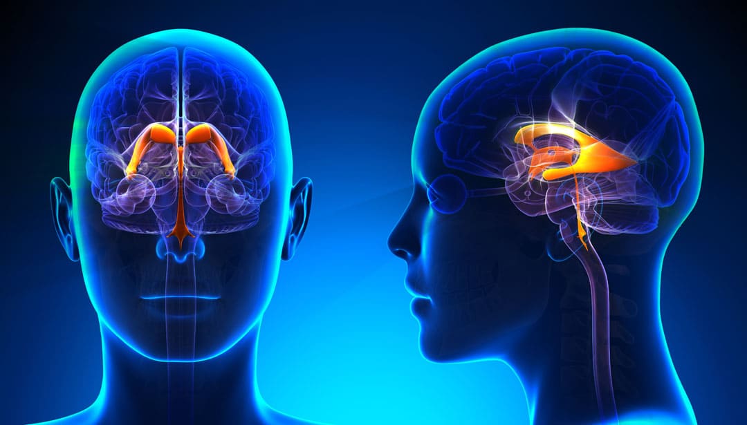
Neurofibromatosis (NF) ही अशी परिस्थिती आहे ज्यामध्ये मज्जासंस्थेमध्ये ट्यूमर वाढतात. न्यूरोफिब्रोमॅटोसिसचे तीन प्रकार आहेत, प्रकार I (NF1), प्रकार II (NF2) आणि श्वानोमॅटोसिस. NF मधील ट्यूमर सामान्यतः कर्करोग नसलेल्या असतात.

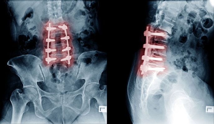
कर्करोगाच्या गाठींच्या तुलनेत कर्करोग नसलेल्या ट्यूमर अर्थातच चांगले असतात, कारण ते वेगाने वाढत नाहीत आणि शरीराच्या इतर भागात पसरत नाहीत. पण महांतेशचे दुर्दैव असे की त्याला पाठीच्या कण्यामध्ये NF ट्यूमर होता. 13 वर्षांपूर्वी NF ट्यूमर काढण्यासाठी त्याच्या कमरेच्या मणक्यामध्ये शस्त्रक्रिया करण्यात आली होती. शल्यचिकित्सकाने दुस-या लंबर कशेरुका [L2] च्या स्तरावरील ट्यूमर काढला होता. पण महांतेशसाठी दुःखाची गोष्ट म्हणजे, न्यूरोफिब्रोमेटोसिस ट्यूमर पुनरावृत्ती झाली! यावेळी ते 12 व्या थोरॅसिक कशेरुका [T12] आणि प्रथम लंबर मणक्यांच्या [L1] वर होते.

ट्यूमर पुन्हा वाढल्यामुळे, महांतेशला खालच्या अंगात दोन्ही बाजुला अशक्तपणा आला. गाठी वाढत गेल्याने हळूहळू त्याला चालता येत नव्हते. त्याच्या एम.आर.आय. स्कॅनमध्ये T12, L1 आणि L2 मणक्यांच्या स्तरावर एकापेक्षा जास्त स्पाइनल ट्यूमर दिसून आले. त्यामुळे त्याला न्यूरोसर्जनकडे पाठवण्यात आले. न्यूरोसर्जनने स्पष्ट केले की शस्त्रक्रिया अत्यंत जोखमीची होती कारण ट्यूमर लहान होते आणि ते अचूकपणे शोधणे कठीण होते आणि ते काढताना महांतेशला त्याच्या खालच्या अवयवांना कायमचा अशक्तपणा किंवा अर्धांगवायू आणि मूत्राशय आणि आतड्यांवरील नियंत्रण आंशिक किंवा पूर्ण नुकसान होण्याची शक्यता होती.

आणि जर महांतेशवर लवकर शस्त्रक्रिया केली नसती, तरीही त्याला कायमचा अशक्तपणा किंवा खालच्या अंगाचा अर्धांगवायू आणि मूत्राशय आणि आतड्यांवरील नियंत्रण अर्धवट किंवा पूर्ण गमावण्याची शक्यता होती!

इकडे आड आणि तिकडे विहीर अशी परिस्थिती महांतेशवर आली होती.

त्यांच्या जीवनात आणखी एक गुंतागुंत होती. महांतेशच्या पत्नीला पोलिओमायलाइटिस आहे. ती दिव्यांग गटात आहे. चालताना तिला काठी लागते. त्यामुळे जर महांतेशला पॅराप्लेजिया झाला असता तर ती त्याला जगण्यास मदत करण्यास सक्षम नव्हती.

याशिवाय, शस्त्रक्रियेचा खर्च हा मोठा प्रश्न होता.

त्यामुळे महांतेश शेल्लिकेरी आणि त्यांची पत्नी खरोखरच हताश परिस्थितीत होते. त्यांच्या असहाय्य परिस्थितीत त्यांच्या कुटुंबाने त्यांना खूप मदत केली.

महांतेशच्या उपचारासाठी अनेक न्यूरोसर्जनचा सल्ला घेण्यात आला. सर्वांनी त्यांना समजावून सांगितले की शस्त्रक्रिया ही एक धोकादायक पायरी आहे कारण महांतेशला पूर्ण पॅराप्लेजिया आणि मूत्राशय तसेच आतड्यांसंबंधी असंयम होण्याची दाट शक्यता होती.

योगायोग आणि संयोग

महांतेशच्या नातेवाईकांपैकी एक जण डॉक्टर आहेत. ते डॉ. सोमनाथ खेराडकर यांना भेटले, त्यांनी दुसरे मत (सेकंड ओपिनियन) घेण्यासाठी माझे नाव सुचवले.

जेव्हा महांतेश आणि कुटुंब माझ्याकडे आले तेव्हा ते खूप घाबरलेले आणि हताश होते. त्याची तब्बेत सुधारेल अशी त्याची फारशी आशा नव्हती. म्हणून सर्वप्रथम मी त्यांना धीर दिला. रोगाबद्दल समजावून सांगितले.

एक गोष्ट स्पष्ट झाली होती – जर महांतेशवर लवकर शस्त्रक्रिया केली गेली नसती नाही तर त्याचे खालच्या अंगांवर, मूत्राशयावर आणि आतड्यांवरील नियंत्रण जाणार होते हे नक्की होते. शस्त्रक्रिया धोक्याची होती पण केल्याशिवाय इलाज नव्हता.

मोठे वरदान

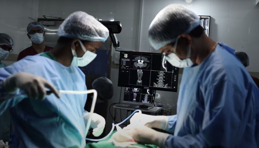
पण आमच्या कडे एक मोठे वरदान होते. मी काम करतो त्या समर्थ न्यूरो आणि सुपरस्पेशालिटी हॉस्पिटलमध्ये सर्जिकल नेव्हिगेशन सिस्टीम आहे जी मेंदू आणि मणक्याच्या शस्त्रक्रियांमध्ये खूप उपयुक्त असते.. महांतेशच्या गाठी लहान होत्या आणि त्यांना अचूक पणे काढण्याची गरज होती. सर्जिकल नेव्हिगेशन उपकरणे मला त्याच्या ट्यूमर शोधण्यात आणि त्याच्या सभोवतालच्या रीढ़ की हड्डीच्या ऊतींना इजा न करता फक्त ट्यूमर काढण्यास मदत करणार होती. मी कमीतकमी कशेरुकाची हाडे काढून टाकून अचूकपणे सर्जरी करू शकलो असतो.

मी महांतेश आणि कुटुंबीयांना समर्थ हॉस्पिटलमध्ये असलेल्या सर्जिकल नेव्हिगेशनच्या अत्याधुनिक साधनांबद्दल सांगितले. सर्जिकल नेव्हिगेशन मला ट्यूमर शोधण्यात आणि त्यांना अचूकपणे काढण्यात कशी मदत करेल याबद्दल समजावून सांगितले. या साधनांमुळे ऑपरेशन यशस्वी होण्याची शक्यता खूप जास्त होती हे पण सांगितले.

महांतेश आणि त्याची पत्नी यांना अनेक प्रश्न होते. मी त्यांना उत्तरे दिली आणि त्यांच्या सर्व शंका दूर केल्या. मग ते आशावादी झाले. ते शस्त्रक्रियेसाठी इच्छुक होते. पण खर्चाबाबत प्रश्न होताच.

माझ्यासाठी हे ऑपरेशन करणे एक खास बाब होती. जवळजवळ सर्व न्यूरोसर्जन्सनी ऑपरेशन करण्यास नकार दिला होता आणि जवळजवळ सर्वांनी सल्ला दिला होता की शस्त्रक्रियेचा धोका फायद्यांपेक्षा कितीतरी जास्त आहे. पण तरीही मी ऑपरेशन करायला तयार होतो त्याचे एकच कारण म्हणजे सर्जिकल नेव्हिगेशन.

मी काम करत असलेल्या समर्थ न्यूरो आणि सुपर स्पेशालिटी हॉस्पिटलमधील सर्जिकल नेव्हिगेशन मार्गदर्शन प्रणालीमुळेच मी शस्त्रक्रियेचा धोका पत्करण्यास तयार होतो. ती प्रणाली मला इतर भागांना नुकसान न पोहोचवता अचूक शस्त्रक्रिया करण्यास मदत करणार होती.  

सर्जिकल नेव्हिगेशनमुळे मी चीरा लहानात लहान घेऊ शकत होतो, म्हणून ऑपरेशनची जखम अगदी लहान होणार होती. नेमके कुठे कापायचे आणि काय काढायचे हे मला सर्जिकल नेव्हिगेशनचा संगणक व स्क्रीन सांगणार होता. सर्व माहिती अचूक मिळाल्यास शस्त्रक्रियेला वेळही खूपच कमी लागणार होता. याचा सर्वाचा परिणाम शस्त्रक्रियेनंतर कमी वेदना होणार होत्या आणि जलद गतीने जखम बरी होणार होती. संसर्गाची शक्यताही काहीशी कमी होती.

नेव्हिगेशनशिवाय…

शस्त्रक्रिया खूप जोखमीची होती. जर मी नेव्हिगेशनशिवाय ऑपरेशन केले असते, तर मला त्याच्या मणक्याचे एक किंवा अधिक स्तर काढून टाकण्यास भाग पडले असते ज्यामुळे शस्त्रक्रियेदरम्यान आणि नंतर गुंतागुंत होण्याची शक्यता होती.

शस्त्रक्रियेचा दिवस

अंतेश शेलीकेरी यांची आर्थिक स्थिती कमकुवत होती म्हणून आम्ही जेवढे शक्य होते तेवढ्या कमी शुल्कात शस्त्रक्रिया करण्याचे ठरवून त्याला लवकरात लवकर शस्त्रक्रियेसाठी तयार केले.

आमच्याकडे महांतेशच्या न्यूरोफिब्रोमाच्या सीटी स्कॅन आणि एमआरआय स्कॅन प्रतिमा होत्या. सी.टी. आणि एम.आर.आय. प्रतिमा संगणक प्रणालीमध्ये फीड केल्यानंतर आम्ही सर्जिकल नेव्हिगेशन प्रणाली सुरू केली. आम्ही नेव्हिगेशन सिस्टमचे ट्रॅकर्स महांतेशच्या पाठीवर अचूक ठिकाणी बसवले. आमच्या काही सर्जिकल उपकरणांमध्ये मार्कर असतात. अशा प्रकारे रुग्णांवर निश्चित केलेले मार्कर आणि आमच्या काही शस्त्रक्रिया उपकरणांवरील मार्कर सर्जिकल नेव्हिगेशन सिस्टमच्या 3D स्क्रीनवर स्पष्टपणे दिसू लागले. त्यामुळे आता नक्की कुठे कापायचे आणि कसे कापायचे हे कळत होते. या प्रतिमा आणि आमच्या कौशल्याच्या मदतीने आम्ही ऑपरेशन केले.

आम्ही ट्यूमरची पातळी अचूकपणे स्थानिकीकृत केली. मग ट्यूमर काढण्यासाठी आम्ही हाडांचा अचूक तुकडा काढला. आम्ही ड्युरामेटर [मेंदू आणि पाठीचा कणा यांचे कडक आवरण] उघडले आणि मी ती गाठ पाठीच्या कण्यापासून काळजीपूर्वक विच्छेदित केली. त्यानंतर मी T-12 आणि L-1 स्तरांमधील दोन्ही ट्यूमर तसेच L-2 पातळीच्या खाली असलेले ट्यूमर काढले.

आम्ही एकाच शस्त्रक्रियेत दोन्ही गाठी काढू शकलो. सर्वात महत्त्वाचे म्हणजे शस्त्रक्रिया यशस्वी झाली.

त्यानंतर शस्त्रक्रियेनंतरची काळजी आली. नर्सिंग केर आणि डॉक्टरांच्या राउंड व्यतिरिक्त फिजिओथेरपिस्ट हे एक अतिशय महत्त्वाचे काळजीवाहक होते. तिने महांतेशला हातपाय हलवायला प्रवृत्त केले आणि मार्गदर्शन केले कारण नसांवरील गाठींचा दाब कमी झाला होता. महांतेशला आश्चर्य वाटले की तो पाय हलवू शकतो. हे लक्षात घ्या की जेव्हा तो माझ्याकडे आला तेव्हा त्याने त्याचे खालचे हातपाय हलवण्याची क्षमता गमावली होती.

केवळ खालचे अंग हलवण्यापासून ते चालता येण्यापर्यंतची प्रगती महांतेशसाठी अवघड पण गरजेची होती. पण त्याने ते पूर्ण केली आणि वॉकरचा आधार घेऊन त्याला चालता आले. पण आपण खूप पुढच्या काळात जात आहोत…

त्या आधी समर्थ हॉस्पिटलमधून महांतेशला डिस्चार्ज मिळाला. शस्त्रक्रियेनंतर सातव्या दिवशी आम्ही त्याला डिस्चार्ज दिला. नेव्हिगेशनच्या सहाय्याने केलेल्या शस्त्रक्रियेमुळे त्याची जखम खूपच लहान होती. त्यामुळे त्याला जखमेच्या ड्रेसिंगची फारशी गरज नव्हती. त्याच्या शस्त्रक्रियेने झालेल्या जखमा लवकर बऱ्या झाल्या.

हे लिहितोय तेव्हा महांतेशला डिस्चार्ज होऊन बरेच महिने उलटले आहेत. शेलीकेरी दाम्पत्य बेळगाव येथे राहतात आणि त्यामुळे त्यांना भेटीसाठी येणे खूप दूर पडते. बेळगावमधील स्थानिक डॉक्टर महांतेशच्या रोजच्या समस्यांची काळजी घेतात.

पण महांतेशच्या पत्नी सौ. शेलीकेरी यांनी मला अनेक महिन्यांनी फोन करून महांतेशची प्रगती चांगली होत असल्याचे सांगितले.

आमच्या प्रयत्नांमुळे आम्ही महांतेशला पुन्हा चालण्यास मदत करू शकलो. मी देवाला आशा करतो की त्याला त्याच्या मणक्यावर किंवा इतर कोठेही न्यूरोफिब्रोमेटोसिसच्या गाठी पुन्हा येऊ नयेत.

शल्यक्रियेमुळे शेलीकेरी कुटुंब आत्ता सुखात आहे.

Had tumours in his spine twice: Surgical Navigation saved him

Surgical Navigation - Ravindra patil

Had tumours in his spine twice: Surgical Navigation saved him

By Dr.Ravindra Patil

Chief Neurosurgeon of Samarth Neuro and Multispeciality Hospital [Miraj, South Maharashtra] Dr Ravindra Patil writes about how he could operate a difficult case with the help of surgical navigation.

Mr Mahantesh Shellikeri is from Belgaum. He had a rare disease called Neurofibromatosis. He had two tumours pressing on the spinal cord in his back and as a result he had lost the ability to walk in the past two months.

Table of Contents

Neurofibromatosis

Neurofibromatosis (NF) is a conditions in which tumours grow in the nervous system. There are three types of neurofibromatosis, type I (NF1), type II (NF2) and Schwannomatosis. The tumours in NF are generally non-cancerous.

Non-cancerous tumours are of course good as compared to cancerous tumours, of course, because they don’t grow rapidly and spread to other parts of the body. But Mahantesh’s bad luck was that he had NF tumours in in spinal cord. He had been previously operated in the lumbar spine for the removal of an NF tumour 13 years ago. The surgeon had removed the tumour at the level of the second lumbar vertebra [L2]. But sadly for Mahantesh, the neurofibromatosis tumours recurred! This time it was at the 12th thoracic vertebra [T12] and first lumbar vertebra [L1].

Due to the regrowth of the tumour, Mahantesh developed weakness in both lower limbs. As the tumours grew, gradually he could not walk. His MRI scan showed multiple spinal tumours at the level of the T12, L1 and L2 vertebrae. So he was referred to a neurosurgeon. The neurosurgeon explained that the surgery was very risky as the tumours were small and would be difficult to locate exactly and during their removal Mahantesh was likely to suffer permanent weakness or paralysis of his lower limbs and partial or complete loss of bladder and bowel control.

And if Mahantesh was not operated soon, then too he was likely to suffer permanent weakness or paralysis of his lower limbs and partial or complete loss of bladder and bowel control!

There was another complication. Mahantesh’s wife has poliomyelitis. She is a differently abled and challenged person. She needs a stick to support herself when she walks. So if Mahantesh suffered paraplegia, she would not be able to help him survive.

Besides, the cost of surgery, if it were attempted, would be high.

Thus Mahantesh Shellikeri and his wife were indeed in a desperate situation. Their extended family helped them a lot during their helpless condition.

Many neurosurgeons were consulted for Mahantesh’s treatment. All explained to them that surgery is a risky step because there was a high chance of Mahantesh developing complete paraplegia and bladder plus bowel incontinence.

Chance referral

One of Mahantesh’s relatives is a doctor. He met Dr Somnath Kheradkar, who suggested my name for another opinion.

When Mahantesh and family they came to me, they were very apprehensive and did not have much hope. I reassured them, explained about the disease, although they knew so much.

It was clear to me that if Mahantesh was not operated soon, he would loose control over his lower limbs, bladder and bowel. The surgery was risky.

Advantage factor

But we had one advantage. At Samarth Neuro and Superspeciality Hospital where I work, we have surgical navigation system which is very useful in fine brain and spine surgeries. Mahantesh’s tumours were small and would require accurate removal. The surgical navigation equipment would help me pinpoint his tumours and remove only the tumours without damaging his surrounding spinal cord tissue. I would remove the least amount of vertebral bones and leave his spine intact.

I explained Mahantesh and family about the one almost magical and extremely tool that Samarth Hospital has, namely Surgical Navigation and how it would help me pinpoint the tumours and remove them accurately.

Mahantesh and his wife had many. I answered them all and cleared all their doubts. Then they became were hopeful. They were willing for surgery. But they had reservations about the cost.

But it was a special case. Nearly all neurosurgeons had refused to operate and almost all had advised that the risk of the surgery was far more than the benefits.

I was willing to take the risk of surgery only because of surgical navigation guidance system in Samarth Neuro and Super-speciality hospital where I work. That system would give me the almost magical advantage of doing accurate surgery without and permanent aftereffects.

Surgical navigation would help me find the tumour inside the spinal canal. Also, I could reduce the size of the incision. Besides knowing exactly where to cut and what to remove, the time of the surgery would be much reduced. This would result in less post operative pain and faster healing. The possibility of infection would also be somewhat reduced.

Without Navigation…

The surgery would have been very risky. Had I operated without navigation, I would have been forced to remove one or more levels of his vertebrae which in turn would cause complications during and after the surgery.

Day of surgery

Then we posted him for surgery at the earliest. We did the surgery at reduced charges as Mahantesh Shellikeri’s financial condition is weak. We had CT scan and MRI scan images of his neurofibromas. We started the surgical navigation system after feeding the CT and MRI images into its computer system. We fitted the trackers of the navigations system at exact locations.

Now we knew exactly where to cut and how to cut. Some of our surgical instruments have markers. Thus the markers fixed on the patients and some of our surgical instruments showed up clearly on the 3D screen of the surgical navigation system. With the help of these images and our skills we operated.

We localized the level of the tumour exactly. Then we removed the exact amount of the bone to excise the tumour. We opened the duramater [a stiff covering of the brain and the spinal cord] and I dissected that tumour carefully away from the spinal cord. Afterwards I removed both the tumours located between the T-12 and L-1 levels as also the tumour below the L-2 level.

We were able to remove both the tumours in a single surgery most important of all, the surgery was successful.

Then came the post-operative care. Apart from nursing care and doctors’ rounds came in a very important care giver, the physiotherapist. She motivated and guided Mahantesh to move his limbs as the pressure of the tumours on the nerves was relieved. Mahantesh was surprised that he could move his legs. Remember, he had lost the ability to move his lower limbs when he came to me.

From merely moving his lower limbs to being able to walk was a gradual journey for Mahantesh. But he accomplished it and was able to walk with the support of a walker. But we are moving ahead too fast.

First came Mahantesh’s discharge from Samarth Hospital. We discharge him on the seventh day after surgery.

Many months have passed since Mahantesh was discharged. His would was very small, thanks to navigation assisted surgery. So he did not require much wound dressing. His surgical wounds healed fast.

The Shellikeri couple lives in Belgaum and hence it is too far away for them to come for follow up visits. Local doctors in Belgaon take care of Mahantesh’s routine problems.

But Mahantesh’s wife Mrs Shellikeri called me once after many months to tell me that Mahantesh is progressing well.

With our efforts we were able help Mahantesh walk again. I hope to god that he does not get more episodes of Neurofibromatosis either on his spine or anywhere else.

The Shellikeri family is happy about the results.

Spine Fusion Surgery Cost in India

spinal fusion surgery cost

Spine Fusion Surgery Cost in India

By Dr.Ravindra Patil

Any type of spine surgery is thought to be a very expensive surgery. It is true to some extent as spinal fusion surgery cost is high because it needs very highly skilled surgeons, state of the art operation theatres and above all, surgical navigation for accuracy. But all these give fairly good results in patients who could not be operated at all a decade ago, because the skillset and surgical navigation were not available then. So now although back surgery cost is high, results are better.

Table of Contents

Cost of Spinal Fusion Surgery

The average Spinal Fusion Surgery cost is very high in western countries.

India is known worldwide for its advanced medical facilities and promising technology for Low Cost of Spinal Fusion Surgery. Many of the best hospitals for Spinal Fusion Surgery can be found in India.

Spinal Fusion Surgery cost in India is much less when compared to any other countries. Also the cost of Spinal Fusion Surgery in India is substantially lower than other developed countries of the world.

Private back surgery cost is higher because Private back surgery cost means treatment in a private hospital

Various deciding factors could determine the price of Spinal Fusion Surgery in India. These can be broadly classified as Hospital, Medical Team or Patient Dependent factors.

Hospital Factors

Type of the hospital (Government/Trust/Private).

Use of insurance, type of insurance or self paid.

Accreditation of the facility

Reputation and brand value of the hospital.

Medical Team Factors

Technology / Approach Used

Surgery Type

Type of Anesthesia or Sedation

Qualification / Expertise of the specialist

Extent of the surgery needed

Patient Factors

Patient’s diagnosis

Patient’s general health

Room Category selected by the patient

Other treatment required by the patient in conjunction

What is the definition of spinal fusion?

Spinal fusion is a surgical procedure wherein one or more vertebrae are united to eliminate any motion between them. The approach is similar to welding. Spinal Fusion Surgery, only does not weld the vertebrae immediately during surgery. The bone grafts are placed around the spine during surgery. The body then heals the grafts over several months.

When are the indications of spinal fusion surgery?

There are several potential reasons to consider spinal fusion. These include treatment of a fractured vertebra, rectifying deformity, elimination of pain caused during motion, treatment of instability, and treatment of cervical disc herniations.

Not all spinal fractures need surgery, some fractures, particularly those associated with spinal cord or nerve injury, generally require fusion as part of the surgical treatment.

Another condition treated is instability, i.e. the abnormal or excessive motion between two or more vertebrae. It is commonly believed that instability can be a source of back or neck pain or cause potential irritation or damage to close by nerves.

Cervical disc herniations that require surgery usually need fusion as well. In this technique, the disc is removed by an incision in the front of the neck and a small piece of bone is inserted in its place.

How is the Spinal Fusion Surgery performed?

There are many surgical methods available to fuse the spine that involve placement of a bone graft between the vertebrae. The spine may be treated from the back (posterior), from the front (anterior) or by a combination of both. The anterior approach is more common. The ultimate objective of fusion is to obtain a solid union between two or more vertebrae.

A fusion may or may not involve use of supplemental tools like plates, screws and cages. Instrumentation is sometimes used to rectify a deformity, but is usually used to hold the vertebrae together to while the bone grafts heal. Whether the hardware is used or not, it is important that bone or bone substitutes be used to get the vertebrae to fuse together.

The bone may be taken either from another bone in the patient or from a bone bank. Fusion using bone from the patient has a long history of use and results in predictable healing. Smoking, medications you are taking for other conditions and your overall health can affect the rate of healing and fusion, too.

How long does it take to recover from spinal fusion surgery?

Immediate discomfort following a spinal fusion is generally greater than with other types of surgeries. Fortunately, there are excellent methods of postoperative pain control available, including pain medications and intravenous injections.

Another option is a patient-controlled postoperative pain control pump. Here the patient presses a button that delivers a predetermined amount of narcotic pain medication through an intravenous line.

Patients generally stay in the hospital for three to four days, but a longer stay after more extensive surgery is also possible. A short stay in a rehabilitation centre after discharge from the hospital is recommended for patients who underwent extensive surgery, or for elderly or debilitated patients.

The fusion process varies in each patient. The healing process after fusion surgery is similar to that after a skeletal fracture. During this time, the patient’s activity is generally restricted. Substantial bone healing does not usually take place until three or four months after surgery.

Many a times spinal decompression surgery cost and healing time is compared with spinal fusion surgery cost. It must be understood that both are far different. Spinal decompression surgery does not need bone fusion and healing like spinal fusion surgery.

How many patients underwent Spinal Fusion Surgery in India in the last 5 years?

Private back surgery prices will vary from place to place. Scoliosis surgery cost is also of interest to many patients who have suffered kyphosis or scoliosis. Cost of spinal decompression surgery is also queried.

In recent years, India has emerged as a medical hub for patients looking for affordable, accessible and efficient low cost Spinal Fusion Surgery Treatment. Some of the best Spinal Fusion Surgery hospitals in the world are found in India. The country is known for offering advanced medical facilities at the most reasonable cost. An average increase of 15 to 20 percent annually has been observed in the number of patients in the last 5 years. The Indian Spinal Fusion Surgery hospitals deliver advanced health care and highest quality services backed by elaborate infrastructure and lower treatment cost

Here are the approximate figures of the patients underwent Spinal Fusion Surgery in the last 5 years in India

Year

2013

2014

2015

2016

2017

Surgeries

4800

6000

7500

9600

10800

What are our special services offered to international patients?

At Samarth Neuro and Super Speciality Hospital we help arranging your treatment in a way that will suit you and be affordable for you. Chief neurosurgeon at Samarth Neuro and Super Speciality Hospital Dr Ravindra Patil is vastly experienced in all types of spine and brain surgeries. Spine fusion surgeries are done there as required.

Congenital Brain and Spine Anomalies

congenital brain and spine malformations

Congenital Brain and Spine Anomalies

By Dr. Ravindra Patil

Table of Contents

What are congenital brain and spine malformations?

Congenital means present at birth.

These conditions may be mild and without symptoms or may be serious, requiring treatment. In some cases, surgery may be recommended to:

  1. Address the child’s or the patient’s symptoms.
  2. Correct the form and function of the brain and spine structures.
  3. Maximize cognitive and motor
  4. Prevent development of neurological deficits.

Congenital abnormalities, called malformations, are conditions affecting the form and function of the nervous system. There are numerous variations of congenital malformations of the bone and soft tissue of the head and spine, including neural tube defects, such as spina bifida, encephaloceles, Chiari malformations and arachnoid cysts.

Some congenital malformations are mild, and some are severe but correctable with surgery by a pediatric neurosurgeon.

Types of some of the Congenital Brain and Spine Malformations are as follows:

Chiari Malformations

This is a condition in which portions of the brain, called the cerebellar tonsils, protrude through the bottom opening of the skull into the upper spine, which can put pressure on the brain or spinal cord. Chiari malformations may block the flow of cerebrospinal fluid, leading to hydrocephalus.

Treatments often focus on removing portions of the bone and soft tissue to relieve pressure on the spinal cord and brain, as well as providing new pathways to drain cerebrospinal fluid. Surgeons use different methods for treating these malformations, including decompression, with or without cutting open a small part of the dura mater [thick membrane covering the brain].

Encephaloceles

Encephaloceles are a type of neural tube defect characterized by the brain being exposed to the outside instead of being covered by the skull and skin. It can lead to infections and hydrocephalus.

Surgical treatment of this condition involves removing bone and soft tissue, draining cerebrospinal fluid, and surgically repairing or closing the encephalocele.

Children who have developed hydrocephalus as a result of an encephalocele will require treatment for that condition, often with a cerebrospinal fluid shunt. Shunting is the placement of a tube into the open area (ventricle) of the brain that allows cerebrospinal fluid to drain to the child’s abdomen or other location where it can be safely reabsorbed into the body.

Arachnoid Cysts

Arachnoid cysts are the most common type of brain cyst. They are congenital (present at birth) lesions that occur as a result of the splitting of the arachnoid membrane. The cysts are fluid-filled sacs, not tumors, appearing in one of the three layers of tissue covering the central nervous system.

Surgical treatment of this condition involves draining the cyst by drilling a small hole or by opening the skull and making small openings in the cyst to open the natural fluid pathways in the brain. This process is called fenestrating.

What are Spinal Deformities?

A spine deformity occurs when your spine varies by more than 10 degrees from ‘healthy’ curvature. But, what does this mean exactly?

Contrary to popular belief, your spine isn’t 100% straight and vertical. The spinal shape has curvatures, but the end result is that it is vertical! Our spine consists of a series of gentle arcs. Our lumbar spine, or lower back, swoops slightly to the back, and our thoracic spine, or upper back, bends subtly forward. The backward curve of your lower back is known as lordosis and the forward stoop that runs between our shoulder blades is known as kyphosis. Lordosis and kyphosis are spinal curvature deformities. Both are abnormal curvatures of the spine.

But, when viewed head-on, our backbone should look like a straight pillar. Hence, it is also called the ‘vertebral column’.

Moreover, the curves and straight stretches of your spine make symmetry possible. Your head sits directly over your pelvis because the lordosis of your lower back and the kyphosis of your upper spine balance each other out. If one of these curves becomes greater or lesser than the other, then problems can occur. We refer to this as sagittal imbalance, because the head and pelvis no longer fall within the same, or sagittal, plane.

Too much swaying backwards can be thought of as ‘lordosis’, and too much forward stooping in the upper back is ‘kyphosis’.

Likewise, when the spine tilts away from the midline of the body, doctors refer to this problem as coronal imbalance or scoliosis. Unevenness in the ‘coronal’ plane (the view from head-on) causes asymmetry in the trunk of the body. This can include uneven hips and shoulders or one-sided bulging of the ribs.

Diagnosis of Congenital Brain and Spine Malformations

If a child is born with any of the above malformations, a thorough evaluation by a paediatrician or neurologist is needed to diagnose the problem and recommend a plan for addressing it.

After a thorough physical and detailed family and patient history, your doctor may order imaging of the brain and/or spine through an MRI scan. If the MRI scan shows any evidence of these malformations, a neurosurgical consultation is a must to plan the best treatment.

Congenital Brain and Spine Malformations Treatment

A multidisciplinary approach is often beneficial for addressing children with congenital brain and spine malformations. Neurosurgeons, craniofacial plastic specialists and geneticists, among others, may be called upon to develop your child’s treatment plan and determine what kind of surgery may be appropriate.

If a congenital brain or spine malformation is mild and not causing any signs or symptoms in your child, the neurosurgeon may recommend observation, which means regular visits and testing to monitor your child’s condition.

If your child does undergo surgery, follow-up care is extremely important in tracking the progress of your child’s recovery. Your paediatric neurosurgeon will schedule follow-up appointments to ensure your child is making the best recovery possible.

Spinal treatments are focused on both correcting the functional shortcomings as well as the structural defects of the spine.

Congenital brain and spine defects treatment in Miraj, Maharashtra

Samarth Neuro and Super Speciality hospital has facilities to surgically treat some of the above brain and spine deformities present since birth. Abnormal curvature of the spine can be corrected to some extent.

Chief Neurosurgeon at Samarth Hospital Dr Ravindra Patil has considerable experience in treating such defects. Besides, as his hospital is located in a tier two city, the cost of treatment is considerably less than in major cities of India. Patients from foreign countries may find going to Miraj for treatment of brain and spine diseases from Dr Ravindra Patil considerably cost effective.

जन्मजात मेंदू आणि मणक्याचे विसंगती

जन्मजात मेंदू आणि मणक्याचे विसंगती

जन्मजात मेंदू आणि मणक्याचे विसंगती

By Dr. Ravindra Patil

Table of Contents

जन्मजात मेंदू आणि मणक्याचे विकृती काय आहेत?

जन्मजात म्हणजे जन्मत: उपस्थित.

या विकृती सौम्य आणि लक्षणे नसलेल्या असू शकतात किंवा गंभीर असू शकतात ज्यांचे उपचार करणे आवश्यक असते. काही केस मध्ये, शस्त्रक्रियेची जरूर असू शकते. सर्जरीचे हेतु असे असतात –

  1. मुलाची किंवा रुग्णाची लक्षणे समजून घ्यावी.
  2. मेंदू आणि मणक्याच्या संरचनांचे स्वरूप आणि कार्य दुरुस्त करणे.
  3. संज्ञानात्मक (म्हणजे विचार, अनुभव आणि इंद्रियांद्वारे ज्ञान आणि समज प्राप्त करण्याची मानसिक क्रिया किंवा प्रक्रिया) आणि मोटर (म्हणजे हालचालीशी संबंधीत) कार्य वाढवा.
  4. न्यूरोलॉजिकल कमतरता वाढू नयेत त्यासाठी प्रतिबंध करा.

जन्मजात उणपा किंवा त्रुट्या, ज्याला विकृती म्हणतात, मज्जासंस्थेच्या स्वरूपावर आणि कार्यावर दुष्परिणाम करणाऱ्या स्थित्या आहेत. डोके आणि मणक्याचे हाडे आणि मऊ ऊतकांच्या जन्मजात विकृती अनेक असतात. त्यामध्ये मज्जासंस्थेतील नळीच्या दोषांचा (न्यूरल ट्यूबचा) पण समावेश आहे. याचे उदाहरण म्हणजे स्पायना बिफिडा, एन्सेफॅलोसेल्स, चियारी विकृती आणि अर्कनॉइड सिस्ट.

काही जन्मजात विकृती सौम्य असतात, आणि काही गंभीर असतात परंतु बालरोग न्यूरोसर्जनद्वारे शस्त्रक्रियेने सुधारता येतात.

काही जन्मजात मेंदू आणि मणक्याच्या विकृतींचे प्रकार खालीलप्रमाणे आहेत:

चियारी विकृती

ही अशी स्थिती आहे ज्यामध्ये मेंदूचे काही भाग, ज्याला सेरेबेलर टॉन्सिल म्हणतात, कवटीच्या खालच्या भागातून वरच्या मणक्यामध्ये पसरतात, ज्यामुळे मेंदू किंवा पाठीच्या कण्यावर दबाव येऊ शकतो. चियारी विकृती सेरेब्रोस्पाइनल फ्लुइडचा प्रवाह रोखू शकते, ज्यामुळे हायड्रोसेफलस (म्हणजे मेंदूतील द्रव वाढल्यामुळे डोके मोठे होणे) होते.

पाठीचा कणा आणि मेंदूवरील दबाव कमी करण्यासाठी हाडे आणि मऊ ऊतींचे काही भाग काढून टाकण्यावर उपचार तसेच सेरेब्रोस्पाइनल द्रवपदार्थ काढून टाकण्यासाठी नवीन मार्ग बनवण्यावर लक्ष केंद्रित केले जाते. या विकृतींवर उपचार करण्यासाठी शल्यचिकित्सक वेगवेगळ्या पद्धती वापरतात, ज्यामध्ये डीकंप्रेशनचा समावेश असतो. यात ड्युरा मॅटरचा [मेंदूला झाकणारा जाड पडदा] एक छोटासा भाग कापून किंवा न कापता सर्जरी करतात.

एन्सेफॅलोसेल्स

एन्सेफॅलोसेल्स हा एक प्रकारचा न्यूरल ट्यूबचा दोष आहे ज्यामध्ये मेंदू कवटी आणि त्वचेने झाकला जाण्याऐवजी उघडा असतो. यामुळे संक्रमण आणि हायड्रोसेफलस होऊ शकते.

या स्थितीच्या सर्जिकल उपचारांमध्ये हाडे आणि मऊ ऊतक काढून टाकणे, सेरेब्रोस्पाइनल द्रवपदार्थ काढून टाकणे आणि एन्सेफॅलोसेलची शस्त्रक्रिया करून दुरुस्ती करणे किंवा बंद करणे हे करतात.

एन्सेफॅलोसेलच्या परिणामी हायड्रोसेफलस विकसित झालेल्या मुलांना त्या स्थितीसाठी सेरेब्रोस्पाइनल फ्लुइड च्या प्रवाहा साठी शंट लावून उपचार आवश्यक असतात. शंट लावणे म्हणजे मेंदूच्या खुल्या भागात (वेंट्रिकल मध्ये) नळी बसवणे. या नळीमुळे सेरेब्रोस्पाइनल फ्लुइड मुलाच्या-रूग्णाच्या ओटीपोटात किंवा शरीरात सुरक्षितपणे पुन्हा शोषले जाऊ शकते अशा ठिकाणी वाहून जाते.

अरॅक्नॉइड सिस्ट्स

अरॅक्नॉइड सिस्ट हे ब्रेन सिस्टचे सर्वाधीक आढळणारे प्रकार आहेत. ही अशी जन्मजात (जन्माच्या वेळी उपस्थित) विकृती असते की जी जे अरक्नोइड मेम्ब्रेनच्या विभाजनामुळे उद्भवतात. या सिस्ट द्रवाने भरलेल्या पिशव्या असतात. या सिस्ट ट्यूमर नसून मध्यवर्ती मज्जासंस्थेला (सेन्ट्रल नर्व्हस सिस्टमला) झाकणाऱ्या ऊतींच्या तीन थरांपैकी एकामध्ये दिसतात.

या अवस्थेच्या सर्जिकल उपचारामध्ये मेंदूतील नैसर्गिक द्रवपदार्थाचे मार्ग उघडण्यासाठी लहान छिद्र पाडून किंवा कवटी उघडून सिस्ट काढून टाकणे आणि सिस्ट ध्ये लहान छिद्र करणे असे करतात. या प्रक्रियेला फेनेस्ट्रेटिंग म्हणतात.

स्पाइनल विकृती म्हणजे काय?

जेव्हा तुमची स्पायनल कॉर्ड म्हणजे पाठीचा कणा ‘निरोगी’ वक्रतेपेक्षा 10 अंशांपेक्षा जास्त बदलतो तेव्हा पाठीच्या कण्याची विकृती उद्भवते. पण, याचा नेमका अर्थ काय? पाठीच्या कण्याची निरोगी वक्रता म्हणजे काय?

सर्वसाधारण कल्पन प्रमाणे तुमचा पाठीचा कणा 100% सरळ आणि उभा नसते. पाठीच्या कण्यात वक्रता असते, परंतु एकूण पाठीचा कणा उभा असतो! आपल्या मणक्यामध्ये सौम्य अर्धगोलांची (आर्क्सची) मालिका असते. आपला कमरेचा पाठीचा कणा, किंवा पाठीचा खालचा भाग, किंचित पाठीमागे झुकतो आणि आपला वक्षस्थळाचा कणा किंवा पाठीचा वरचा भाग पुढे वाकतो. तुमच्या पाठीच्या खालच्या बाजूचा मागचा वक्र भाग लॉर्डोसिस म्हणून ओळखला जातो आणि तुमच्या खांद्या मधील भाग किंचित पुढे झुकलेला असतो व त्या कायफोसिस म्हणतात. लॉर्डोसिस आणि कायफोसिस हे पाठीच्या कण्याची नैसर्गिक वक्रता असेत, ती वाढली की ती विकृती होते.

पण, डोके वर पाहिले असता, आपला पाठीचा कणा सरळ खांबासारखा दिसला पाहिजे. म्हणून, त्याला ‘कशेरुकी स्तंभ’ (वर्टीब्रल कॉलम) असे म्हणतात.

शिवाय, तुमच्या मणक्याचे वक्र आणि सरळ ताणणे सममिती शक्य करतात. वरून पाहिले तर तुमचे डोके थेट तुमच्या ओटीपोटावर असते कारण तुमच्या पाठीच्या खालच्या भागाचा लॉर्डोसिस आणि तुमच्या वरच्या मणक्याचा कायफोसिस एकमेकांना संतुलित करतात. जर यापैकी एक वक्र दुसऱ्यापेक्षा मोठा किंवा कमी झाला तर समस्या उद्भवू शकतात. याला सजायटल असंतुलन म्हणूतात, कारण डोके आणि नितंबांचा भाग समतोल नसतात.

पाठीचा कणा पाठीमागे खूप डोलणे म्हणजे ‘लॉर्डोसिस’ आणि पाठीच्या वरच्या बाजूला खूप पुढे वाकणे म्हणजे ‘कायफोसिस’.

त्याचप्रमाणे, जेव्हा पाठीचा कणा शरीराच्या मध्यरेषेपासून डावीकडे किंवा उजवीकडे झुकतो, तेव्हा डॉक्टर या समस्येस कोरोनल असंतुलन किंवा स्कोलियोसिस म्हणतात. ‘कोरोनल’ पातळीतील असमानता (डोक्यावरून दिसणारे दृश्य) शरीराच्या खोडात विषमता निर्माण करते. यामध्ये असमान कूल्ले आणि खांदे किंवा बरगड्यांचा एकतर्फी फुगवटा असे असू शकते.

जन्मजात मेंदू आणि मणक्याच्या विकृतींचे निदान

जर एखाद्या मुलाचा जन्म वरीलपैकी कोणत्याही विकृतीसह झाला असेल, तर समस्येचे निदान करण्यासाठी बालरोगतज्ञ किंवा न्यूरोलॉजिस्टद्वारे सखोल मूल्यांकन करणे आवश्यक आहे आणि त्यानंतर संगळ्यात चांगला उपचार योजनेची निवड करणे आवश्यक असते.

संपूर्ण शारीरिक आणि तपशीलवार कौटुंबिक आणि रुग्णाच्या इतिहासानंतर, डॉक्टर एमआरआय स्कॅनद्वारे मेंदू आणि/किंवा मणक्याचे इमेजिंग परीक्षणे करतील. एम.आर.आय. स्कॅनमध्ये या विकृतींचे कोणतेही पुरावे आढळल्यास, सर्वोत्तम उपचारांची योजना करण्यासाठी न्यूरोसर्जनची सल्लामसलत आवश्यक आहे.

जन्मजात मेंदू आणि मणक्याच्या विकृतीचे उपचार

जन्मजात मेंदू आणि मणक्याचे विकृती असलेल्या मुलांचे उपचार करण्यासाठी अनेक वैद्यकिय तज्ञांनी मिळून काम करणे आवश्यक असते. मुलांची उपचार योजना विकसित करण्यासाठी आणि कोणत्या प्रकारची शस्त्रक्रिया योग्य असू शकते हे निर्धारित करण्यासाठी न्यूरोसर्जन, क्रॅनिओफेशियल प्लास्टिक सर्जन आणि अनुवांशिक रोगांच्या तज्ञांना बोलावणे जरूरी होऊ शकते.

जर जन्मजात मेंदू किंवा मणक्याचे विकृती सौम्य असेल आणि तुमच्या मुलामध्ये कोणतीही चिन्हे किंवा लक्षणे उद्भवत नसतील, तर न्यूरोसर्जन कडून नियमित तपासणी करणे आवश्यक असते.  मुलाच्या किंवा रूग्णाच्या स्थितीचे नियमित निरीक्षण आणि चाचण्या करून लक्षणे पाहून इलाज करता येतो. 

जर मुलावर शस्त्रक्रिया होत असल्यास, पुनर्प्राप्तीच्या प्रगतीचा मागोवा घेण्यासाठी फॉलो-अप काळजी अत्यंत महत्त्वाची आहे. बालरोग न्यूरोसर्जन त्यासाठी फॉलो-अप अपॉइंटमेंट देतील.

स्पाइनल उपचारांमध्ये कार्यात्मक उणीवा तसेच मणक्याचे संरचनात्मक दोष सुधारण्यावर लक्ष केंद्रित केले जाते.

मिरज, महाराष्ट्र येथे जन्मजात मेंदू आणि मणक्यातील दोषांवर उपचार

समर्थ न्यूरो आणि सुपर स्पेशालिटी हॉस्पिटलमध्ये वरील अनेक मेंदू आणि मणक्याच्या विकृतींवर शस्त्रक्रिया करून उपचार करण्याची सुविधा आहे. मणक्याची असामान्य वक्रता काही प्रमाणात दुरुस्त केली जाऊ शकते.

समर्थ हॉस्पिटलचे मुख्य न्यूरोसर्जन डॉ. रवींद्र पाटील यांना अशा दोषांवर उपचार करण्याचा पुरेसा अनुभव आहे. याशिवाय, त्याचे रुग्णालय लहान शहरात असल्याने, उपचारांचा खर्च भारतातील मोठ्या शहरांपेक्षा खूपच कमी आहे. डॉक्टर रवींद्र पाटील यांच्याकडून मेंदू आणि मणक्याच्या आजारांवर उपचारासाठी परदेशातील रुग्णांना मिरजला जाणे अत्यंत किफायतशीर ठरू शकते.

भारतात स्पाइन फ्यूजन सर्जरीचा खर्च

स्पाइन फ्यूजन सर्जरीचा खर्च

भारतात स्पाइन फ्यूजन सर्जरीचा खर्च

By Dr.Ravindra Patil

कोणत्याही प्रकारची मणक्याची शस्त्रक्रिया ही खूप महागडी शस्त्रक्रिया मानली जाते. हे काही प्रमाणात खरे ही आहे कारण स्पाइन ट्यूमरच्या शस्त्रक्रियेसाठी अत्यंत कुशल सर्जन, अत्याधुनिक ऑपरेशन थिएटर्स आणि सर्वात महत्त्वाचे म्हणजे अचूकतेसाठी शस्त्रक्रियेत वापरले जाणारे नेव्हिगेशन या सर्वांची आवश्यकता असते. परंतु या सर्व सोईंमुळे, काही दशकांपूर्वी ज्या शस्त्रक्रिया करता येत नव्हत्या अशाही शस्त्रक्रिया आजच्या काळात करता येतात आणि त्यांचे परिणामही चांगले येतात. याचे कारण म्हणजे पूर्वीच्या काळी सर्जरीचे कौशल्य आणि सर्जिकल नेव्हिगेशन उपलब्ध नव्हते.

Table of Contents

स्पाइनल फ्यूजन शस्त्रक्रिया

पाश्चात्य देशांमध्ये स्पाइनल फ्यूजन शस्त्रक्रियेची सरासरी किंमत खूप जास्त असते.

त्या मानाने भारतात प्रगत वैद्यकीय सुविधा आणि कमी किमतीच्या स्पाइनल फ्यूजन सर्जरीसाठी आश्वासक तंत्रज्ञानासाठी जगभरात ओळखला जातो. स्पाइनल फ्यूजन सर्जरीसाठी अनेक रुग्णालये भारतात आहेत.

इतर कोणत्याही देशांच्या तुलनेत भारतात स्पाइनल फ्यूजन सर्जरीची किंमत खूपच कमी आहे. भारतात स्पाइनल फ्यूजन सर्जरीचा खर्च जगातील इतर विकसित देशांच्या तुलनेत खूपच कमी आहे.

भारतातील स्पाइनल फ्यूजन सर्जरीची किंमत विविध निर्णायक घटक ठरवू शकतात. ते घटक म्हणजे हॉस्पिटल चे घटक, मेडिकल टीमचे घटक किंवा पेशंट वर आधारीत घटक म्हणून वर्गीकृत केले जाऊ शकतात.

हॉस्पिटलचे घटक

  • रुग्णालयाचा प्रकार (सरकारी / खाजगी)
  • विमा असलेले रूग्ण
  • स्वतः हॉस्पिटलची फी देणारे रूग्ण
  • हॉस्पिटलची एन.ए.बी.एच. अथवा जे.सी.आय. कडून मान्यता
  • रुग्णालयाची प्रतिष्ठा आणि ब्रँड मूल्य.

.

वैद्यकीय संघ (मेडिकल टीमचे) घटक

  • तंत्रज्ञान
  • शस्त्रक्रियेचा प्रकार
  • ऍनेस्थेसिया किंवा सेडेशनचा प्रकार
  • तज्ञाची पात्रता / कौशल्य
  • शस्त्रक्रियेची आवश्यक व्याप्ती

रुग्ण घटक

  • रुग्णाचे निदान
  • रुग्णाचे सामान्य आरोग्य
  • रुग्णाने निवडलेली खोली श्रेणी
  • रुग्णाला आवश्यक इतर उपचार

स्पाइनल फ्यूजन म्हणजे काय?

स्पाइनल फ्यूजन ही एक शस्त्रक्रिया प्रक्रिया आहे ज्यामध्ये एक किंवा अधिक कशेरुक (मणके) त्यांच्यातील एकमेंकाशी जोडले जातात व त्यांच्यामधील हालचाल पूर्णपणे बंद होते. हे काही अंशी वेल्डिंग सारखेच असते. पण फरक असा असतो की शस्त्रक्रियेदरम्यान मणके ताबडतोब एकमेकांशी जोडले जात नाहीत. शस्त्रक्रिये नंतर अनेक महिन्यांत मणके कायमचे जोडले जातात.

स्पाइनल फ्यूजन शस्त्रक्रिया कोणत्या रोगांत करतात?

स्पाइनल फ्यूजन शस्त्रक्रिया विचारात घेण्याची अनेक संभाव्य कारणे आहेत. यामध्ये फ्रॅक्चर झालेल्या कशेरुकाचे उपचार, विकृती सुधारणे, हालचालीदरम्यान होणार्‍या वेदना दूर करणे, अस्थिरतेवर उपचार करणे आणि मणक्यामधील डिस्क हर्निएशनवर उपचार करणे अशी कारणे असतात.

सर्व पाठीच्या फ्रॅक्चरला शस्त्रक्रियेची आवश्यकता नसते. काही फ्रॅक्चर मध्ये, विशेषत: पाठीचा कणा किंवा मज्जातंतूच्या दुखापतीशी संबंधित केसमध्ये उपचारांचा भाग म्हणून फ्यूजन शस्त्रक्रिया आवश्यक असते.

उपचार लागतो अशी दुसरी स्थिती म्हणजे पाठीच्या कण्याची अस्थिरता, म्हणजे दोन किंवा अधिक कशेरुकांमधील असामान्य किंवा जास्त हालचाल होणे. सामान्यतः असे मानले जाते की अशी मणक्यांमधील अस्थिरता पाठीच्या किंवा मानेच्या वेदनांचे स्त्रोत असू शकते किंवा त्यामुळे मज्जातंतु बिघडू शकतात किंवा मज्जातंतूंना नुकसान होऊ शकते.

दोन मणक्यांमधील चकती चे (डिस्क चे) जेव्हा हर्निएशन तेव्हा त्या रूग्णांना सहसा फ्यूजन शस्त्रक्रियेची आवश्यकता असते. या तंत्रात मानेच्या पुढच्या भागात चीर टाकून डिस्क काढून त्या जागी हाडाचा छोटा तुकडा बसवला जातो.

स्पाइनल फ्यूजन शस्त्रक्रिया कशी केली जाते?

दोन किंवा अधीक मणक्याचे फ्यूजन करण्यासाठी अनेक शस्त्रक्रिया पद्धती उपलब्ध आहेत ज्यात कशेरुकाच्या दरम्यान हाडांचे कलम (ग्राफ्ट) लावणे समाविष्ट आहे. फ्यूजन मणक्याचा मागच्या बाजूने पुढील किंवा दोन्हीच्या मिश्रणाने केले जाऊ शकते. फ्यूजनचे अंतिम उद्दिष्ट दोन किंवा अधिक कशेरुकांमध्‍ये एक ठोस मिलन मिळवणे आहे. यानंतर अर्थात आधी नमूद केल्या प्रमाण त्या दोन कशेरुकांमध्ये हालचाल होऊ शकत नाही.

फ्यूजनमध्ये प्लेट्स, स्क्रू आणि पिंजरे यांसारख्या पूरक साधनांचा वापर केला जातो. सर्जीकल साधंनांचा वापर कधीकधी विकृती सुधारण्यासाठी केला जातो, परंतु सामान्यतः हाडांच्या कलम बरे होईपर्यंत मणक्यांना एकत्र ठेवण्यासाठी केला जातो. हार्डवेअर वापरले असो किंवा नसो, हे महत्त्वाचे असते की हाड किंवा हाडांचा पर्याय मणक्यांना एकत्र जोडण्यासाठी वापरला जाणे आवश्यक आहे.

हाडाचे कलम करायला एकतर रुग्णाच्या दुसर्या हाडातून घेतले जाते किंवा हाडांच्या बँकेतून (बोन बॅंक मधून) घेतले जाऊ शकते. रुग्णाच्या हाडांचा वापर करून फ्यूजन करण्याचा इतिहास मोठा आहे आणि त्याचा परिणाम चांगले होतात. धूम्रपान, इतर परिस्थितींसाठी घेत असलेली औषधे आणि एकूण आरोग्याचाही बरे होण्याच्या दरावर आणि संलयनाच्या दरावर परिणाम होऊ शकतो.

स्पाइनल फ्यूजन शस्त्रक्रियेतून बरे होण्यासाठी किती काळ लागतो?

स्पाइनल फ्यूजननंतर बराच काळ रूग्णाला बरे वाटत नसते. सुदैवाने, वेदनाशामक औषधे आणि अंतस्नायु इंजेक्शन्ससह पोस्टऑपरेटिव्ह वेदना खूप कमी करता येतात.

दुसरा पर्याय म्हणजे रुग्ण-नियंत्रित पोस्टऑपरेटिव्ह वेदना नियंत्रण पंप. येथे रुग्णाला दुखणे सुरू झाले की रूग्ण एक बटण दाबतो / दाबते, की ज्यामुळे इंट्राव्हेनस लाइनद्वारे दुखणं कमी करणारे अमली वेदना शामक औषध पूर्व निर्धारित डोसने रक्तात वितरीत होते.

रूग्ण साधारणपणे तीन ते चार दिवस हॉस्पिटलमध्ये राहतात, परंतु अधिक व्यापक शस्त्रक्रियेनंतर जास्त काळ राहवे लागते. रूग्णालयातून डिस्चार्ज झाल्यानंतर पुनर्वसन केंद्रात थोडे दिवस राहण्याचा सल्ला दिला जाऊ शकतो. प्रत्येक रुग्णामध्ये फ्यूजन वेगळी असते. फ्यूजन शस्त्रक्रियेनंतर बरे होण्याची प्रक्रिया हाडांच्या फ्रॅक्चर नंतरच्या प्रक्रिये सारखीच असते. या काळात रुग्णाची सर्व हालचाल प्रतिबंधित असते. शस्त्रक्रियेनंतर तीन किंवा चार महिन्यांनंतर हाडांचे भरीव बरे होतात.

गेल्या 5 वर्षांत भारतात किती रुग्णांवर स्पाइनल फ्यूजन शस्त्रक्रिया झाली असेल?

गेल्या काही वर्षांत भारत म्हणजे परवडणारे, सुलभ आणि कार्यक्षम कमी खर्चात स्पाइनल फ्यूजन शस्त्रक्रिया उपचारांसाठी विख्यात झाला आहे. स्पाइनल फ्यूजन शस्त्रक्रिया करून घेण्यास हॉस्पिटल शोधत असलेल्या रुग्णांसाठी वैद्यकीय केंद्र म्हणून भारत उदयास आला आहे. जगातील काही सर्वोत्तम स्पाइनल फ्यूजन शस्त्रक्रिया रुग्णालये भारतात आहेत. अत्यंत वाजवी दरात प्रगत वैद्यकीय सुविधा उपलब्ध करून देण्यासाठी हल्ली भारत देश ओळखला जातो. गेल्या 5 वर्षांत स्पाइनल फ्यूजन शस्त्रक्रिया करून घेणार्‍या रुग्णांच्या संख्येत दरवर्षी सरासरी 15 ते 20 टक्के वाढ दिसून आली आहे. भारतीय स्पाइनल फ्यूजन सर्जरी रुग्णालये प्रगत पायाभूत सुविधा आणि कमी उपचार खर्चाद्वारे समर्थित प्रगत आरोग्य सेवा आणि उच्च दर्जाच्या सेवा देतात.

भारतात गेल्या 5 वर्षात स्पाइनल फ्यूजन शस्त्रक्रिया केलेल्या रुग्णांची अंदाजे आकडेवारी अशी आहे

वर्ष

2013

2014

2015

2016

2017

शस्त्रक्रिया

4,800

6,000

7,500

9,600

10,800

मिरजेच्या एका हॉस्पिटलमध्ये स्पाईन फ्यूजन सर्जरीची उत्तम सेवा

मिरजेतील समर्थ न्युरो एन्ड सुपरस्पेश्यालिटी हॉस्पिटल मध्ये वाजवी खर्चात स्पाईन फ्यूजन, ब्रेन सर्जरी, बॅक सर्जरी वगैरे तेथील मुख्य ब्रेन व स्पाईन सर्जन डॉ रवींद्र पाटील यांच्या देखरेखीखाली होतात. मिरज लहान शहर असल्याने तेथे ऑपरेशनचा एकूण खर्च मोठ्या शहरांच्या मानाने खूपच कमी होतो.

कान आणि जबड्या मध्ये वेदना

कान आणि जबड्या मध्ये वेदना

By Dr. Ravindra Patil

आपल्या कानाजवळील भागात, आपला जबडा किंवा आपल्या चेहऱ्याच्या बाजूला असलेल्या स्नायूंमध्ये वेदना, काहीवेळा क्लिक किंवा पॉपिंग आवाज आणि/किंवा जबडयाच्या हालचाली पूर्ण पणे करता न येणे या सर्व लक्षणांना टेम्पोरो-मँडिब्युलर डिसऑर्डर किंवा थोडक्यात TMD म्हणतात. जबड्याच्या सांध्याशी संबंधित आणखी एक शॉर्ट फॉर्म आहे आणि तो म्हणजे टीएमजे (टेम्पोरो-मँडिबुलर जॉइंट). आपल्या चेहऱ्याच्या दोन्ही बाजूला दोन TMJ असतात. हे सांधे कवटी व खालील जबड्याला जोडतात.

जबडा दुखण्याची कारणे अनेक आहेत आणि ती TMJ रोग किंवा मानसिक तणावामुळे असू शकतात!

TMD म्हणजे TMJ आणि/किंवा त्याच्या सभोवतालच्या स्नायूंच्या वेदना आणि बिघडलेले कार्य हे होय. या लक्षणांचे कारण शोधणे नेहमी सोपे नसते. बहुतेक टीएमडी च्या रूग्णांचे साध्या उपायांनी उपचार केले जाऊ शकतात. पण जर हे उपाय उपयुक्त नसतील तर मात्र दंत उपचार किंवा शस्त्रक्रिया यासारख्या मोठ्या प्रक्रिया केल्या पाहिजेत.

खालचा जबडा, ज्याच्या हाडाला मॅन्डिबल म्हणतात, दोन्ही बाजूंच्या कवटीच्या टेम्पोरल हाडांशी जोडलेले असते. हे दोन सांधे अतिशय गुंतागुंतीचे सांधे आहेत. हे सांधे तीन आयामांमध्ये हालचाल करू शकतात. TMJची रचना अशी आहे की खालचा जबडा आणि कवटीचे टेंपोरल हाड बॉल आणि सॉकेट सांध्याने जोडलेले असते. या दोन हाडांमध्ये उशी सारखी कार्टिलेजची चकती असते. गाल आणि कपाळाच्या दोन बाजू या मधील स्नायूंच्या मोठ्या जोड्या खालच्या जबड्याला हलवतात. यापैकी कोणताही भाग – डिस्क, स्नायू किंवा सांधे स्वतःच – TMD समस्येचे कारण बनू शकतात.

TMD च्या वेदनांनी ग्रस्त असलेल्या लोकांसाठी किंवा ज्यांना त्यांचा जबडा उघडण्यास किंवा बंद करण्यात अडचण येते अशा लोकांच्या केसेस मध्ये जबडा उघडण्यात आणि बंद करण्यात वेदना किंवा अडचण का येते याचे कारण शोधण्यासाठी सखोल वैद्यकिय तपासणी आवश्यक आहे.

Table of Contents

TMD कशामुळे होतो?

टेम्पोरो-मॅन्डिब्युलर जॉइंट किंवा टीएमजेला इतर कोणत्याही सांध्याप्रमाणेच ऑर्थोपेडिक रोग होऊ शकतात. या रोगांमुळे सूज येणे, स्नायू दुखणे, अस्थिबंधन आणि उपास्थि दुखणे वगैरे होते.

TMD होण्यात आनुवंशिकता देखील भूमिका बजावते. पुरुषांच्या तुलनेत स्त्रिया या रोगाला जास्त बळी पडतात. जसजसे आपले वय वाढते तसतशी TMD होण्याची शक्यता जास्त असते. शारीरिक आणि मानसिक ताण यामुळे देखील TMD होऊ शकतो. काही प्रकरणांमध्ये, जबड्याचे दुखणे फायब्रोमायल्जियासारख्या अधिक व्यापक, वेदना-प्रेरक वैद्यकीय स्थितीशी संबंधित असू शकते. फायब्रोमायल्जिया हे TMJ [किंवा इतर कोणत्याही सांध्याच्या] सभोवतालचे स्नायू आणि संयोजी ऊतींचे दुखणे आहे.

चिन्हे आणि लक्षणे

आपल्याला चेहेर्‍याच्या दोन बाजुला दोन टेंपोरो मँडिब्युलर सांधे असल्यामुळे डोक्याच्या डाव्या बाजूला दुखणे, डाव्या बाजूला जबडा दुखणे, कानाच्या वरच्या डोक्याच्या डाव्या बाजूला दुखणे, डोक्याच्या उजव्या बाजूला दुखणे, कानाजवळ डाव्या बाजूला जबडा दुखणे, किंवा एकाच बाजूला जबडा दुखणे, अशा सर्व प्रकारच्या वेदनांचे संयोजन शक्य आहे.

क्लिक सारखा आवाज: TMD असलेल्या काही लोकांना तोंड उघडताना किंवा बंद करताना TMJ मधून येणारा क्लिक, पॉप किंवा कर्कश्श आवाज ऐकू येतो. हे सहसा संयुक्त आतील उपास्थि चकतीच्या अचानक स्थलांतरामुळे होते. हा क्लिक किंवा पॉपिंग आवाज कधीकधी रुग्णाच्या शेजारी उभ्या असलेल्या कोणालाही ऐकू येतो. क्लिक आवाज येथे हे खरेतर गंभीर लक्षण नाही, कारण असे आढळून आले आहे की सर्व लोकसंख्येपैकी एक तृतीयांश लोकांच्या जबड्याचे सांधे क्लिक करतात. तथापि, क्लिक करताना वेदना होत असल्यास किंवा जबड्याची मर्यादित हालचाल आणि कार्य होत असल्यास किंवा जबडा अनेकदा उघड्या किंवा बंद स्थितीत ‘अडकला’ जात असल्यास, हे निश्चितपणे TMD आहे.

TMD स्नायू वेदना चक्र

प्रत्येक सांधा स्नायूंद्वारे चालतो. जबडा खूप शक्तिशाली असलेल्या दोन स्नायूंच्या जोड्या, म्हणजे टेम्पोरलिस स्नायू आणि मासेटर स्नायूं – यांच्या द्वारे बंद होतो. टेम्पोरलिस स्नायू आणि मासेटर स्नायूं आपल्या चेहऱ्याच्या दोन्ही बाजूला स्थित आहेत. या स्नायूंमध्ये वेदना होत असल्यात काळजी घेतली पाहिजे. जबड्याचे दुखणे अनेक लोगांना होते.

डोक्याच्या उजव्या बाजूला वेदना किंवा डाव्या बाजूला डोके दुखणे, कोणत्या बाजूला TMJ प्रभावित आहे यावर अवलंबून असते. अनेकदा दोन्ही बाजूंना जबडा दुखू शकतो.

जबडा दुखण्याचे कारण टेम्पोरलिस आणि मासेटर स्नायूंचे स्नायू उबळ असू शकते.

स्नायुंचे दुखणे गाल (मासेटर स्नायू) आणि कपाळाच्या दोन्ही बाजू (टेम्पोरलिस स्नायू) मध्ये जाणवू शकते, जेथे जबडा-बंद करणार्या स्नायूंच्या दोन मोठ्या जोड्या असतात. जर एखाद्या व्यक्तीला सकाळी उठल्यावर वेदना आणि जडपणा जाणवत असेल, तर तो अनेकदा रात्री दात घासण्याच्या आणि/किंवा दात एकमेकावर घासण्याच्या सवयीमुळे असते. जर तुम्हाला अशा प्रकारची रात्री दात एकमेकांवर घासण्याची संवय असेल, तर त्यावर खास बनवलेल्या गार्डद्वारे उपचार केले जाऊ शकतात. यामुळे उगीचच जबड्याच्या स्नायुंची शक्ती वापरली जात नाही. यामुळे तुमच्या स्नायूंना आराम मिळतो आणि वेदना कमी होतात. यामुळे तुमच्या जबड्याच्या सांध्यावरील दबाव कमी होईल. इतर काही स्वयं-काळजी उपायांची खाली चर्चा केली आहे.

टीएमजे डोकेदुखी म्हणजे टीएमडीमुळे डोकेदुखी किंवा जबडा दुखणे.

सांधे दुखी

एक किंवा दोन्ही जबड्याच्या सांध्यातून उद्भवणाऱ्या वेदनांना TMJs चा संधिवात म्हणतात. जेव्हा आपण TM सांध्यांच्या क्ष-किरण प्रतिमा (एक्स-रे) पाहतो, तेव्हा आपल्याला आर्थराईटीसचे बदल दिसुं शकतात. गंमतीचा भाग म्हणजे, काही लोकांमध्ये सांधेदुखीसारखे दिसणारे TMJ असतात परंतु वेदना किंवा बिघडलेली कोणतीही लक्षणे दिसत नाहीत; इतरांमध्ये वेदना आणि बिघडलेली लक्षणे असतात परंतु त्यांचे सांधे एक्स-रे प्रतिमांमध्ये तंदुरुस्त दिसतात. खेदाची गोष्ट म्हणजे शरीरात कुठेही सांधेदुखीवर कायमस्वरूपी इलाज नाही, पण सांधेदुखी आणि सूज दूर करण्यासाठी औषधे नक्कीच मदत करतात.

वेदनांपासून आराम

नमूद केल्याप्रमाणे, TMD रूग्णावर सखोल तपासणी केल्याने वेदनांचे कारण शोधण्यात आणि नंतर त्यावर उपचार करण्यात किंवा कमी करण्यात मदत होईल. काहीवेळा मऊ आहाराकडे जाण्याइतका साधा उपाय पण मदत करते. जे अन्न चाऊन चाऊन खावे लागत नाही आणि त्यामुळे स्नायू आणि सांध्यावरील ताण कमी होतो आणि वेदना कमी होतात असे अन्न घेऊन पण दुखणे कमी होते.

बर्फ आणि/किंवा ओलसर उष्णतेने शेक घेतल्यास सूज आणि वेदना कमी होण्यास मदत होते. स्नायूंच्या स्पाझम मुळे जर वेदना होत असल्या तर हलक्या स्ट्रेचिंग व्यायामाने आराम मिळू शकतो. नॉन-स्टेरॉइडल अँटी-इंफ्लेमेटरी ड्रग्स [NSAIDs] आणि स्नायू शिथिल करणारी औषधे देखील आराम देऊ शकतात. NSAIDs सूज कमी करण्याव्यतिरिक्त, वेदना देखील कमी करतात.

इतर उपचार

गंभीर TMD प्रकरणांमध्ये अधिक जटिल उपचारांची आवश्यकता असू शकते, ज्यामध्ये ऑर्थोडॉन्टिक्स, ब्रिजवर्क सारख्या दंत पुनर्संचयित करणे किंवा सांध्यात कॉर्टिसोन इंजेक्शन्स देणे किंवा सांध्याच्या आत लॅव्हेज (फ्लशिंग) करणे यांसारख्या किरकोळ प्रक्रियांनी पण बरे वाटते. टीएमडीच्या बाबतीत मोठ्या शस्त्रक्रियेची आवश्यकता दुर्मिळ केसेस मध्येच होते. साधे करता येण्याजोग्या उपचारांनी प्रयत्न करून पाहणे आणि त्यांना कार्य करण्यासाठी पुरेसा वेळ देणे महत्वाचे आहे, कारण ते जवळजवळ नेहमीच प्रभावी ठरतात.

तंबाखू चघळणे आणि TMD

तंबाखू चघळल्याने तोंडाचा कर्करोग होतो हे सर्वांना माहित असते. पण तंबाखू चघळल्याने गिळण्यात, बोलण्यात किंवा जीभ आणि जबडा हलवण्यातही त्रास होतो. तंबाखू सेवन करणाऱ्यांना त्यांचे तोंड उघडणे फार कठीण जाते आणि त्यामुळे त्यांना नीट खाणे अशक्य होते. तंबाखू चघळल्याने कर्करोगाचा धोका सर्वात मोठा असला तरी जबडा कडक होणे आणि जबडा पूर्णपणे उघडता न येणे हेही एक प्रमुख गंभीर लक्षण आहे. गालावर सूज देखील येऊ शकते.

काही वर्षांपासून तंबाखू आणि गुटखा चघळणाऱ्या अनेकांना तोंड उघडता येत नाही. तोंड 30 मिमी पेक्षा जास्त उघडते आणि काही काळानंतर उघडणे कमी होते. जबडे कडक होतात. अशा रुग्णांना तोंड लहान असल्यामुळे जेवता येत नाही. याशिवाय, त्यांना मसालेदार अन्न खाणे अशक्य होते. या स्थितीला ओरल सब म्यूकस फायब्रोसिस म्हणतात.

तंबाखू चघळणे थांबवणे ही ओरल सब म्यूकस फायब्रोसिसच्या उपचारातील पहिली पायरी आहे. सर्जिकल उपचार देखील जबडा रुंद उघडण्यास मदत करतात.

थोडक्यात…

टीएमडी, किंवा टेम्पोरोमंडिब्युलर डिसऑर्डर, जबड्याच्या सांध्यावर परिणाम करणाऱ्या विविध वेदनादायक परिस्थितींसाठी एक संज्ञा आहे. TMD समस्यांवर उपचार करण्याचे वेगवेगळे पध्दती आहेत, परंतु सर्वच विज्ञानावर आधारित नाहीत. नवीनतम माहिती जाणून घेणे आणि एक सुशिक्षित रुग्ण असणे महत्वाचे आहे. आम्ही या लेखात टीएमडी बद्दल उपयुक्त माहिती देण्याचा प्रयत्न केला आहे.

Pituitary Gland Disorders

Pituitary Gland Disorders

Pituitary Gland Disorders

By Dr. Ravindra Patil

What is the pituitary gland and where is it located?

The pituitary gland is a tiny pea-sized organ located at the base of the brain. Barely a cubic centimetre in volume, it produces and stores so many hormones of our body that it is known as the ‘master gland’. These pituitary hormones control the activity of other hormones in the body.

The pituitary gland has 2 parts: the anterior (front) and the posterior (back) parts, each producing a different set of hormones. Each of these hormones acts on different parts of the body and controls their proper functioning. The pituitary hormones are:

  • Prolactin
  • Growth hormone
  • Adrenocorticotrophic hormone [ACTH]
  • Thyroid stimulating hormone [TSH]
  • Luteinizing hormone
  • Follicle stimulating hormone
  • Melanocyte stimulating hormone
  • Antidiuretic hormone [ADH]
  • Oxytocin

Function of pituitary hormones are vital for human survival. The deficiency or overproduction of each and every of the above hormones results in major medical disorders. For example, a shortage of growth hormone during childhood leads to reduced growth. The child does not grow like a normal adult but remains a dwarf. Conversely, excess of growth hormone leads to gigantism, a condition where the person keeps on growing abnormally.

Table of Contents

Hormones and Hormone Diseases

Hormones are the body’s chemical messengers, sending signals into the bloodstream and tissues. Hormones work slowly, over time, and affect many different processes, including growth and development, blood sugar, sexual functions, reproduction, urine output, managing stress and mood. Hormone diseases occur when the level of any hormone is reduced or increased because of any reason. The Pituitary Gland produces many hormones and any pituitary gland problems lead to major illnesses.

What are common pituitary disorders?

Apart from a deficiency or excess production of the above hormones, disorders of pituitary gland are as follows:

  • Tumours
  • Pituitary damage because of Head injury
  • Birth defects
  • Inherited genetic defects
  • Low blood supply to the pituitary gland
  • Previous history of pituitary disorders
  • Iron overload
  • Medication
  • Radiation therapy in the head and neck region

What are the different types of anterior pituitary disorders?

Anterior pituitary disorders occur as a result of overproduction or underproduction of hormones secreted by the anterior lobes.

     Disorders caused by over-production of anterior pituitary hormones

  • Acromegaly & gigantism
  • Prolactemia
  • Cushing’s disease


      Disorders caused by under-secretion of anterior pituitary hormones

  • Dwarfism
  • Central adrenal insufficiency
  • Gonadotropin deficiency
  • Hypothyroidism


What are the different types of posterior pituitary disorders?

Posterior pituitary disorders are caused by either under-production or overproduction of the ADH hormone. ADH helps the kidneys to prevent excess water loss through urine. Imbalance in ADH production can lead to the following disorders:

Pituitary disorders symptoms: Hypopituitarism

Hypopituitarism is a condition that results in a partial or complete loss of the anterior pituitary gland functions. Panhypopituitarism is a condition, characterized by damage to the entire pituitary gland. In such cases, the production of all pituitary hormones stops. 

Causes of hypopituitarism

  • Growth of a pituitary tumour
  • Head injuries
  • Brain surgery
  • Medication
  • Radiation therapy

Signs and symptoms of hypopituitarism

  • Tumours may cause eyesight problems.
  • The symptoms of hypopituitarism depend upon which hormones are no longer being produced.
  • Symptoms vary depending upon the age of the patients.

Diagnosis

  • Blood tests and stimulation or dynamic testing to identify low levels of pituitary hormones
  • Brain imaging using an MRI or CT scan to detect tumour or other pituitary gland problems
  • Vision tests to determine if the pituitary gland is affecting eyesight

Pituitary gland disorders treatment

  • Hormone replacement therapy: Patients need to take life-long medications to control the symptoms. The doctor monitors and adjusts the medication dose periodically based on the patient condition
  • Surgery or radiation treatment may be required in some patients suffering from pituitary tumours

What are pituitary tumours?

Pituitary tumours are abnormal growths in the pituitary glands. They may cause overproduction or low levels of pituitary hormones.  Most pituitary tumours are noncancerous growth and remain confined within the pituitary gland. These are known as an adenomas.

When the tumour size is less than one cm, it is called a Microadenoma. Most pituitary adenomas are microadenomas. When the tumour is greater than one cm in size, it is called a Macroadenoma.

Malignant Pituitary tumours (cancers) are rare and mostly affects old aged persons. Pituitary carcinomas can spread to the brain, spinal cord or the bone surrounding the pituitary glands. In some patients, the pituitary adenoma can turn cancerous and spread to other parts of the body.


What is the prevalence of pituitary tumours?

A pituitary adenoma is the third largest cause of brain tumours, accounting for 10% of all cases. The global prevalence of pituitary adenomas is approximately 17%. Studies have reported the risk of pituitary tumours to increase with age, with maximum cases being diagnosed between 30 and 60 years.

What causes pituitary tumours? Are there any risk factors?

The cause of pituitary adenoma remains unknown. Genetic alteration is thought to play an important role in adenoma development. Studies have shown that people with certain genetic factors such as multiple endocrine neoplasia, type 1 (MEN 1) are at increased risk of pituitary tumours.

What are the signs and symptoms of pituitary adenomas?

  • Tumours can put pressure on the pituitary gland and the nearby structures. This can cause headaches and loss of side-visions
  • As the tumour grows in size, it can damage the normal functioning of the gland and interfere with hormone production. Overproduction or hormonal deficiencies can cause specific signs and symptoms or sometimes a combination of them.
  • For example, ACTH tumours exhibit signs and symptoms of Cushing’s syndrome.

Are pituitary tumours life-threatening?

If diagnosed early, pituitary tumours can be managed well. However, if left undiagnosed and untreated for a long period, such tumours can become large and affect the functioning of several organs of the body, causing blindness, hypertension, diabetes, osteoporosis, heart disease and death.

Evidence suggests the 5-year survival rate of pituitary gland tumours be about 82%. 

Survival rate also depends upon factors like:

  • Type of tumour
  • Person’s age
  • How far the tumour has spread in the brain or to other parts of the body

Complications of pituitary tumours

  • Blindness
  • Permanent hormone deficiency
  • Pituitary apoplexy is a rare and serious complication

How are pituitary tumours diagnosed?

  • Blood and urine tests help to determine whether there is an overproduction of deficiency of hormones
  • Biopsy: This involves removing a small number of cells and examining them under a microscope to check for abnormal growth
  • Brain imaging: A CT scan or brain MRI scan can help the doctor to determine the size and location of the tumour
  • Vision testing to understand if the tumour has caused eyesight problem
  • Neurological exam: A set of questions and answers to check a person’s mental status and appropriate brain functioning controlling movement and coordination.

Pituitary gland diseases treatment

Not all pituitary tumours require treatment. Treatment depends upon the following:

  • Type and size of the tumour
  • If the tumour is making hormones
  • If the tumour is obstructing vision or associated with other signs or symptoms
  • If the tumour is localized or has spread to other parts of the body
  • If the tumour has occurred for the first time or has recurred
  • The patient’s overall healt

    The treatment is usually given by a brain surgeon (neurosurgeon), endocrine system specialist (endocrinologist) and a radiation oncologist. Doctors use a combination of surgery, radiation therapy and medications to normalize hormone levels.

Surgery for pituitary

Surgery is needed when the tumour presses on the optic nerve or leads to the overproduction of hormones. The Endoscopic transnasal transsphenoidal approach involves removing the tumour through the nose without making an incision. This is usually done when the tumour is small in size and no other part of the brain is affected by it. The Transcranial approach (craniotomy) is used for removing large tumours by making an incision in the scalp.

Radiation therapy

This technique uses high-energy radiation to destroy tumour cells. It can be used alone or in combination with surgery.

Medicines

They help to limit excess hormone production and shrink certain types of pituitary tumours.

Hormone replacement therapy is done in patients with reduced hormone production after surgery.

Watchful waiting: Many patients do not exhibit any symptoms and function normally without any treatment. In such cases, patients are advised only regular tests to keep a check on the tumour growth.

To summarise, a disease of pituitary gland can be very harmful. It must be diagnosed and treated as soon as possible. Such complex disease treatment is available in Samarth Neuro and Superspeciality Hospital in Miraj, Maharashtra. Dr. Ravindra Patil, the chief neurosurgeon there, is an expert in performing such operations.

Ventricles of the Human Brain

Ventricles of the Human Brain

Ventricles of the Human Brain

By Dr. Ravindra Patil

A ventricle is a hollow part or cavity in an organ. The word ventricle originates from Latin, the language which has given most of the words used in anatomy and medicine. Ventricles of brain are cavities within the brain. Ventricles of the brain have distinctive functions, which we will see in this article.

Two major organs of the major organs of the human body which have ventricles are the heart and the brain. The ventricles of the heart pump blood through our body, while the ventricles of the brain are cavities within the brain that produce and store a liquid called cerebrospinal fluid (CSF). This liquid surrounds our brain and spinal cord, cushions them and protects them from injury. The CSF is also responsible for removing waste and delivering nutrients to our brain.

Table of Contents

Where in the brain are the ventricles?

There are four ventricles in the brain. They are interconnected cavities. They extend throughout the brain. As has been mentioned, they produce, store and circulate the cerebrospinal fluid (CSF).

The two lateral ventricles of brain are C-shaped chambers found in the cerebral hemispheres, which is one in each hemisphere.

Lateral ventricles are connected to the third ventricle by an opening called the inter-ventricular foramen. The third ventricle is a very narrow cavity that runs along the midline of the diencephalon.

What structure connects the third and fourth ventricles? The third ventricle communicates with the fourth ventricle via the cerebral aqueduct. The fourth ventricle is wedged between the cerebellum on one side and the brainstem on the other; it extends to the central canal of the spinal cord. Which means in effect, the four ventricles extend from the brain to the end of the spinal cord. The CSF surrounds the brain and is also within the ventricles. The CSF is a liquid cushion in which the brain literally floats. The brain ventricles essentially supply, store and circulate the CSF around the brain.

The CSF is a thick viscous fluid which serves to cushion the brain, regulate its temperature and supply the brain with nutrients.

More about the brain ventricles

When looking at the interior of the brain, the four hollow ventricular cavities stand out in contrast to the rest of the organ, which is mostly composed of brain tissue, which in turn is made of grey matter and white matter. The main ventricle function is of course, producing CSF.

As the ventricles contained nothing except fluid, in ancient times it was thought that the ventricles contained ‘animal spirits’, a mysterious substance that allowed the immortal soul to exert control over the physical body. Later it was thought that the ventricles were credited with functions like the generation of imagination and memory. However, it was in 1764 that it was discovered that the ventricles were filled with CSF, fluid, and not animal spirits. And the CSF flowed around and inside the brain through the connections between ventricles. The ventricles gave the CSF a route to flow throughout the brain. Gradually it was discovered that it is the production, storage and circulation of CSF which is the main role of the ventricles.

How is CSF produced?

The production of CSF is done by a specialized membrane called the choroid plexus, which is made up of ependymal cells. This choroid plexus lines the ventricles in brain. Ependymal cells are glial cells tailored to produce CSF, and they secrete the fluid into the ventricles at a relatively constant rate; about a half a litre of CSF is produced by ependymal cells every day. CSF passes through the ventricular system and circulates around the brain and spinal cord in a small area between the meninges called the subarachnoid space.

Functions of the CSF

CSF is thought to play many important roles in the brain. The CSF and the brain ventricular system together keep the brain buoyant, reducing its weight and hence the physical stress it would otherwise experience from the forces of gravity and movement. The brain is a very delicate and soft organ. If the  brain would not have been suspended in the CSF, it would have been distorted with its own weight! While organs like the kidney and liver and heart are much sturdier, the brain is very soft. Without being suspended in a fluid the delicate tissue of the brain can tear.

There is about 125–150 mL of CSF at any one time. The actual mass of the human brain is about 1400–1500 grams; however, the net weight of the brain suspended in CSF is equivalent to a mass of 25-50 grams. The brain therefore exists in neutral buoyancy, which allows the brain to maintain its density without being impaired by its own weight. Had the brain been suspended in the skull without floating in the CSF, its blood supply would have been cut off and its neurons would be killed in the lower sections of the brain!

The layer of CSF surrounding the brain also acts as a buffer against potential injuries that can be caused by mechanical pressure or force (e.g. being hit hard in the head). Additionally, as the CSF circulates over the brain it carries away toxins and other waste matter and empties these into the bloodstream where they can eventually be removed by mechanisms like kidney filtration.

Ventricles and the CSF

The rate of CSF production in the ventricles is fairly constant regardless of changes in pressure within the ventricles (i.e. inter-ventricular pressure). This can be problematic if the passage of CSF is blocked somewhere within the ventricular system. In the ventricular system of brain, CSF will continue to be produced, but it will have no means of exiting the system. This will cause pressure within the ventricles to increase, and the rising pressure may eventually force the ventricles to expand. The expanding ventricles can then put pressure upon other brain structures, and cause a variety of complications depending on where exactly the blockage has occurred and which structures are most affected by the blockage.

When such a blockage of the brain ventricles occurs in children whose skull has not completely become bony [generally under age of 2 years], it results in the enlargement of the head. It is known as hydrocephalus. Hydrocephalus can be cause by blockage in the passage of CSF as well as by excess CSF production. In lay language, hydrocephalus is referred as ‘Water in the Brain’.

There can be a number of causes of a blockage that leads to hydrocephalus, such as a tumour, infection, or congenital malformation, which impedes the function of ventricles. Hydrocephalus can often be treated by surgically implanting a shunt that drains the ventricles of excess CSF and empties it into the abdominal cavity. This approach can be successful, but if the reason for the blockage isn’t resolved additional surgeries may be required in the future (e.g. to replace an outgrown shunt, treat an infected shunt, etc…).

How is blockage in ventricles diagnosed?

A contrast CT Scan can show the size and shape of the ventricles in brain. Either a radiopaque dye is injected in the patient’s veins or the CSF is replaced with air and then CT Scan of brain ventricles is done. In either case, a good contrast will be seen and the ventricles can be visualised. While the radio opaque dyes shows up as white colour, air shows up as black colour in CT scans and that helps doctors view the ventricular shape and size and reach a diagnosis about tumours, raised CSF pressure or blockage in the brain ventricular system.

Samarth Neuro and Superspeciality Hospital in Miraj has all the infrastructure and staff to treat hydrocephalus in children and other reasons of CSF blockage. The chief neurosurgeon Dr Ravindra Patil has operated upon many children suffering from hydrocephalus who needed shunts. These children are now grown up. Dr Ravindra Patil has also successfully operated on many adult patients with brain tumours.

पिट्यूटरी ग्रंथी आणि त्याचे विकार

पिट्यूटरी ग्रंथी आणि त्याचे विकार

पिट्यूटरी ग्रंथी आणि त्याचे विकार

By Dr. Ravindra Patil

पिट्यूटरी ग्रंथी म्हणजे काय आणि ती कुठे आहे?

पिट्यूटरी ग्रंथी मेंदूच्या पायथ्याशी स्थित एक लहान वाटाण्या एवढा अवयव आहे. त्याचे घनफळ जेमतेम एक क्यूबिक सेंटीमीटर असले तरी पिट्यूटरी ग्रंथी आपल्या शरीरात इतके संप्रेरक (हॉर्मोन्स) तयार करते आणि साठवते की त्याला ‘मास्टर ग्रंथी’ म्हणतात. हे पिट्यूटरी हार्मोन्स शरीरातील इतर संप्रेरकांची क्रिया नियंत्रित करतात.

पिट्यूटरी ग्रंथीमध्ये 2 भाग असतात: पुढचा भाग आणि मागचा भाग. प्रत्येक भाग संप्रेरकांचा भिन्न संच तयार करतो. यातील प्रत्येक संप्रेरक शरीराच्या वेगवेगळ्या भागांवर कार्य करतो आणि त्या भागांचे कार्य योग्य प्रकारे नियंत्रित करतो. पिट्यूटरी ग्रंथीचे खालील हॉर्मोन्स आहेत:

  • प्रोलॅक्टिन
  • ग्रोथ हॉर्मोन
  • एड्रेनोकॉर्टिकोट्रॉफिक हॉर्मोन [ACTH]
  • थायरॉईड उत्तेजक संप्रेरक [TSH]
  • ल्युटेनाइझिंग हॉर्मोन
  • फॉलिकल उत्तेजक हॉर्मोन
  • मेलानोसाइट उत्तेजक हॉर्मोन
  • अँटीडाययुरेटिक हॉर्मोन [ADH]
  • ऑक्सिटोसिन

पिट्यूटरी संप्रेरकांचे कार्य मानवी जीवनासाठी अत्यावश्यक आहेत. वरील प्रत्येक संप्रेरकांची कमतरता किंवा अतिउत्पादनामुळे मोठे विकार होतात. उदाहरणार्थ, बालपणात ग्रोथ हॉर्मोनच्या कमतरतेमुळे वाढ कमी होते. मूल सामान्य प्रौढांसारखे वाढत नाही परंतु त्यांची उंची व वाढ खुंटते व ते आकाराने लहानच राहते. याउलट, वाढीव संप्रेरकांच्या अतिरेकामुळे महाकायपणा येतो, अशी स्थिती जिथे व्यक्ती असामान्यपणे वाढत राहते.

Table of Contents

संप्रेरक आणि संप्रेरक रोग

हार्मोन्स हे शरीराचे रासायनिक संदेशवाहक आहेत, जे रक्तप्रवाहात आणि ऊतींमध्ये सिग्नल पाठवतात. हार्मोन्स हळूहळू, कालांतराने कार्य करतात आणि वाढ आणि विकास, रक्तातील साखर, लैंगिक कार्ये, पुनरुत्पादन, मूत्र उत्पादन, मानसिक तणाव आणि मूड व्यवस्थापित करणे यासह अनेक भिन्न प्रक्रियांवर परिणाम करतात. संप्रेरक रोग उद्भवतात जेव्हा कोणत्याही हॉर्मोनची पातळी कोणत्याही कारणाने कमी होते किंवा वाढते. पिट्यूटरी ग्रंथी अनेक संप्रेरकांची निर्मिती करते आणि पिट्यूटरी ग्रंथीची कोणतीही समस्या मोठ्या आजारांना कारणीभूत ठरते.

सामान्य पिट्यूटरी विकार कोणते?

वरील संप्रेरकांची कमतरता किंवा जास्त उत्पादन व्यतिरिक्त, पिट्यूटरी ग्रंथीचे विकार खालीलप्रमाणे आहेत:

  • ट्यूमर (गाठ)
  • डोक्याच्या दुखापतीमुळे पिट्यूटरी नुकसान
  • जन्मजात दोष
  • अनुवांशिक दोष
  • पिट्यूटरी ग्रंथीला कमी रक्तपुरवठा
  • पिट्यूटरी विकारांचा पूर्वीचा इतिहास
  • लोह (आयर्न) ओव्हरलोड
  • औषधोपचारांचे दुष्परिणाम
  • डोके आणि मान क्षेत्रामध्ये रेडिएशन थेरपी

पुढच्या भागातील पिट्यूटरी ग्रंथी विकार कोणते?

पुढच्या लोब्सद्वारे स्रावित हार्मोन्सच्या अतिउत्पादनामुळे किंवा कमी उत्पादनामुळे उद्भवतात.

हार्मोन्सच्या जास्त उत्पादनामुळे होणारे विकारः

  • एक्रोमेगाली आणि विशालता
  • प्रोलॅक्टेमिया
  • कुशिंग रोग

पुढच्या लोबद्वारे उत्पन्न होणार्‍या पिट्यूटरी हार्मोन्सच्या कमी स्रावामुळे होणारे विकार

  • बटु अथवा ठेंगु पणा
  • मध्य अधिवृक्क अपुरेपणा
  • गोनाडोट्रॉपिनची कमतरता
  • हायपोथायरॉईडीझम

पोस्टरियर पिट्यूटरीच्या विकारांचे प्रकार कोणते?

ए.डी.एच. संप्रेरकाच्या कमी उत्पादनामुळे किंवा जास्त उत्पादनामुळे पोस्टरियर पिट्यूटरी विकार होतात. ए.डी.एच. मूत्रपिंडांना मूत्राद्वारे अतिरिक्त पाणी कमी होण्यास मदत करते. ए.डी.एच. उत्पादनातील असंतुलन खालील विकारांना कारणीभूत ठरू शकते:

पिट्यूटरी विकारांची लक्षणे: हायपोपिट्युटारिझम

हायपो म्हणजे कमी. हायपोपिट्यूटरीझम ही अशी स्थिती आहे ज्याचा परिणाम पुढच्या पिट्यूटरी ग्रंथीच्या कार्यांचे आंशिक किंवा पूर्ण नुकसान होते. पॅन- हायपोपिट्यूटरीझम ही एक स्थिती आहे, जी संपूर्ण पिट्यूटरी ग्रंथीचे (पुढच्या व मागच्या) नुकसान होते. अशा परिस्थितीत, सर्व पिट्यूटरी हार्मोन्सचे उत्पादन थांबते

हायपोपिट्युटारिझमची कारणे

  • पिट्यूटरी ट्यूमरची वाढ
  • डोक्याला दुखापत
  • मेंदूची शस्त्रक्रिया
  • औषधोपचाराचे दुष्परिणाम
  • रेडिएशन थेरपी

हायपोपिट्युटारिझमची चिन्हे आणि लक्षणे

  • ट्यूमरमुळे दृष्टी समस्या उद्भवू शकतात.
  • हायपोपिट्युटारिझमची लक्षणे यापुढे कोणते हार्मोन्स तयार होत नाहीत यावर अवलंबून असतात.
  • रुग्णांच्या वयानुसार लक्षणे बदलतात.

निदान

  • पिट्यूटरी संप्रेरकांची कमी पातळी ओळखण्यासाठी रक्त चाचण्या आणि डायनॅमिक चाचण्या
  • ट्यूमर किंवा इतर पिट्यूटरी ग्रंथी समस्या शोधण्यासाठी एम.आर.आय. किंवा सी.टी. स्कॅन वापरून ब्रेन इमेजिंग
  • पिट्यूटरी ग्रंथीचा दृष्टीवर परिणाम होत आहे की नाही हे निर्धारित करण्यासाठी दृष्टी चाचण्या

पिट्यूटरी ग्रंथीच्या विकारांचे उपचार

  • हॉर्मोन रिप्लेसमेंट थेरपी: लक्षणे नियंत्रित करण्यासाठी रुग्णांना आयुष्यभर औषधे घ्यावी लागतात. डॉक्टर रुग्णाच्या स्थितीवर आधारित वेळोवेळी औषधांच्या डोसचे नियंत्रण करतात
  • पिट्यूटरी ट्यूमरने ग्रस्त असलेल्या काही रुग्णांमध्ये शस्त्रक्रिया किंवा रेडिएशन उपचार आवश्यक असू शकतात

पिट्यूटरी ट्यूमर म्हणजे काय?

पिट्यूटरी ट्यूमर म्हणजे पिट्यूटरी ग्रंथींमध्ये असामान्य वाढ. पिट्यूटरी ट्यूमर हॉर्मोन्सचे जास्त उत्पादन किंवा कमी उत्पादनाचे कारण होऊ शकतो. बहुतेक पिट्यूटरी ट्यूमर कर्करोगरहित असतात (कॅंसरचे नसतात) आणि पिट्यूटरी ग्रंथीमध्येच मर्यादित राहतात, पसरत नाहीत. यांना एडेनोमा म्हणतात.

जेव्हा ट्यूमरचा आकार एक सेमीपेक्षा कमी असतो तेव्हा त्याला मायक्रोएडेनोमा म्हणतात. बहुतेक पिट्यूटरी एडेनोमा मायक्रोएडेनोमा असतात. जेव्हा ट्यूमरचा आकार एक सेमीपेक्षा जास्त असतो तेव्हा त्याला मॅक्रोएडेनोमा म्हणतात.

घातक पिट्यूटरी ट्यूमर (कर्करोग किंवा कॅंसर) दुर्मिळ असतात आणि बहुतेक वृद्ध व्यक्तींना होतात. पिट्यूटरी कार्सिनोमा मेंदू, पाठीचा कणा किंवा पिट्यूटरी ग्रंथींच्या सभोवतालच्या हाडांमध्ये पसरू शकतो. काही रुग्णांमध्ये, पिट्यूटरी एडेनोमा कर्करोगात बदलू शकतो आणि शरीराच्या इतर भागांमध्ये पसरतो.

पिट्यूटरी ट्यूमरचे प्रमाण काय आहे?

पिट्यूटरी एडेनोमा हे ब्रेन ट्यूमरचे तिसरे सर्वात मोठे कारण आहे, जे सर्व प्रकरणांपैकी 10% आहे. पिट्यूटरी एडेनोमाचा जागतिक प्रसार अंदाजे 17% आहे. पिट्यूटरी ट्यूमरचा धोका वयानुसार वाढण्याची शक्यता अभ्यासांनी नोंदवली आहे, जास्तीत जास्त प्रकरणे 30 ते 60 वर्षांच्या दरम्यान आढळतात.

पिट्यूटरी ट्यूमर कशामुळे होतात? काही जोखीम घटक आहेत का?

पिट्यूटरी एडेनोमाचे कारण अज्ञात आहे. एडेनोमाच्या विकासामध्ये अनुवांशिक बदल महत्त्वपूर्ण भूमिका बजावतात असे मानले जाते. अभ्यासातून असे दिसून आले आहे की काही अनुवांशिक घटक जसे की एकाधिक अंतःस्रावी निओप्लाझिया, प्रकार 1 (मेन 1) पिट्यूटरी ट्यूमरचा धोका वाढतो.

पिट्यूटरी एडेनोमाची चिन्हे आणि लक्षणे काय आहेत?

  • ट्यूमर पिट्यूटरी ग्रंथी आणि जवळच्या संरचनेवर दबाव आणू शकतात. यामुळे डोकेदुखी आणि साइड-व्हिजन नष्ट होऊ शकतात
  • जसजसा ट्यूमरचा आकार वाढतो तसतसा तो ग्रंथीच्या सामान्य कार्यास हानी पोहोचवू शकतो आणि संप्रेरकांच्या निर्मितीमध्ये व्यत्यय आणू शकतो. अतिउत्पादन किंवा हॉर्मोनल कमतरतेमुळे विशिष्ट चिन्हे आणि लक्षणे किंवा कधीकधी त्यांचे संयोजन होऊ शकते.
  • उदाहरणार्थ, ACTH ट्यूमर कुशिंग सिंड्रोमची चिन्हे आणि लक्षणे प्रदर्शित करतात.

पिट्यूटरी ट्यूमर जीवघेणे आहेत का?

लवकर निदान झाल्यास, पिट्यूटरी ट्यूमरचे व्यवस्थापन चांगले केले जाऊ शकते. तथापि, निदान न झाल्यास आणि दीर्घकाळ उपचार न केल्यास, असे ट्यूमर मोठे होतात आणि शरीराच्या अनेक अवयवांच्या कार्यावर परिणाम करतात, ज्यामुळे अंधत्व, उच्च रक्तदाब, मधुमेह, ऑस्टियोपोरोसिस, हृदयरोग आणि मृत्यू होऊ शकतात.

पुराव्यांवरून असे सूचित होते की पिट्यूटरी ग्रंथी ट्यूमरचा 5 वर्षांचा जगण्याचा दर सुमारे 82% आहे.

जगण्याचा दर देखील यासारख्या घटकांवर अवलंबून असतो:

  • ट्यूमरचा प्रकार
  • व्यक्तीचे वय
  • मेंदूमध्ये किंवा शरीराच्या इतर भागांमध्ये ट्यूमर किती दूर पसरला आहे

पिट्यूटरी ट्यूमरची दुष्परिणाम

  • अंधत्व
  • कायमस्वरूपी हॉर्मोनची कमतरता
  • पिट्यूटरी अपोप्लेक्सी ही एक दुर्मिळ आणि गंभीर गुंतागुंत आहे

पिट्यूटरी ट्यूमरचे निदान कसे केले जाते?

  • रक्त आणि लघवीच्या चाचण्या हार्मोन्सच्या कमतरतेचे जास्त उत्पादन आहे की नाही हे निर्धारित करण्यात मदत करतात

  • बायोप्सी: यामध्ये पेशींची एक लहान भाग काढून आणि त्यांची असामान्य वाढ तपासण्यासाठी सूक्ष्मदर्शकाखाली तपासणी करतात.

  • ब्रेन इमेजिंग: सी.टी. स्कॅन किंवा ब्रेन एम.आर.आय. स्कॅन डॉक्टरांना ट्यूमरचा आकार आणि स्थान निर्धारित करण्यात मदत करू शकते.
  • ट्यूमरमुळे दृष्टी समस्या झाली आहे का हे समजून घेण्यासाठी दृष्टी चाचणी
  • न्यूरोलॉजिकल परीक्षा: एखाद्या व्यक्तीची मानसिक स्थिती तपासण्यासाठी प्रश्न आणि उत्तरांचा संच आणि मेंदूच्या हालचाली आणि समन्वय नियंत्रित करणारे योग्य कार्य.

पिट्यूटरी ग्रंथी रोग उपचार

सर्व पिट्यूटरी ट्यूमरला उपचारांची आवश्यकता नसते. उपचार खालील गोष्टींवर अवलंबून आहे:

  • ट्यूमरचा प्रकार आणि आकार
  • जर ट्यूमर हार्मोन्स बनवत असेल
  • जर ट्यूमर दृष्टीस अडथळा आणत असेल किंवा इतर चिन्हे किंवा लक्षणांशी संबंधित असेल
  • जर ट्यूमर स्थानिकीकृत असेल किंवा शरीराच्या इतर भागात पसरला असेल
  • जर ट्यूमर पहिल्यांदाच झाला असेल किंवा पुनरावृत्ती झाला असेल
  • रुग्णाचे एकूण आरोग्य

उपचार सामान्यतः मेंदू सर्जन (न्यूरोसर्जन), अंतःस्रावी प्रणाली विशेषज्ञ (एंडोक्रिनोलॉजिस्ट) आणि रेडिएशन ऑन्कोलॉजिस्टद्वारे दिले जातात. संप्रेरक पातळी सामान्य करण्यासाठी डॉक्टर शस्त्रक्रिया, रेडिएशन थेरपी आणि औषधे यांचे संयोजन वापरतात.

पिट्यूटरी साठी शस्त्रक्रिया

जेव्हा ट्यूमर ऑप्टिक नर्व्ह दाबतो किंवा हॉर्मोन्सच्या जास्त उत्पादनास कारणीभूत ठरतो तेव्हा शस्त्रक्रिया आवश्यक असते. एन्डोस्कोपिक ट्रान्सनेसझल ट्रान्सफेनॉइडल पध्दतीमध्ये चीरा न लावता नाकातून ट्यूमर काढतात. हे सहसा तेव्हा करतात जेव्हा ट्यूमर आकाराने लहान असतो आणि मेंदूच्या इतर कोणत्याही भागावर त्याचा दुष्परिणाम होत नसतो. ट्रान्सक्रॅनियल अ‍ॅप्रोच (क्रॅनिओटॉमी) टाळूमध्ये चीरा बनवून मोठ्या गाठी काढण्यासाठी वापरला जातो.

रेडिएशन थेरपी

हे तंत्र ट्यूमर पेशी नष्ट करण्यासाठी उच्च-ऊर्जा रेडिएशन वापरते. हे नुसते किंवा शस्त्रक्रियेच्या बरोबर वापरले जाऊ शकते.

औषधोपचार

ते अतिरिक्त संप्रेरक उत्पादन मर्यादित करण्यास आणि विशिष्ट प्रकारचे पिट्यूटरी ट्यूमर कमी करण्यास मदत करतात.

शस्त्रक्रियेनंतर हॉर्मोनचे उत्पादन कमी झालेल्या रुग्णांमध्ये हॉर्मोन रिप्लेसमेंट थेरपी केली जाते.

सावध प्रतीक्षा: अनेक रुग्ण कोणतीही लक्षणे दाखवत नाहीत आणि कोणत्याही उपचाराशिवाय सामान्यपणे कार्य करतात. अशा परिस्थितीत, ट्यूमरच्या वाढीवर नियंत्रण ठेवण्यासाठी रुग्णांना फक्त नियमित चाचण्यांचा सल्ला दिला जातो.

थोडक्यात, पिट्यूटरी ग्रंथीचा रोग खूप हानिकारक असू शकतो. शक्य तितक्या लवकर निदान आणि उपचार करणे आवश्यक आहे. महाराष्ट्रातील मिरज येथील समर्थ न्युरो आणि सूपरस्पेशालिटी हॉस्पिटलमध्ये अशा प्रकारच्या जटिल आजारावरील उपचार उपलब्ध आहेत. तेथील मुख्य न्युरोसर्जन डॉ रवींद्र पाटील असली ऑपरेशने करण्यात तरबेज आहेत.