Headache behind the Eyes

Headache behind the eyes

Headache behind the Eyes


By Dr. Ravindra Patil

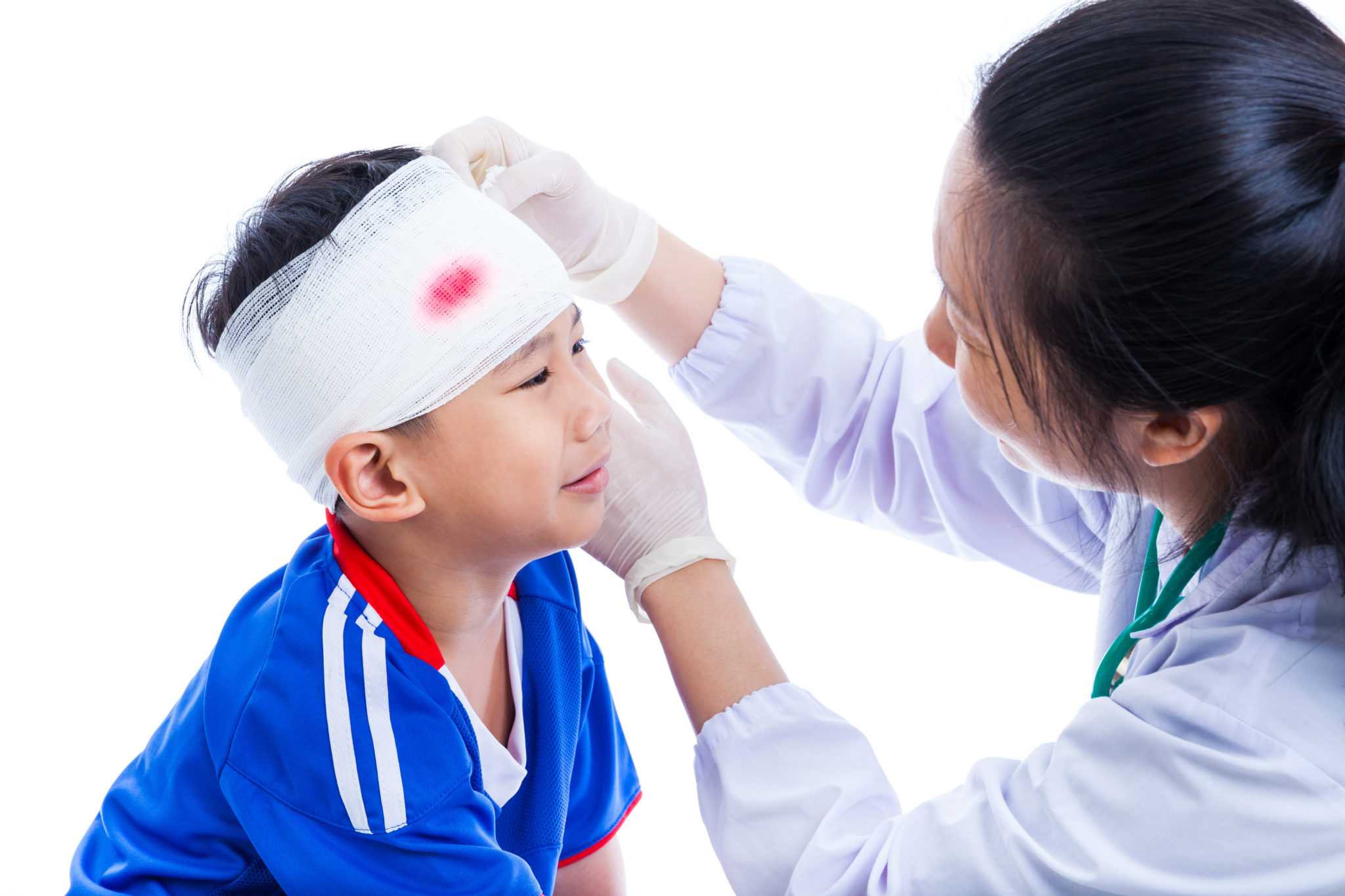
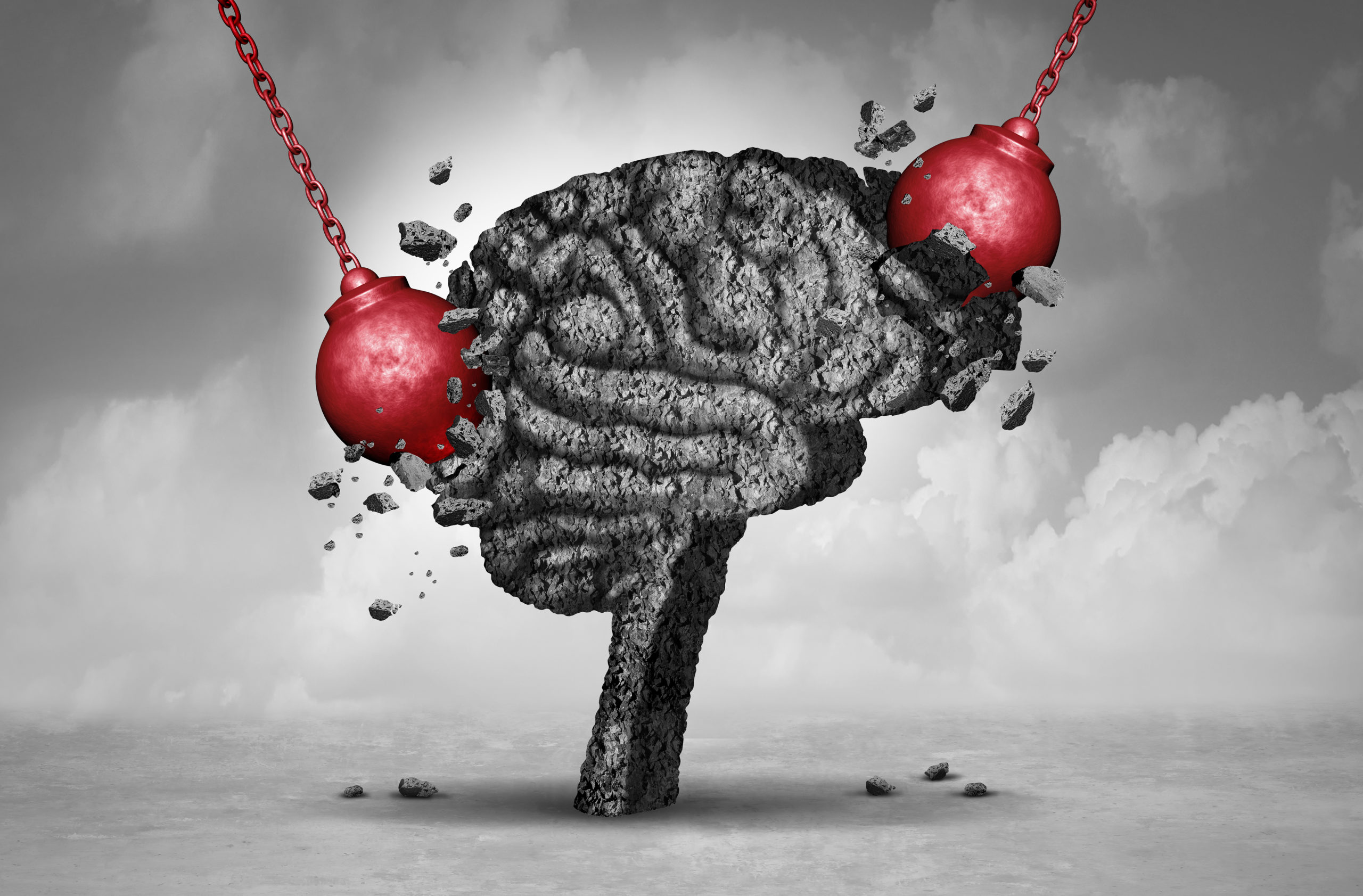
Headache never feels good, but a headache behind the eyes can make you miserable. Eye pain and Headaches can occur together. There are many reasons for such headaches.

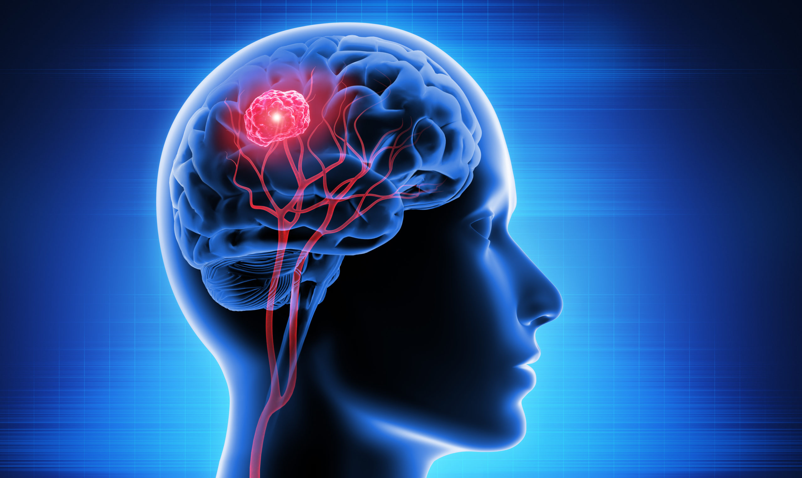
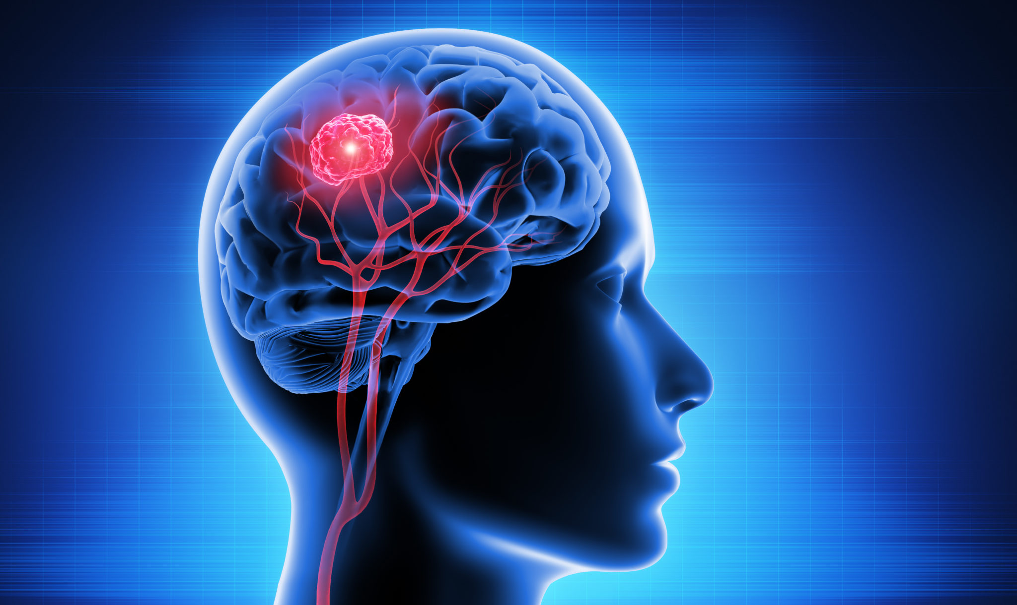
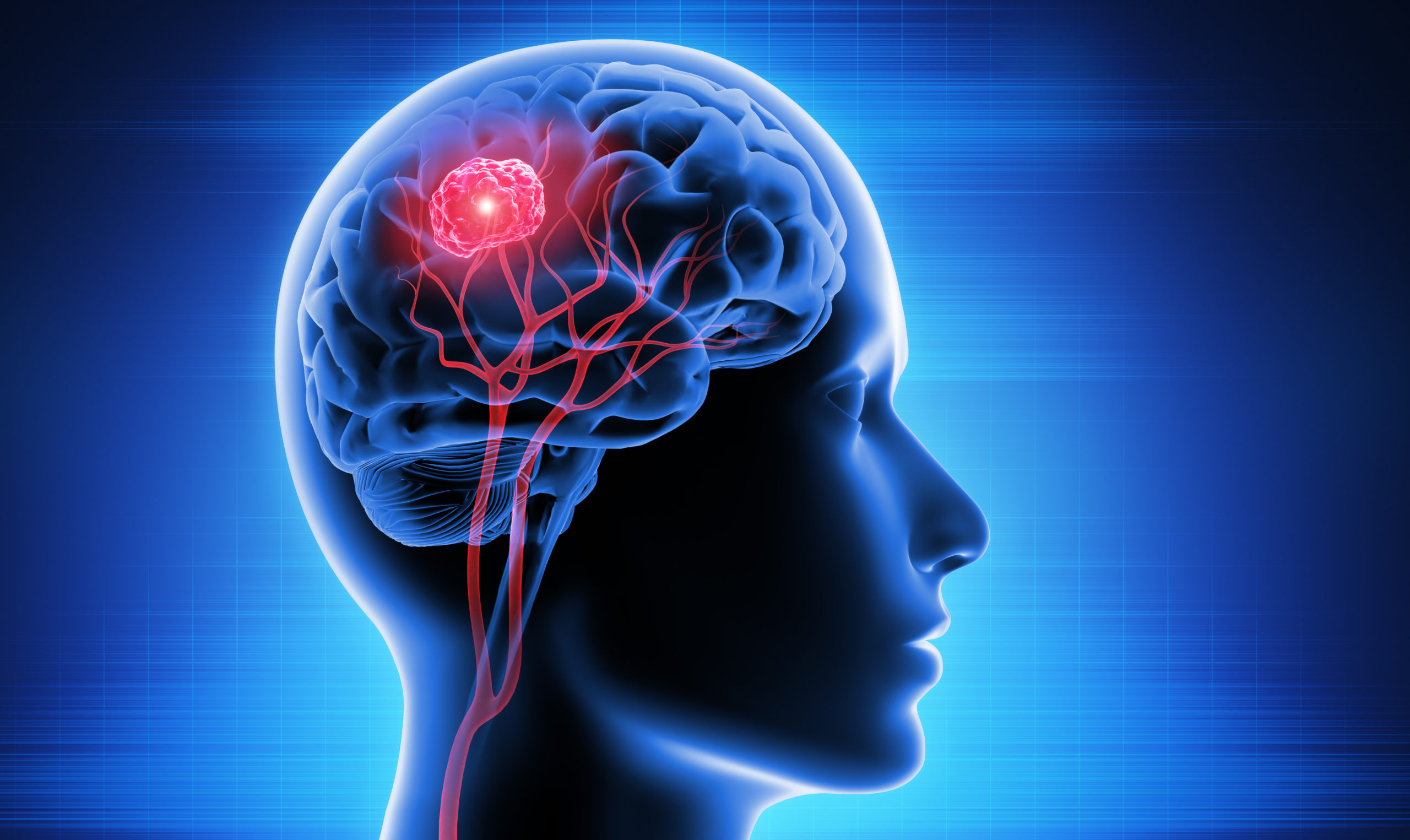
Please note that if you feel a headache behind your eyes, it does not mean that you have an eye problem. Remember, there is a part of the brain behind our eyes, and headache behind the eyes may be something serious like a brain tumour.

Headache on right side of head and eye and Headache on left side of head and eye may be cause of the same diseases, but on the opposite sides.

Some types of headaches are continuous for days, while others just come and go. While some times eye pain causes headache.  

Also, it’s also very much possible that this type of headaches could cause vision problems too.

Headache is perhaps the commonest symptom in human beings all through out the world. But the good news is that most headaches are caused by trivial reasons and can be cured by a cup of tea or coffee or over the counter medicines. However, if a headache persists, it is time to see a specialist neurosurgeon and get yourself checked. Remember, howsoever rare it may be, there may be a possibility of a brain disease and it is very wise to get it checked, diagnosed and treated at an early stage.

Headache associated with the eyes may have many reasons. Here are some reasons you may be feeling pain behind your eyes.

Table of Contents

Sinus or Pressure Headaches

Sinuses are empty spaces inside the skull bones. These cavities make the skull very light. At the same time these sinus cavities are places where infection occurs and causes a lot of pain and discomfort. Sinus headaches are diagnosed by the fact that they occur exactly at the site of the sinus, which could be in the forehead, around the eyes, in the cheeks and so on. The openings into the sinuses are blocked and so the air inside the sinuses is absorbed creating partial vacuum. The air pressure outside presses on the sinuses and causes ‘pressure pain’ because of the vacuum inside the sinus.

Pain in forehead above eyes is usually because of frontal sinusitis, meaning infection inside the frontal sinuses located in the forehead.

Triggers include sinusitis (including chronic sinusitis), colds, or allergies.

Treatment: Using a humidifier, warm compress, or breathing in warm, moist air from a vaporizer or pot of boiling water can help. the water vapour goes inside the sinuses and helps open the chocked sinus openings. Some people get relief by taking cold medications.

Tension Headaches

These is the most common type of headache, and may cause pain behind your eyes, as well as on one or both sides of your head and from your neck down to your shoulders. Mental stress, sedentary work, too much focus on any type of screen, be it a mobile phone, computer or TV, may lead to such headaches. No doubt all three, that is phones, computers and TVs are not only essential but a necessity of life. However, it is up to us to manage both, using these devices and not getting tension headaches.

Triggers for tension headaches include poor posture, lack of sleep, being hungry, being dehydrated or eye strain from staring too long at a computer screen.

Treatments: If you have an occasional tension headache, doctors may recommend an over-the-counter headache medicine, such as paracetamol or aspirin. You can also try a warm compress, a cold compress, sitting or lying down in a dark room without flickering lights or merely resting with your eyes closed. If you have these types of headaches too frequently, it is time that you consulted a specialist for investigations and treatment.

Migraine Headaches

These types of headaches are known for being accompanied by both pain and visual symptoms, such as seeing a halo or flashing lights, or being extra sensitive to light. Nausea and a runny nose can also sometimes accompany the pain. There are many more symptoms of migraine headaches, like Constipation, Mood swings, Food cravings, Increased thirst and urination, Frequent yawning, Aura- symptoms of the nervous system experienced before or during the migraine and Changes in vision.

Triggers include not sleeping well, feeling stressed, bright lights, certain food and drink (such as alcohol or chocolate) or certain smells.

For treating migraines, a cup of tea or coffee is sometimes enough. Other people may need prescription medicines for the treatment of migraine.

Glaucoma

One of the eye pain causes is Glaucoma. It is a leading cause of vision loss in people older than 60. Pressure inside the eyeball increases and damages the optic nerve. Severe, throbbing eye pain combined with headache are two common symptoms of Glaucoma. Blurry vision, eye redness, seeing halos, and nausea and vomiting may also occur. Acute Glaucoma is a medical emergency, it is best to seek urgent treatment.

Cluster Headaches

If you have this kind of headache, you may feel extreme pain around your eyes (and particularly around just one eye) and your eyes may also feel watery. The name “cluster” has to do with the fact that these headaches usually show up several times for days or weeks, then disappear for a time before you have them again. Cluster headaches, which occur in cyclical patterns or cluster periods, are one of the most painful types of headache. A cluster headache commonly awakens you in the middle of the night with intense pain in or around one eye on one side of your head.

Bouts of frequent attacks, known as cluster periods, can last from weeks to months, usually followed by remission periods when the headaches stop. During remission, no headaches occur for months and sometimes even years.

Fortunately, cluster headache is rare and not life-threatening. Treatments can make cluster headache attacks shorter and less severe. In addition, medications can reduce the number of cluster headaches you have.

Triggers include smoking, alcohol, and certain medications.

Treatments are by medications like verapamil or prednisone, or injections of triptans or lidocaine nose drops. Some people find relief from breathing pure oxygen.

Optic Neuritis

It is the inflammation of the optic nerve, and it can cause left eye pain and headache and right eye pain and headache. Blurred vision, dim vision, and painful eye movements also occur.

Back of the Head Pain

The back side of the head is called the Occipital region and pain there is called occipital neuralgia. It is a painful condition. It comes all of a sudden, lasts for a few seconds to a few minutes. It is paramount that physicians understand the differential diagnosis for this condition and specific diagnostic criteria. There are multiple treatment modalities, several of which have well-established efficacy in treating this condition.

Special medicines like NSAIDs and antidepressants may help reduce such pain. Nerve blocks can be done to alleviate pain.

Dr. Ravindra Patil Neurosurgeon at Samarth Neuro and Superspeciality Hospital has good experience in this field.
NCP of Head Injury

NCP of Head Injury

NCP of Head Injury

By Dr. Ravindra Patil

NCP means nursing care plan.

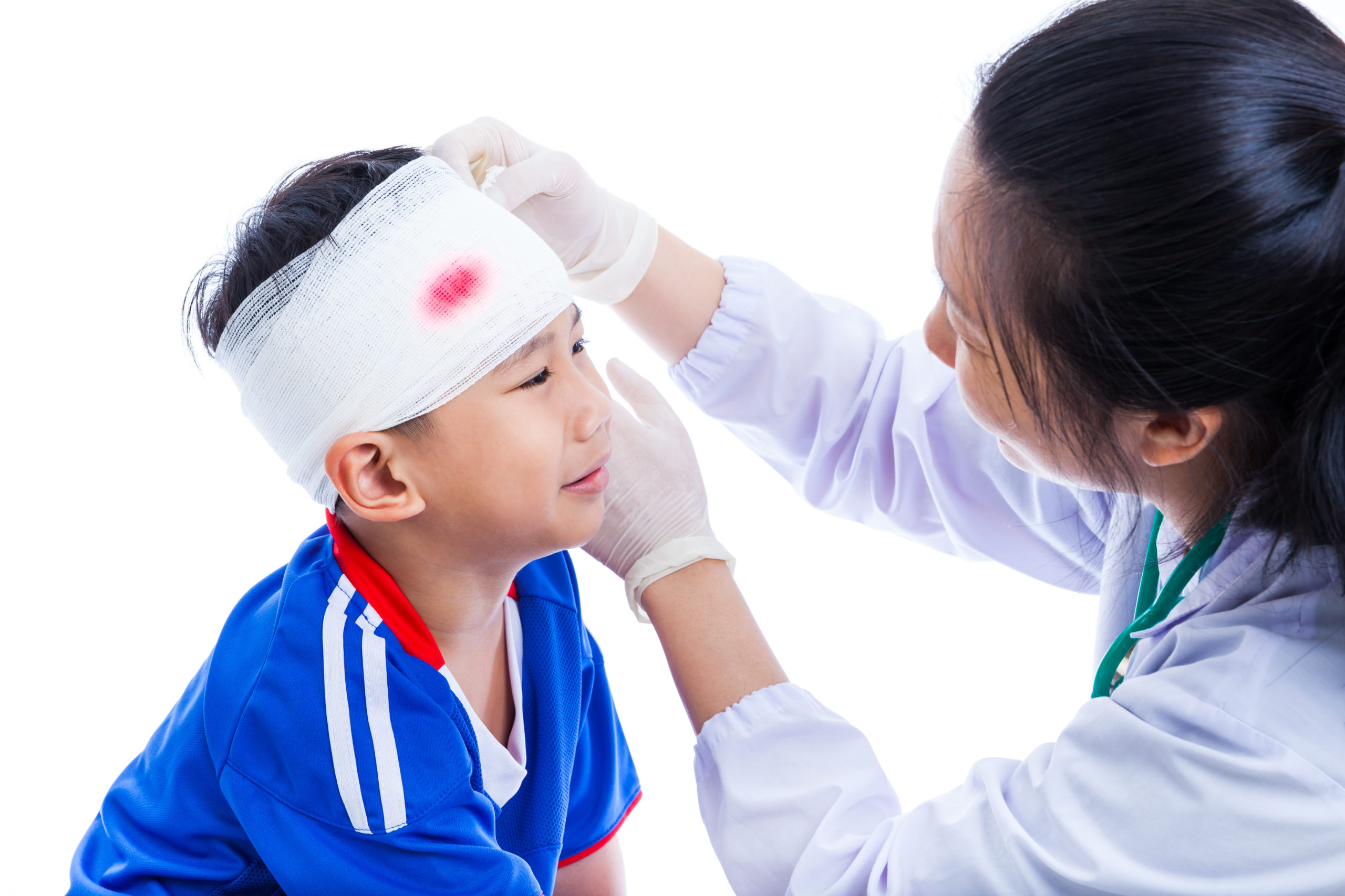
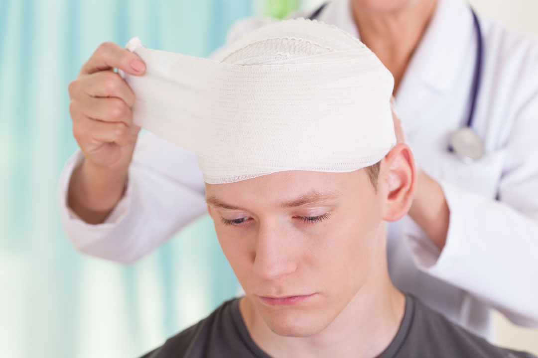
Any severe blow to the brain, skull or the scalp is considered a head injury. It ranges from a minor bump to a fractured skull.Along with the neurosurgeons and critical care specialists, nurses have a major role in managing head injury and traumatic brain injury. Nurses have a nursing care plan for managing every type of patients, and similarly they have a nursing care plan or NCP for managing head injury patients.

Table of Contents

Primary Goal

  • The primary goal of nursing care plan head injury is to maintain adequate blood supply and improve cerebral blood flow in order to prevent cerebral ischaemia and secondary injury to the brain.
  • The intracranial pressure of CSF must also be under control.
  • Blood oxygen and CO2 saturation must be maintained in every nursing care plan of head injury.
  • Nursing care plan on head injury must ensure that the patient has no seizures.
  • If the patient is unconscious, the NCP for unconscious patient will come into play.

Nursing Diagnosis

Nurses have their own head injury nursing diagnosis. While surgeons operate and physicians decide the medicines, a proper nursing care plan of head injury helps administer medicines and take due patient care. Remember, a critically ill head injury patient needs constant care from his or her head to his or her toes. For a nurse, the head injury is a just one part.

In head injury nursing management, most important is general nursing care which includes eye care, hair care, nail care, intubation and ventilator care, urinary catheter care, intravenous lines care, central line care, feeding and feeding tube care and so on. Over and above all this the nurse will care for the head injury with her nursing care plan for head injury.

If a patient is unable to move or is unconscious, he/she has to be turned over every two hours to prevent pressure sores. Such patients are on diapers and the diapers need to be changed and the area sponged and cleaned. The patient’s body must be sponged at least once or twice a day. Head injury nursing management also includes urinary catheter care.o.

Raised Intracranial pressure [ICP]

Because of various reasons, the pressure of the cerebrospinal fluid [CSF] may increase in head injury patients. The patient will have an optimal cerebral tissue perfusion only with a normal ICP. To maintain normal ICP, in every nursing care plan for head injury, the nurse will monitor the patient’s neurological status by checking the pupils, Level of Consciousness [LOC] and Glasgow Coma Scale [GCS] scores continuously.

Remember, it is the nursing staff who is with the patient 24×7. Only the nurses can observe such changes and report them to the physician, who in turn can take appropriate action.

Any changes in vital signs may be a sign of increased pressure in the brain. An increased ICP causes slowing heart rate, a widening pulse pressure, and irregular respiration.

Every nursing care plan on head injury will check for fluid leakage from the ears and nose. Leakage of a clear liquid from the nose (rhinorrhoea) and ears (otorrhea) might be the cerebrospinal fluid leaking after head injury caused by fractures. Because there is no accumulation of fluid in the brain, there might be no signs of raised ICP.

O2 and CO2 Levels

In a typical ICU patient’s file will be written:

“Keep PO2 between 80 and 100 mmHg and PCO2 between 35 and 38 mmHg.”

These are levels of blood oxygen and carbon-dioxide which are to be maintained by the oxygen administered through nasal prongs, mask, or a tube inside the trachea, a process called “Intubation”. When a patient is intubated, a machine called the “ventilator” controls the patient’s breathing.

In every NCP of head injury, the goal is to prevent prolonged states of hypoxemia (decreased blood level of oxygen) and hypercarbia (increased amount of carbon dioxide). 

Besides all that, look after…

A competent ICU nurse’s nursing management of head injury will avoid any activities and symptoms that increase ICP. They are:

  • Keep patient’s head straight
  • Do endotracheal suctioning
  • Prevent patient’s coughing, vomiting
  • Head injury nursing management must prevent bending at the waist
  • Reduce Pain
  • Prevent Fever
  • Don’t allow shivering in the patient

In a good nursing care plan of head injury, although it is essential, it is good to limit suctioning and do hyperoxygenation before suctioning to help keep ICP at bay.

Intracranial monitoring system

Head injury nursing management may have equipment to measure ICP continuously. An ICP that is greater than 15 mmHg should be reported right away.

A nurse must administer medication as ordered to decrease ICP. Medicines used to reduce ICP are:

  • Hyperosmotic agents (Mannitol)
  • Steroids
  • Barbiturates
  • Antipyretics
  • Muscle relaxants
  • Anticonvulsants

Thus, all the above types of medicines are a vital necessity in a nursing care plan for head injury.

Control Seizures

Seizures are caused by many reasons, some of which are:

  • Intracranial Bleeding
  • Contusion
  • Hyponatremia
  • Open and closed brain injuries
  • Hypoxia
  • Protect the patient’s airway during seizure activity.

Every nurse trained in a nursing care plan on head injury must know how to look the above. A good nurse must have her own head injury nursing diagnosis. The above characteristics of seizures must be noted by nurses and recorded:

The worst that can happen during head injury nursing management is patient’s self-injury. To prevent injuries, do this:

  • Reduce disturbance
  • Pad side rails of the patient’s bed
  • Place the bed in the lowest position
  • Provide head protection by extra pillows

Every nursing care plan of head injury must assist the patient during the seizure by:

  • Turning the patient’s head to the side
  • Suctioning if necessary

These measures protect the patient’s airway during and after the seizure.

Anticonvulsants medicines must be given as ordered. Phenytoin is given as an anti-seizure medicine. But it requires close monitoring or toxicity may occur.

Acute Confusion due to increased ICP

ICU nursing care plan for head injury includes very close constant monitoring of the patient’s consciousness. The nurse must check for:

  • Assess the patient’s level of consciousness frequently as ordered.
  • A change in mental status might indicate an increase in ICP.
  • Reorient the patient to person, time, place, and situation frequently.

Memory might be affected. Hence requires frequent repetition of the same information. Informing the patient about their situation might reduce anxiety levels and bring their cognitive status back to baseline.

Talk to the patient

Only in the NCP for unconscious patient will communication with the patient be not be possible. Otherwise in every nursing care plan for head injury, the nurse must talk to the patient often and explain things in short and simple sentences before and throughout the process. Also;

  • Promote continuity of care.
  • A good nursing care plan of head injury will prevent frequent changes in staff
  • Don’t change environment as it might further worsen the patient’s confused state.
  • If possible, have the family communicate with the patient via a smartphone.
  • Seeing familiar faces and recognizing familiar voices might stimulate memory and help with reorientation.

Deficient Knowledge, Difficult Healing

No patient knows about his head injury. Hence head injury patients are very, very confused. It is a sudden event. Hence the nursing care plan on head injury must factor this in.

Brain injury might affect short-term memory and cause behaviour and mood changes.  Ability to focus and learn new information might be difficult and take more time. Most patients and families have no prior experience with head trauma injuries. In most cases, head injuries arise from very sudden and unexpected events.

Managing the relatives

Whatever good care is taken, it must be communicated to the patients and family members daily. Family members and caregivers are a vital part of the healthcare team.

Head injury nursing management will prepare the patient and family for the need for physical therapy, occupational therapy, speech therapy and home nursing care. This does not raise expectations greatly and helps the family adjust themselves to the new situation.

All in all, along with the surgeon, only a good head injury nursing care plan implemented by committed nurses will make the head injury patient better.

And at Samarth Neuro and Superspeciality Hospital, run by neurosurgeon Dr. Ravindra Patil, this combination of a competent neurosurgeon and the best nursing care is accessible around-the-clock.

Special Note

In the above essay, nurses are mentioned as women only for the sake of convenience. Male nurses are also equally dedicated and capable.

डोक्याला दुखापत झालेल्या रूग्णाची नर्सिंग काळजी

डोक्याला दुखापत झालेल्या रूग्णाची नर्सिंग काळजी

डोक्याला दुखापत झालेल्या रूग्णाची नर्सिंग काळजी

By Dr. Ravindra Patil

कोणत्याही रूग्णाच्या उपचारात डॉक्टरांइतका, कदाचित डॉक्टरांहूनही जास्त, नर्सेसचे (परिचारिकांचे) योगदान असते. कारण नर्सेस सतत रूग्णाच्या बरोबर असतात.

नर्सेस कोणत्याही रूग्णाची काळजी घेतांना एक प्लॅन बनवतात. याला नर्सिंग केर प्लॅन असे म्हणतात. डोक्याला गंभीर इजा असलेल्या रूग्णांसाठी पण नर्सेसचा नर्सिंग केर प्लॅन असतो. पाहूया अशा गंभीर परिस्थितीत असलेल्या रोग्यांची परिचारिका किंवा नर्सेस कशी काळजी घेतात.

Table of Contents

डोक्याची गंभीर इजा (हेड इन्जुरी) म्हणजे नक्की काय

मेंदू, कवटी किंवा टाळूला कोणताही गंभीर आघात हा डोक्याची दुखापत मानली जाते.अशा इजा किरकोळ धक्क्यापासून फ्रॅक्चर झालेल्या कवटीपर्यंत असतात. न्यूरोसर्जन आणि क्रिटीकल केर तज्ञांसोबत, हेड इन्जुरी आणि मेंदूला झालेल्या दुखापतीची काळजीघेण्यात परिचारिकांची प्रमुख भूमिका असते. प्रत्येक प्रकारच्या रूग्णांचे व्यवस्थापन करण्यासाठी परिचारिकांकडे नर्सिंग केअर योजना असते आणि त्याचप्रमाणे त्यांच्याकडे डोक्याला दुखापत झालेल्या रूग्णांचे व्यवस्थापन करण्यासाठी नर्सिंग केअर योजना किंवा NCP असते.

प्राथमिक ध्येय

  • सेरेब्रल इस्केमिया(रक्त पुरवठा बंद होणे) आणि मेंदूला होणारी दुय्यम इजा टाळण्यासाठी मेंदूत पुरेसा रक्तपुरवठा राखणे आणि सेरेब्रल रक्त प्रवाह सुधारणे हे नर्सिंग केअर प्लॅनचे मुख्य उद्दिष्ट आहे.
  • सेरेब्रोस्पायनल फ्लुइड (सी.एस.एफ.) चा इंट्राक्रॅनियल (कवटीच्या आतील) दबाव वाढू न देणे.
  • डोक्याच्या दुखापतीच्या प्रत्येक नर्सिंग काळजी योजनेमध्ये रक्तातील ऑक्सिजन आणि कार्बन-डायऑक्सइडच्या मात्रा योग्य प्रमाणात राखल्या पाहिजेत.
  • डोक्याच्या दुखापतीमुळे रुग्णाला कोणतेही दौरे येत नाहीत इकडे नर्सिंग केअर प्लॅनमध्ये विशेष लक्ष देण्यात येते.
  • जर रुग्ण बेशुद्ध असेल, तर बेशुद्ध रुग्णासाठी वेगळा “नर्सिंग केर प्लॅन” वापरण्यात येईल.

नर्सिंग निदान

डॉक्टर निदान करतात त्याच प्रकारे परिचारिकांचे पण “नर्सिंग निदान”असते. शल्यचिकित्सक ऑपरेट करत असताना आणि डॉक्टर औषधे ठरवत असताना, परिचारिकाडोक्याच्या दुखापतीची योग्य नर्सिंग काळजी योजना आखतात, औषधे व्यवस्थापित करतात आणि रुग्णाची योग्य काळजी घेण्यास मदत करतात. लक्षात ठेवा, गंभीरपणे आजारी असलेल्या डोक्याला दुखापत झालेल्या रुग्णाची त्याच्या डोक्यापासून ते त्याच्या पायाच्या बोटांपर्यंत अशी त्याच्या अख्ख्या शरीराची सतत काळजी घ्यावी लागते. नर्ससाठीडोक्याची दुखापत हा फक्त एक छोटा भाग आहे.

हेड इन्जुरी नर्सिंग मॅनेजमेंटमध्येसर्वात महत्वाचे असते सामान्य नर्सिंग केअर. यामध्ये डोळ्यांची काळजी, केसांची काळजी, नखांची काळजी, इंट्यूबेशन आणि व्हेंटिलेटर केअर, युरिनरी कॅथेटर केअर, इंट्राव्हेनस लाईन्स केअर, सेंट्रल लाइन केअर, फीडिंग आणि फीडिंग ट्यूब केअर इत्यादींचा समावेश होतो.हे सर्व संभाळून नर्स डोक्याच्या दुखापतीची पण तिच्या नर्सिंग केअर प्लॅन मध्ये योग्य काळजी घेईल.

जर एखादा रुग्ण हालचाल करू शकत नसेल किंवा बेशुद्ध असेल, तर त्याला “प्रेशर सोर्स”(हालचाल नसल्याने व सतत दाबाने कातडी घासली जाऊन आतले मास दिसणे)  टाळण्यासाठी दर दोन तासांनी एक कुशीवरून दुसर्‍या कुशीवर उलटावे लागते. जे रुग्ण डायपरवर आहेत त्यांचे डायपर बदलणे आवश्यक असते आणि तिथला भाग स्पंज करून स्वच्छ करणे आवश्यक असते. रुग्णाच्या आख्ख्या शरीराला दिवसातून किमान एकदा किंवा दोनदा स्पंज करणे आवश्यक आहे. डोके दुखापत नर्सिंग व्यवस्थापनामध्ये मूत्र कॅथेटरची काळजी देखील समाविष्ट आहे.

वाढलेले इंट्रा क्रॅनियल प्रेशर [ICP]

विविध कारणांमुळे, डोक्याला दुखापत झालेल्या रुग्णांमध्ये सेरेब्रोस्पाइनल फ्लुइड [CSF] चा दाब वाढू शकतो. रुग्णाच्या मेंदूला आदर्श परिस्थिती म्हणजे नॉर्मल इंट्रा क्रेनियल प्रेशर, जेव्हा सेरेब्रल टिश्यू परफ्यूजन इष्टतम असते.ICP नॉर्मल राखण्यासाठीडोक्याच्या दुखापतीच्या प्रत्येक नर्सिंग केअर प्लॅनमध्ये, नर्स रुग्णाची न्यूरोलॉजिकल स्थिती, चेतनेची पातळी [शुद्ध] आणि ग्लासगो कोमा स्केल [GCS] स्कोअर यांचे सतत निरीक्षण करेल.

लक्षात ठेवा, फक्त नर्सिंग स्टाफच रुग्णासोबत 24×7 असतो. केवळ परिचारिकाच अशा बदलांचे निरीक्षण करू शकतात आणि डॉक्टरांना ही माहिती देऊ शकतात. ते बघून डॉक्टरयोग्य कारवाई सुरू करू शकतात.

महत्त्वाच्या लक्षणांमधील कोणतेही बदल हे मेंदूतील दबाव वाढण्याचे लक्षण असू शकतात. वाढलेल्या इंट्रा क्रेनियल प्रेशरमुळे हृदय गती मंदावते, नाडीचा दाब वाढतो आणि श्वासोच्छ्वास अनियमित होतो.

डोक्याच्या दुखापतीवरील प्रत्येक नर्सिंग काळजी योजना कान आणि नाकातून द्रव गळतीची तपासणी करतेच. कारण कवटीच्या फ्रॅक्चरमुळे डोक्याला दुखापत झाल्यानंतर नाकातून सेरेब्रोस्पायनल फ्लुइडचा द्रव बाहेर पडणे (राइनोरिया) आणि कानातून पडणे (ओटोरिया) असे होऊ शकते. अशा गळती मुळे मेंदूमध्ये द्रवपदार्थ साचत नसल्यामुळे, सेरेब्रोस्पायनल फ्लुइडचा दाबवाढूनही तो वाढल्याची चिन्हे दिसत नाहीत.

O2 आणि CO2 पातळी

कुठल्याही ICU मध्ये रुग्णाच्या फाईल मध्ये असे लिहिलेले असेल:

“PO2 80 आणि 100 mmHg या मध्ये आणि PCO2 35 आणि 38 mmHg या दरम्यान ठेवा.”

हे रक्तातील ऑक्सिजन आणि कार्बन-डायऑक्साइडचे स्तर आहेत. ते रूग्णाला नाकातून नळ्या लावून, मास्क लावून किंवा श्वासनलिकेच्या आत असलेल्या नळीद्वारे देण्यात येणार्‍या ऑक्सिजनद्वारे राखले जातात. तोंडातून किंवा गळ्यावर भोक पाडून श्वास नलिकेत नळी घालायच्या प्रक्रियेला “इंट्युबेशन” म्हणतात. जेव्हा एखाद्या रुग्णाला इंट्यूबेट केले जाते तेव्हा नळीच्या दुसर्‍या टोकाला “व्हेंटिलेटर” नावाचे मशीन रुग्णाच्या श्वासोच्छवासावर नियंत्रण ठेवते.

डोक्याच्या दुखापतीच्या प्रत्येक नर्सिंग केअर प्लॅन मध्ये, हायपोक्सिमिया (रक्तातील ऑक्सिजनची घटलेली पातळी) आणि हायपरकार्बिया (कार्बन डायऑक्साइडचे वाढलेले प्रमाण) यांचे दीर्घकाळापर्यंत नियंत्रण करणे हे लक्ष्य असते.

त्याशिवायच्याइतर अनेक बाबी…

सक्षम ICU परिचारिकेचे हेड इन्जुरी नर्सिंग व्यवस्थापन कशाही प्रकारे इंट्रा क्रेनियल प्रेशर (ICP) वाढू देणार नाहीत. ते करण्यास खालील गोष्टी करण्यात येतात:

  • रुग्णाचे डोके सरळ ठेवा
  • एंडोट्रॅकियल सक्शन करा
  • रुग्णाचा खोकला, उलट्या थांबवा
  • डोके दुखापत नर्सिंग व्यवस्थापनात रूग्णाला कंबरेत वाकू देणे प्रतिबंधित आहे
  • वेदना कमी करा
  • ताप टाळा
  • रुग्णाला थरथर कापू देऊ नका

डोक्याच्या दुखापतीच्या चांगल्या नर्सिंग केअर योजनेत गळ्यातून सक्शन मर्यादित प्रमाणात करतात आणि सक्शन करण्यापूर्वी हायपरऑक्सिजनेशन करणे आवश्यक असते.

इंट्राक्रॅनियल मॉनिटरिंग सिस्टम

डोके दुखापत नर्सिंग व्यवस्थापनाकडे सतत ICP मोजण्यासाठी उपकरणे असू शकतात. 15 mmHg पेक्षा जास्त ICP असेक तर लगेच डॉक्टरांना कळवणे आवश्यक असते.

ICP कमी करण्याच्या आदेशानुसार परिचारिकाने औषधोपचार करणे आवश्यक आहे. ICP कमी करण्यासाठी वापरलेली औषधे आहेत:

  • हायपरस्मोटिक एजंट (मॅनिटोल)
  • स्टिरॉइड्स
  • बार्बिट्युरेट्स
  • अँटीपायरेटिक्स
  • स्नायू शिथिल करणारी औषधे
  • अँटीकन्व्हल्संट्स

अशाप्रकारे, वरील सर्व प्रकारची औषधे डोक्याच्या दुखापतीच्या नर्सिंग केअर प्लॅनमध्ये एक अत्यावश्यक घटक असतात.

जप्ती नियंत्रित करा

जप्तीचे दौरे अनेक कारणांमुळे होतात, त्यापैकी काही खालीलप्रमाणे आहेत:

  • इंट्राक्रॅनियल रक्तस्त्राव
  • कवटीच्या आत मेंदूची इजा
  • हायपोनेट्रेमिया
  • उघड्या आणि बंद मेंदूच्या दुखापती
  • हायपोक्सिया
  • जप्तीदरम्यान रुग्णाच्या श्वासमार्गाचे रक्षण करा.

डोक्याच्या दुखापतीवर नर्सिंग केअर प्लॅनप्रमाणे कार्य करणार्या प्रत्येक प्रशिक्षित परिचारिकेला वरील गोष्टी कशा तपासाव्या हे माहित असणे आवश्यक आहे. कोणत्याही सक्षम नर्सला डोक्याच्या दुखापतीचे नर्सिंग निदान करता येणे अति आवश्यक असते.जर जप्तीची वरील वैशिष्ट्ये परिचारिकांनी लक्षात घेतली पाहिजेत आणि रेकॉर्ड केली पाहिजेत:

डोके दुखापत नर्सिंग मॅनेजमेंट दरम्यान घडणारी सर्वात वाईट गोष्ट म्हणजे रुग्णाची स्वत: ची दुखापत. ते टाळण्यासाठी, हे करा:

  • रूग्णाला कोणत्याही प्रकारचा त्रास होऊ देऊ नका
  • रुग्णाच्या पलंगाच्या साइड रेलना पॅडींग करा
  • बेडची उंची कमी ठेवा
  • जास्त उशांद्वारे डोक्याला इजा होऊ देऊ नका

डोक्याच्या दुखापतीच्या प्रत्येक नर्सिंग केअर प्लॅनमध्ये रुग्णाला जप्ती दरम्यान मदत करणे आवश्यक आहे. त्यासाठी:

  • रुग्णाचे डोके बाजूला वळवणे
  • आवश्यक असल्यास सक्शन करणे

हे उपाय जप्ती दरम्यान आणि नंतर रुग्णाच्या वायुमार्गाचे संरक्षण करतात.

अँटीकॉन्व्हल्संट औषधे सूचने प्रमाणे दिली पाहिजेत. फेनिटोइन हे जप्तीविरोधी औषध म्हणून दिले जाते. परंतु यासाठी रूग्णचे सूक्ष्म निरीक्षण आवश्यक आहे, नाहीतर विपरीत परिणाम होऊ शकतो.

ICP वाढल्यामुळे तीव्र मानसिक गोंधळ

डोक्याच्या दुखापतीसाठी आयसीयू नर्सिंग केअर योजनेमध्ये रुग्णाच्या चेतनेचे सतत आणि सूक्ष्म निरीक्षण जरूरी असते. नर्सने हे सर्व तपासले पाहिजे:

  • आदेशानुसार वारंवार रुग्णाच्या चेतनेच्या पातळीचे मूल्यांकन करा.
  • मानसिक स्थितीतील बदल ICP मध्ये वाढ दर्शवू शकतो.
  • रुग्णाला व्यक्ती, वेळ, ठिकाण आणि परिस्थिती या विषयी वारंवार सांगा.

रुग्णाच्या स्मरणशक्तीवर दुष्परिणाम होऊ शकतो. त्यामुळे सामान्य माहितीची वारंवार पुनरावृत्ती करणे आवश्यक आहे. रुग्णाला त्यांच्या परिस्थितीबद्दल माहिती दिल्यास त्याची चिंता कमी होऊ शकते आणि त्यांची संज्ञानात्मक स्थिती पुन्हा योग्य पातळीवर येऊ शकते.

रुग्णाशी बोला

केवळ बेशुद्ध रुग्णासाठी रुग्णाशी संवाद साधता येणार नाही. अन्यथा डोक्याच्या दुखापतीसाठी प्रत्येक नर्सिंग केअर योजनेमध्ये, परिचारिकेने रुग्णाशी अनेकदा बोलले पाहिजे आणि रुग्णावर कोणतीही प्रक्रिया करण्या पूर्वी आणि प्रक्रियेदरम्यान लहान आणि सोप्या वाक्यांमध्ये रुग्णाशी स्पष्ट संपर्क ठेवला पाहिजे. तसेच;

  • रुग्णाची सतत काळजी घेणे.
  • डोक्याच्या दुखापतीची चांगली नर्सिंग केअर योजना कर्मचाऱ्यांमध्ये वारंवार होणारे बदल न करणे. नवीन नर्स किंवा अटेन्डन्ट आले की रूग्णाचा गोंधळ उडतो.
  • वातावरण बदलू नका कारण यामुळे रुग्णाची गोंधळलेली स्थिती आणखी बिघडू शकते.
  • शक्य असल्यास, कुटुंबाला रुग्णाशी स्मार्टफोनद्वारे संवाद साधण्यास सांगा.
  • परिचित चेहरे पाहणे आणि परिचित आवाज ओळखणे हे स्मृती उत्तेजित करू शकतात आणि पुनर्स्थित करण्यात मदत करू शकतात.

कमी ज्ञान, कठीण उपचार

जसे तापाचे किंवा ब्लड प्रेशर अथवा डायाबिटीसची सर्वसामान्य लोकांना माहिती असते तशी डोक्याच्या गंभीर दुखापतीबद्दल आणि त्याच्या उपचारा बद्दल कोणत्याही रुग्णाला माहिती नसते. त्यामुळे डोक्याला दुखापत झालेले रुग्ण खूप गोंधळलेले असतात. हेड इन्जुरी ही अचानक घडलेली घटना असते. त्यामुळे डोक्याच्या दुखापतीवरील नर्सिंग केअर योजनेत हे लक्षात घेणे आवश्यक आहे.

मेंदूच्या दुखापतीमुळे अल्पकालीन स्मरणशक्तीवर परिणाम होऊ शकतो आणि वर्तन आणि मूड बदलू शकतो. लक्ष केंद्रित करण्याची आणि नवीन माहिती शिकण्याची क्षमता कठीण असू शकते आणि काहीपण शिकण्यास अधिक वेळ लागू शकतो. बहुतेक रूग्ण आणि कुटुंबांना डोक्याला झालेल्या दुखापतींचा कोणताही पूर्वीचा अनुभव नसतो. बहुतेक प्रकरणांमध्ये, डोके दुखापत अगदी अचानक आणि अनपेक्षित घटनांमुळे उद्भवते.

नातेवाईकांना सांभाळणे

जी काही चांगली काळजी घेतली जाते, ती दररोज रुग्णांना आणि कुटुंबियांना कळवली पाहिजे. कौटुंबिक सदस्य आणि परिचारिका हे आरोग्य सेवा देणार्यांचा एक महत्त्वाचा भाग आहेत.

हेड इंजरी नर्सिंग मॅनेजमेंटच्या नर्सेसनी रुग्ण आणि त्याच्या कुटुंबाची हॉस्पिटलमधून घरी गेल्यावर पुढच्या उपचाराची मानसिक तयारी करून देणे आवश्यक असते. त्यात शारीरिक उपचार (फिझियथेरपी), व्यावसायिक (ऑक्युपेशनल) थेरपी, स्पीच थेरपी आणि होम नर्सिंग केअर देणे गरजेचे असते. डोक्याच्या गंभीर इजेनंतर व ऑपरेशननंतर लगेच काही रूग्ण बरा होत नाही. पूर्ण बरे होणे अवघड असते व त्याला खूप वेळ लागतो हे रूग्ण व त्याच्या परिवाराला माहित हवे. त्या साठी त्यांची मानसिक तयारी हवी. हे केल्याने यामुळे अपेक्षा मोठ्या प्रमाणात वाढत नाहीत आणि कुटुंबाला नवीन परिस्थितीशी जुळवून घेण्यास मदत होते.

एकंदरीत, शल्यचिकित्सकासह, कुशल परिचारिकांनी अंमलात आणलेली एक चांगली डोके दुखापत नर्सिंग केअर योजना अत्यावश्यक असते आणि डोके दुखापतग्रस्त रुग्णाला बरे होण्यास खूप जरूरी असते. आणि समर्थ न्यूरो आणि सुपरस्पेशालिटी हॉस्पिटलमध्ये, न्यूरोसर्जन डॉ. रवींद्र पाटील यांनी चालवत असलेले , सक्षम न्यूरोसर्जन आणि सर्वोत्तम नर्सिंग केअरचे हे संयोजन चोवीस तास उपलब्ध आहे.

विशेष नोंद

नर्स म्हणजे फक्त स्त्रिया नसतात. पुरुषही उत्कृष्ट नर्स असतात. वरील लेखात प्रत्येक वेळी परिचारिका किंवा नर्स हे लिहिण्यास सोपे जावे म्हणून त्यांचा स्त्रीलिंगी उल्लेख केला आहे. मुलेही नर्सेस असतात आणि मुलीं इतकेच छान व कुशल काम करतात.

भारतात ब्रेन ट्यूमर शस्त्रक्रियेचाअंदाजे खर्च

ब्रेन ट्यूमर शस्त्रक्रियेचा खर्च

भारतात ब्रेन ट्यूमर शस्त्रक्रियेचाअंदाजे खर्च

By Dr. Ravindra Patil

जेव्हा ब्रेन ट्यूमरचे निदान होते, तेव्हा पहिली भीती मृत्यूची असते आणि दुसरी भीती उपचारांच्या खर्चाची असते.

ट्यूमरचे उपचार सामान्यतः तीन प्रकारचे असतात, औषधे [केमोथेरपी म्हणतात], ब्रेन ट्यूमर शस्त्रक्रिया आणि रेडिएशन थेरपी.

प्रत्येक व्यक्ती शस्त्रक्रियेशिवाय ब्रेन ट्यूमरचा उपचार घेण्याचा प्रयत्न करेल कारण प्रत्येकजण शस्त्रक्रियेला घाबरतो. होय, ब्रेन ट्यूमरवर शस्त्रक्रियेशिवाय उपचार करणे शक्य आहे, परंतु सौम्य ट्यूमरचा [कर्करोग नसलेला ट्यूमरचा] शस्त्रक्रियेद्वारेच सर्वोत्तम उपचार होतो कारण सर्जन सौम्य ट्यूमर पूर्णपणे काढून टाकू शकतो.

ब्रेन ट्यूमरवर शस्त्रक्रियेशिवाय उपचार करणे खूप आकर्शक आहे. कारण त्यात कवटी कापून शस्त्रक्रियेची जरूर नसते. कवटी कापतांना मेंदूला इजा होण्याच्या शक्यता असते ती टळते.


शस्त्रक्रियेशिवाय ब्रेन ट्यूमरचे उपचार कोणत्या प्रकारचे आहेत?

Table of Contents

केमोथेरपी

ब्रेन ट्यूमरवर शस्त्रक्रियेशिवाय औषधाच्या उपचारांना केमोथेरपी म्हणतात. यात ब्रेन ट्यूमर पेशी नष्ट करण्यासाठी कर्करोगविरोधी (सायटोटॉक्सिक) औषधे वापरते. पण लक्षात ठेवा, औषधे इन्जेक्शनने टोचली किंवा तोंडी घेतली तरी ती तुमच्या शरीरात रक्तप्रवाहात फिरतात. अशा प्रकारे सायटोटॉक्सिक औषधे शरीराच्या इतर भागांना हानी पोहोचवू शकतात. याशिवाय, काही केमोथेरपी औषधांनी मेंदूच्या गाठींवर उपचार करणे कठीण होऊ शकते कारण औषधे “रक्त मेंदूचा अडथळा” (blood brain barrier) नावाचा अडथळा ओलांडू शकत नाहीत.


केमोथेरपी उपचारांचा एक कोर्स 6-12 महिने टिकू शकतो, ज्यामध्ये 6-12 चक्रे असतात. रुग्णांना दर काही आठवड्यांनी, काही दिवस केमोथेरपी दिली जाते.

रेडिएशन थेरपी

यात तीन मुख्य प्रकारचे उपचार आहेत. तुम्हाला एकापेक्षा जास्त प्रकारच्या उपचारांची आवश्यकता असू शकते.

  • पहिला प्रकार म्हणजे बाह्य बीम रेडिएशन थेरपी (EBRT) आहे. या प्रकारातएक मशीन ट्यूमरमध्ये उर्जेचे किरण पाठवते. हे उपचार सामान्यतः दर रोज देतात व काही आठवडे चालतात.
  • अंतर्गत विकिरण (ब्रेकीथेरपी). याला इंटरस्टिशियल थेरपी देखील म्हणतात. किरणोत्सर्गाच्या लहान बिया थोड्या काळासाठी ट्यूमरच्या आत किंवा जवळ ठेवल्या जातात. काही प्रकारच्या उपचारांमध्ये बिया काढून टाकाव्या लागतात.
  • गामा चाकू (knife) विकिरण. यामध्ये गामा किरण नावाच्या रेडिएशन बीमचा वापर केला जातो. किरण मशीनमधून पाठवले जातात आणि एकाच वेळी शेकडो कोनातून ट्यूमरवर केंद्रित केले जातात. उपचार सहसा एकदाच करावा लागतो. पण याच्यात कोणताही खरा चाकू नसतो.गामा किरणांच्या अचूक हॅमरिंगमुळे कदाचित हे नाव दिले गेले आहे: गामा चाकू (knife).

ब्रेन ट्यूमर ऑपरेशन

अर्थातच सौम्य [कर्करोग नसलेल्या] ब्रेन ट्यूमरमध्ये उपचारांचा हा मुख्य आधार आहे. ट्यूमर काढून टाका आणि रोग विसरा.

परंतु बर्‍याच प्रकरणांमध्ये ब्रेन ट्यूमरवर उपचार करण्यासाठी तीन्ही प्रकारचे उपचार वापरले जातात. सर्जन ट्यूमर काढून टाकतो, रेडिएशन ऑन्कोलॉजिस्ट कोणत्याही अवशिष्ट ट्यूमर पेशींवर उपचार करण्यासाठी रेडिएशन देतो आणि वैद्यकीय ऑन्कोलॉजिस्ट कोणत्याही कर्करोगाच्या पेशी पूर्णपणे नष्ट करण्यासाठी कर्करोग-विरोधी सायटोटॉक्सिक औषधे देतात.

कुठे होते ब्रेन ट्यूमर शस्त्रक्रिया?

ब्रेन ट्युमरच्या शस्त्रक्रिया फक्त मोठ्या शहरांमध्ये आणि मोठ्या कॉर्पोरेट हॉस्पिटलमध्येच करता येतात हे उघड आहे. अशा रुग्णालयांमध्ये सर्वोत्तम उपकरणे आणि सर्वोत्तम डॉक्टर एकाच छताखाली असतात. पण ते खूप पैसे घेतात. भारतात किंवा कोठेही मेंदूच्या शस्त्रक्रियेचा खर्च नेहमीच जास्त असतो. पण त्यासाठी कारणे आहेत.

ब्रेन ट्यूमरच्या उपचारांसाठी अनेक निदानात्मक चाचण्या कराव्या लागतात. सीटी स्कॅन, एमआरआय स्कॅन आणि पीईटी स्कॅन सारखी इमेजिंग आवश्यक आहे. मग ब्रेन ट्यूमर ऑपरेशनसाठी खूप महाग उपकरणे लागतात. ऑपरेशन थिएटर मध्ये खास HEPA फिल्टर्स आणि लॅमिनार एअरफ्लो बसवलेले असतात.

ब्रेन सर्जरी ऑपरेशन थिएटरमधील उपकरणांमध्ये ऑपरेशन थिएटर लाइट्स, ऑपरेशन थिएटर टेबल, ऍनेस्थेसिया ट्रॉली आणि याशिवाय स्टिरिओटॅक्टिक फ्रेम, सर्जिकल नेव्हिगेशन आणि इमेजिंग स्क्रीन असणे आवश्यक आहे. CUSA [Cavitronic Ultrasonic Surgical Aspirator] सारखी विशेष उपकरणे आवश्यक असू शकतात, जे पुन्हा खर्चात भर घालतात.

ब्रेन ट्यूमर ऑपरेशननंतर बरे होण्यासाठी काही दिवसांसाठी अतिदक्षता विभाग [ICU] मध्ये रोग्याला ठेवणे आवश्यक आहे. नंतर रुग्णाला हॉस्पिटलच्या स्पेशल खोलीत हलवले जाऊ शकते. त्याला/तिला सामान्य वॉर्डमध्ये ठेवता येत नाही कारण ब्रेन ट्यूमरच्या रूग्णांना विशेष काळजीची आवश्यकता असते. सांगायचा उद्देश हा की या सर्वांमुळे खर्च वाढत जातो.

ब्रेन ट्यूमरच्या ऑपरेशननंतर फिजिओथेरपी आवश्यक आहे. प्रशिक्षित फिजिओथेरपिस्टच्या देखरेखीखाली योग्य सौम्य व्यायामाने कार्यात्मक पुनर्प्राप्ती हळूहळू प्राप्त करणे आवश्यक आहे.

कधीकधी स्पीच थेरपीची (बोलण्याचे प्रशिक्षण) आवश्यकता असू शकते. ऑक्यूपेशनल थेरपीची आवश्यकता असू शकते.

कधीकधी इम्प्लांट किंवा उपकरणे कवटीत किंवा मेंदूमध्ये टाकली जातात आणि ती कायमस्वरूपी मेंदूमध्येच राहतात. अशी उपकरणे खूप महाग असतात. ते मेंदूच्या शस्त्रक्रियेच्या खर्चात भर घालतात.

शेवटी पण अतिशय महत्त्वाची गोष्ट म्हणजे, महानगर किंवा कॉस्मोपॉलिटन शहरांमधील रुग्णालये अतिशय उच्च किंमतीच्या जमीनीवर बांधलेली असतात.यामुळे पण प्रत्येक ब्रेन ट्यूमर उपचार रूग्णाच्या खर्चात वाढ होते. मुंबईसारख्या शहरात नवीन हॉस्पिटल बांधणे हे मोठ्या कॉर्पोरेट्ससाठीही अकल्पनीय आहे कारण तिथे जमिनीची किंमत अतिशय जास्त आहे.

एका छोट्या गावात चांगलं हॉस्पिटल

एखाद्या लहान शहरात जर वैद्यकीय आणि पॅरामेडिकल व्यावसायिकांची चांगली टीम असलेले एक चांगले रुग्णालय असेल तर ओव्हरहेड खर्च खूप कमी होतो.

समर्थ न्यूरो आणि सुपरस्पेशालिटीहॉस्पिटल हे महाराष्ट्राच्या दक्षिणेकडील मिरज आणि सांगली या छोट्या जुळ्या शहरांमधील कॉर्पोरेट हॉस्पिटलचे एक उत्तम उदाहरण आहे.

समर्थ न्यूरो आणि सुपरस्पेशालिटीहॉस्पिटलचे संस्थापक व प्रमुख म्हणजेसुवर्णपदक विजेते न्यूरो सर्जन डॉ. रवींद्र पाटील. ते तेथे ब्रेन ट्यूमरच्या अनेक शस्त्रक्रिया करतात. त्यांनी केलेली ऑपरेशन्सभारतात इतर कुठल्याही मोठ्या हॉस्पिटल मध्ये केलेल्या ब्रेन ट्यूमरच्या ऑपरेशन इतकीच चांगली होतात.

आणि समर्थ न्यूरो आणि सुपरस्पेशालिटीहॉस्पिटल कॉस्मोपॉलिटन किंवा महानगरांमध्ये ब्रेन ट्यूमर ऑपरेशनच्या खर्चाच्या तुलनेत खूपच कमी शुल्क आकारते.

भारतात ब्रेन ट्यूमर शस्त्रक्रियेचा खर्च लहान शहरे आणि शहरांमध्ये कमी असल्याची अनेक कारणे आहेत. हे जमिनीची कमी किंमत, कमी जमीन महसूल, शहराच्या मध्यभागी पर्यंत जाण्यास कमी अंतर आणि तुलनेने पोहोचण्यासाठी सुलभ.

डॉ. रवींद्र पाटील यांनी मोठ्या शहरांमध्ये प्रशिक्षण घेतले आणि मिरज आणि सांगली या छोट्या शहरांमध्ये स्थायिक होण्याचा निर्णय घेण्यापूर्वी त्यांनी हजारो शस्त्रक्रिया केल्या. आपल्या सहकारी नागरिकांची सेवा करणे आणि गर्दीपासून दूर जाणे हा त्यांचा उद्देश होता. पण सर्वात मोठे कारण म्हणजे त्यांनाया छोट्या टियर टू शहरांमध्ये एक शाश्वत रुग्णालय बांधता आले. अशा प्रकारे डॉ रवींद्र यांच्या शस्त्रक्रिया कौशल्यासह समर्थ न्यूरो आणि सुपरस्पेशालिटी हॉस्पिटलसुरू झाले.

त्यांनीब्रेन ट्यूमरच्या छोट्या ऑपरेशन्सपासून सुरुवात केली आणि लवकरच डोक्याला दुखापत आणि रस्ते अपघात आणि इतर अनेक मेंदूच्या विकारांसाठी शस्त्रक्रिया करण्यासाठी त्याच्या सेवा वाढवल्या. आज डॉ. रवींद्र दक्षिण महाराष्ट्रातील आघाडीच्या न्यूरोसर्जनपैकी एक आहेत आणि समर्थ न्यूरो आणि सुपरस्पेशालिटीहॉस्पिटलमध्ये जवळजवळ प्रत्येक ब्रेन ट्यूमर ऑपरेशनसाठी उत्कृष्ट उपकरणे आहेत. सीटी स्कॅनर आणि लॅमिनार एअर फ्लो आणि हिपा फिल्टरसह सुसज्ज ऑपरेशन थिएटर्स व्यतिरिक्त, त्याच्या हॉस्पिटलमध्ये विविध प्रकारच्या अल्ट्रा-प्रिसिजन ब्रेन ट्यूमर ऑपरेशन्ससाठी सर्जिकल नेव्हिगेशन सिस्टम आहे.

पण खर्च काय?

भारतातील वैद्यकीय पर्यटनाला प्रोत्साहन देणार्‍या एका वेबसाइटने भारतात वेगवेगळ्या शहरांमध्ये ब्रेन ट्यूमर ऑपरेशन किंवा शस्त्रक्रियांचे खालील दर दर्शविले आहेत. लक्षात घ्या की आकडेवारी फक्त जाहिरातींसाठी आहे. ब्रेन ट्यूमर शस्त्रक्रियेसाठी भारतात किंवा कोठेही खर्च ब्रेन ट्यूमर ऑपरेशनच्या प्रकारावर अवलंबून असेल.

  • बेंगळुरू INR 1,25,000 – INR 3,60,000
  • नवी दिल्ली INR 1,20,000 – INR 3,95,000
  • मुंबई INR 1,30,000 – INR 4,90,000
  • गुडगाव INR 1,25,000 – INR 3,60,000
  • चेन्नई INR 1,35,000 – INR 3,10,000
  • हैदराबाद INR 1,15,000 – INR 3,80,000
  • वरील शहरांमध्ये प्रवासाचा खर्च तसेच सोबत असलेल्या व्यक्ती/व्यक्तींचा भोजन आणि राहण्याचा खर्च जोडा

हे पाहिले जाऊ शकते की भारतातील बहुतेक शहरांमध्ये ब्रेन ट्यूमर शस्त्रक्रियेचा खर्च मुंबई वगळता काहीसा समान आहे, जेथे ब्रेन ट्यूमर शस्त्रक्रियेचा खर्च सर्वात जास्त आहे. पण त्याचं कारण म्हणजे मुंबई हे राहण्यासाठी सर्वात महागड्या शहरांपैकी एक आहे.

नमूद केल्याप्रमाणे, पायाभूत सुविधा, जमिनीची किंमत, कर्मचार्‍यांच्या पगाराची किंमत, स्थानिक कर, विजेची किंमत, सीएनजीची किंमत, औषधे, वैद्यकीय वायू आणि उपभोग्य वस्तूंची किंमत या सर्वांमुळे प्रमुख शहरांमध्ये ब्रेन ट्यूमर शस्त्रक्रियेचा खर्च वाढतो. या सर्व खर्चांवर अतिरिक्त अधिभार चढतोच.

Brain Tumour Surgery Cost in India

Brain tumour surgery cost

Brain Tumour Surgery Cost in India

By Dr. Ravindra Patil

When the diagnosis is of a brain tumour, the first fear is of death and the second fear is the cost of treatment.

Treatments of tumours is usually of three types, with medicines [called chemotherapy], brain tumour surgery and radiation therapy.

Everyperson will try to seek brain tumour treatment without surgery first because everyone is afraid of surgery. Yes, brain tumour treatment without surgery is possible, but a benign tumour [non-cancerous tumour] is best treated by surgery as it can be removed totally.

Brain tumour treatment without surgery would seem to be so ideal. No need to cut open the skull with the possibility of damaging the brain. What are the types of brain tumour treatment without surgery?

Table of Contents

Chemotherapy

Medical treatment for brain tumour treatment without surgery is called chemotherapy. it uses anti-cancer (cytotoxic) drugs to destroy brain tumour cells. But remember, medicines whether injected or taken orally circulate throughout your body in the bloodstream. Thus the cytotoxic drugs may damage other parts of the body. Besides, it can be difficult to treat brain tumours with some chemotherapy drugs because the drugs cannot cross what is know as the “blood brain barrier”.

A typical a course of chemotherapy treatment may last 6-12 months, consisting of 6-12 cycles. Patients are given chemotherapy for a few days, every few weeks.

Radiation Therapy

There are three main types of treatments. You may need more than one type of treatment.

  • First is the External Beam Radiation Therapy (EBRT). For this type, a machine sends the rays of energy to the tumour. This treatment is usually done every weekday over several weeks.
  • Internal radiation (brachytherapy). This is also called interstitial therapy. Small seeds of radiation are put inside or near the tumour for a short while. The seeds have to removed in some types of treatments.
  • Gamma Knife radiation. This uses radiation beams called gamma rays. The rays are sent from a machine and focused at the tumour from hundreds of angles at the same time. Treatment is usually done in 1 session.However there is no real knife, the precise hammering of the gamma rays has probably given the name: Gamma Knife.

Brain Tumour Operation

This is of course the mainstay of treatment in benign [non-cancerous] brain tumours. Remove it and forget it.

But in many cases all three types of treatments are used in treating brain tumours. The surgeon removes the tumour, the radiation oncologist gives radiation to treat any residual tumour cells and the medical oncologist gives anti-cancer cytotoxic medicines to completely eradicate any cancer cells.

Where?

It seems obvious that brain tumour surgeries can be done only in major cities and in large corporate hospitals. Such hospitals have the best equipment and the best doctors under one roof. But they also charge a lot of money. Brain surgery cost in India or anywhere is always high. But there are reasons for that.

Brain tumours treatments needs a lot of diagnostic workup. Imaging like CT scan, MRI scan and PET scan are required. Then brain tumour operations require a lot of very expensive equipment. The operation theatre itself must be made specially with HEPA filters and laminar airflow.

The equipment inside a brain surgery operation theatre must ideally have a stereotactic frame, surgical navigation andimaging screens apart from the operation theatre lights, operation theatre table, anaesthesia trolley and so on. Special equipment like CUSA [Cavitronic Ultrasonic Surgical Aspirator] may be required, which again adds to the cost.

For recovery after a brain tumour operation requires an intensive care unit [ICU] for a few days. Later on the patient can be shifted to a special room. He/she cannot be put in a general ward as brain tumour patients need special care.

Physiotherapy is necessary after the brain tumour operation. Functional recovery must be slowly gained by appropriate gentle exercise under the supervision of a trained physiotherapist.

Sometimes speech therapy may be required. Other therapies like occupational therapies may be needed.

Sometimes implants or devices are put into the skull or brain and these stay inside the brain forever. Such devices are extremely expensive. They add to brain surgery cost in India.

Lastly but also very importantly, hospitals in metropolitan or cosmopolitan cities occupy land that is very, veryhigh-priced. That also coverts to higher costs of every brain tumour treatment patient. Building a new hospital in a city like Mumbai is unthinkable even for large corporates because of the high cost of land.

A good hospital in a small town

Not surprisingly, lot of overhead costs are reduced when there is a good hospital with a good team of medical and paramedical professionals in a small town.

Samarth Neuro and Superspeciality Hospital is a good example of a Corporate Hospital in smaller twin towns of Miraj and Sangli in the south of the state of Maharashtra in India.

Dr Ravindra Patil, a gold medallist neurosurgeon heads Samarth Neuro and Superspeciality Hospital. They do lots of brain tumour surgeries there. Their outcomes are comparable to brain tumour operations done anywhere in India.

And Samarth Neuro and Superspeciality Hospital charges much less as compared to the cost of brain tumour operations in cosmopolitan or metropolitan cities.

There are many reasons that the cost of brain tumour surgery in India is lesser in smaller cities and towns. It starts from lower cost of land, less land revenue, smaller distances https://marry.ua/ to the city centre and relatively easier approachability.

Dr Ravindra Patil trained in large cities and did thousands of surgeries  before he decided to settle in his home-town smaller cities of Miraj and Sangli. His aim was to to serve his fellow citizens and also to get away from the crowds. But the biggest reason was that he could build a sustainable hospital in these smaller tier two cities. Thus began the journey of Samarth Neuro and Superspeciality Hospital alongwith Dr Ravindra’s surgical skills.

He began with small brain tumour operations and soon increased his services to head injuries and road traffic accidents and surgeries for many other brain disorders. Today Dr Ravindra is one of the foremost neurosurgeons in south Maharashtra and Samarth Neuro and Superspeciality Hospital has top end equipment for nearly every brain tumour operation. Apart from CT Scanner and well-equipped operation theatres with laminar air flow and HEPA filters, his hospital has Surgical Navigation systems for ultra-precision brain tumour operations of different types.

But what is the cost?

One website promotes medical tourism in India has shown the following rates of brain tumour operation or surgeries in India in different cities. Again, the figures are only general and for advertisement. The cost for brain tumour surgery in India or anywhere will depend on the type of the brain tumour operation.

  • Bengaluru INR 1,25,000 – INR 3,60,000
  • New Delhi INR 1,20,000 – INR 3,95,000
  • Mumbai INR 1,30,000 – INR 4,90,000
  • Gurgaon INR 1,25,000 – INR 3,60,000
  • Chennai INR 1,35,000 – INR 3,10,000
  • Hyderabad INR 1,15,000 – INR 3,80,000
  • Add to the above the cost of travelling plus cost of food and stay of the accompanying person/s in the above cities

It can be seen that the brain tumour surgery cost in Indiain most cities is somewhat same except in Mumbai, where the brain tumour surgery cost is the highest. But that is because Mumbai is one of the costliest cities to live in.

As has been mentioned, the brain tumour surgery cost increases in major cities because of the infrastructure, the land cost, the staff salary cost, the local taxes, cost of electricity, cost of CNG, cost of medicines, medical gases and consumables,  all of which attract additional surcharges.

And not surprisingly, each and every thing of the above comes at a much lower cost in tier two or tier three cities.

However, if the skill of the leaders of the surgical team is the same, automatically the brain tumour surgery cost is reduced!

And that is why Samarth Neuro and Superspeciality Hospital headed by neurosurgeon Dr Ravindra Patil offers treatments like complex brain surgeries at a much reasonable cost in the towns of Miraj and Sangli in India. The bill for a complex surgery on a baby was only Rs 200,000 [rupees two lakhs].

AND REMEMBER, THE BABY’S FAMILY DID NOT HAVE TO INCUR ADDITIONAL COSTS IN TRAVELLING TO AND STAYING IN THE LARGER CITIES, WHICH SOMETIMES DOUBLES THE COST!

ब्रेन ट्यूमरची माहिती

Quick brain tumor

ब्रेन ट्यूमरची माहिती

By Dr. Ravindra Patil

गुगल वर माहिती शोधणे मजेदार असते. हे उपयुक्त माहिती बरोबर निरुपयोगी माहिती पण देते. उपयुक्त माहिती गोळा करणे अर्थातच उपयुक्त आहे. ब्रेन ट्यूमरबद्दल येथे काही गुगल वरून शोधलेली माहिती आहे. पुरुष आणि स्त्रिया यांच्यातील ब्रेन ट्यूमर आणि कर्करोगाच्या आणि कर्करोग नसलेल्या ट्यूमरपासून सुरुवात करूया.

  • सर्व ब्रेन ट्यूमरपैकी अंदाजे 58%ट्यूमरमहिलांमध्ये आढळतात.
  • सर्व ब्रेन ट्यूमरपैकी अंदाजे ४२% ट्यूमरपुरुषांमध्ये आढळतात.
  • सर्व ब्रेन ट्यूमरपैकी अंदाजे 71%ट्यूमरसौम्य [कर्करोग नसलेले] असतात.
  • सर्व ब्रेन ट्यूमरपैकी अंदाजे 29%ट्यूमरघातक असतात.

ब्रेन ट्यूमरबद्दल सर्वांना काळजी असली तरी त्या अनेक गुड न्यूज पण आहेत… त्यासाठी हा लेख शेवटपर्यंत वाचा. शेवटी सगळ्यात बेस्ट न्यूज आहे.

Table of Contents

जर तुम्हाला ब्रेन ट्यूमर असेल तर तुम्ही किती काळ जगाल?

सर्व प्रकारच्या कर्करोगजन्य (घातक) ब्रेन ट्यूमर झाल्यास जगण्याची शक्यता:

  • 100पैकी 40लोक (40%) 1 वर्ष किंवा त्याहून अधिक काळ त्यांच्या कर्करोगात जीवंत राहतात.
  • 100पैकी 10पेक्षा जास्त लोक (10% पेक्षा जास्त) 5 वर्षे किंवा त्याहून अधिक काळ त्यांच्या कर्करोगात जगतात.

ब्रेन ट्यूमर किती गंभीर असतो?

ब्रेन ट्यूमर धोकादायक असतात कारण ते मेंदूच्या निरोगी भागांवर दबाव आणू शकतात किंवा इतर भागात पसरतात. काही ब्रेन ट्यूमर कर्करोगाचे नसूनही ते कर्करोग होऊ शकतात. जर त्यांनी मेंदूभोवती द्रवपदार्थाचा प्रवाह रोखला तर ते समस्या निर्माण करू शकतात, आणि त्यामुळे कवटीच्या आत दाब वाढू शकतो.

ब्रेन ट्यूमरपासून तुम्ही पूर्णपणे जगू शकता का?

काही ब्रेन ट्यूमर खूप हळू वाढतात (कमी दर्जाचे) आणि ते बरे होऊ शकत नाहीत. निदान करताना तुमच्या वयानुसार, ट्यूमरमुळे तुमचा मृत्यू होऊ शकतो. किंवा तुम्ही पूर्ण आयुष्य जगू शकता आणि दुसर्‍या कशामु मृत्यू पावाल. तुमच्या ट्यूमरचा प्रकार, तो मेंदूमध्ये कुठे आहे आणि उपचारांना कसा प्रतिसाद देतो यावर ते अवलंबून असतो.

बहुतेक ब्रेन ट्यूमर घातक असतात का?

सौम्य ट्यूमर असलेल्या रूग्णांचे जगणे सामान्यतः बरेच चांगले असते परंतु, सर्वसाधारणपणे, सर्व प्रकारच्या मेंदूच्या कर्करोगासाठी, सौम्य आणि घातक, जगण्याचे दर आहेत: मुलांमध्ये सुमारे 70%. प्रौढांसाठी, जगणे वयाशी संबंधित आहे.

लोकांना ब्रेन ट्यूमर का होतो?

जेव्हा सामान्य पेशी त्यांच्या डीएनएमध्ये बदल (म्युटेशन) विकसित करतात तेव्हा प्राथमिक मेंदूच्या गाठी सुरू होतात. सेलच्या डीएनएमध्ये सूचना असतात, ज्या सेलला काय करावे हे सांगतात. उत्परिवर्तन पेशींना वेगाने वाढण्यास आणि विभाजित करण्यास सांगतात आणि निरोगी पेशींनी मरायला पाहिजे तेव्हाही जिवंत राहण्यास सांगतात. आणि म्हणून सतत रोगीष्ट वाढ होत राहते.

आनंदाची बातमी क्र. १…
जास्त विचार केल्याने ब्रेन ट्यूमर होऊ शकतो का?

नाही, तणावामुळे कर्करोगाचा धोका वाढत नाही. सर्वोत्तम दर्जाच्या अभ्यासाने अनेक वर्षांपासून अनेक लोकांचा पाठपुरावा केला आहे. ज्यांना जास्त ताण येतो त्यांना कर्करोग होण्याची शक्यता जास्त असते याचा कोणताही पुरावा त्यांना सापडलेला नाही.

त्यामुळे विचार करणे थांबवू नका, आपल्या मनाचा सर्जनशील गोष्टींसाठी वापर करा.

ब्रेन ट्यूमरची पहिली चिन्हे कोणती असतात?

  • डोकेदुखी, जी तीव्र असू शकते आणि क्रियाकलापाने किंवा पहाटे खराब होऊ शकते.
  • दौरे. लोकांना वेगवेगळ्या प्रकारचे दौरे येऊ शकतात. काही औषधे त्यांना रोखण्यात किंवा नियंत्रित करण्यात मदत करू शकतात. …
  • व्यक्तिमत्व किंवा स्मृती बदल.
  • मळमळ किंवा उलट्या.
  • थकवा.
  • तंद्री.
  • झोपेच्या समस्या.
  • स्मरणशक्ती समस्या.

मुलांमध्ये ब्रेन ट्यूमर कोणत्या वयात होऊ शकतो?

ते बहुतेकदा 5 ते 8 वयोगटातील मुलांमध्ये विकसित होतात. त्यांना निम्न-श्रेणीचे ग्लिओमा देखील म्हणतात, हे मुलांमध्ये सर्वात सामान्य ब्रेन ट्यूमर आहेत. मेडुलोब्लास्टोमास हा बालपणातील मेंदूच्या कर्करोगाचा सर्वात सामान्य प्रकार आहे. बहुतेक मेडुलोब्लास्टोमा 10 वर्षापूर्वी होतात.

तुम्हाला ब्रेन ट्यूमर आहे हे जाणवू शकते का?

सुरुवातीच्या टप्प्यात ब्रेन ट्यूमरमध्ये लक्षणीय लक्षणे नसतात. जेव्हा ते मेंदूवर किंवा मेंदूच्या मज्जातंतूंवर दबाव आणण्यासाठी पुरेसे मोठे होते तेव्हा डोकेदुखी होऊ शकते. ब्रेन ट्यूमर डोकेदुखीचे स्वरूप तणाव किंवा मायग्रेन डोकेदुखीपेक्षा काही लक्षणीय मार्गांनी वेगळे असते.

ब्रेन ट्यूमर वेदनादायक असतात का?

त्यांचे वर्णन अनेकदा मंद “दबाव-असल्यासारखी” डोकेदुखी असे केले जाते, जरी काही रुग्णांना तीक्ष्ण किंवा “दाभण घुसल्यासारख्या” वेदना देखील होतात. ते विशिष्ट क्षेत्रामध्ये होते किंवा डोक्याच्या सर्व ठिकाणी होते. खोकणे, शिंकणे किंवा ताणणे यामुळे ही डोकेदुखी आणखी वाईट होऊ शकते.

कोणते पदार्थ ब्रेन ट्यूमर कमी करतात?

गडद, हिरव्या पालेभाज्या. पालक आणि अरुगुला हे सर्व जळजळ कमी करणारे खनिजांचे उत्तम स्रोत आहेत, जे रोगाशी लढणाऱ्या पेशींना तुमच्या रोगप्रतिकारक शक्तीला मदत करण्यासाठी मदत करतात. फॅटी नट्स आणि तेलांसह जोडल्यास, ते आपल्या सिस्टममध्ये द्रुतपणे शोषले जाऊ शकतात. हा चांगला सल्ला असला तरी ब्रेन ट्यूमर रोखण्याची ही खात्रीची पद्धत नाही.

तुम्हाला नकळत अनेक वर्ष ब्रेन ट्यूमर असू शकतो का?

मेंदूचे वेगवेगळे भाग शरीराच्या वेगवेगळ्या कार्यांवर नियंत्रण ठेवतात, जिथे ट्यूमर असतो त्या भागांवर परिणाम होतो. काही ट्यूमर मोठे होईपर्यंत कोणतीही लक्षणे नसतात आणि नंतर आरोग्यामध्ये गंभीर, जलद घट निर्माण करतात. इतर ट्यूमरमध्ये हळूहळू विकसित होणारी लक्षणे असू शकतात.

ब्रेन ट्यूमर बरा होऊ शकतो का?

घातक ब्रेन ट्यूमरचे भविष्य तो मेंदूमध्ये कुठे आहे, त्याचा आकार कसा आहे आणि तो कोणत्या दर्जाचा आहे यासारख्या गोष्टींवर अवलंबून असते. काहीवेळा ट्यूमरचे लवकर निदान झाले तर तो बरा होऊ शकतो, परंतु मेंदूतील ट्यूमर अनेकदा परत होतो आणि काहीवेळा तो काढणे शक्य नसते.

ब्रेन ट्यूमरसाठी सर्वात सामान्य ठिकाण कोठे आहे?

त्यांची सर्वात सामान्य स्थाने म्हणजे कवटीचा पाया आणि मणक्याचा खालचा भाग. जरी हे ट्यूमर सौम्य असले तरी ते जवळच्या हाडांवर आक्रमण करू शकतात आणि जवळच्या न्यूरल टिश्यूवर दबाव आणू शकतात.

एंड-स्टेज ब्रेन कॅन्सरची लक्षणे काय आहेत?

  • वारंवार डोकेदुखी.
  • आंदोलन आणि उन्माद.
  • एगोनल श्वासोच्छ्वास (एखाद्या व्यक्तीला श्वास घेण्यास त्रास होत असताना होणारे श्वासोच्छ्वास)
  • दीर्घकाळ गोंधळ.
  • भ्रम.
  • भूक न लागणे.
  • दृष्टी कमी होणे.
  • अनैच्छिक हालचाली.

चांगली बातमी क्र. २…
फोनमुळे ब्रेन ट्यूमर होऊ शकतो का?

20 वर्षांच्या कालावधीत 420,000 हून अधिक सेल फोन वापरकर्त्यांचे अनुसरण केलेल्या एका अभ्यासात, संशोधकांना सेल फोन आणि ब्रेन ट्यूमर यांच्यातील संबंधाचा कोणताही पुरावा आढळला नाही.

ब्रेन ट्यूमरचा धोका कोणाला आहे?

ब्रेन ट्यूमर कोणत्याही वयात सुरू होऊ शकतो. परंतु जसजसे आपण मोठे होत जातो तसतसे मेंदूतील गाठीसह बहुतेक कर्करोग होण्याचा धोका वाढतो. 85 ते 89 वर्षे वयोगटातील लोकांमध्ये ब्रेन ट्यूमरचा धोका जास्त असतो.

तुम्ही ब्रेन ट्यूमर कसे टाळू शकता?

ब्रेन ट्यूमर टाळता येईल का? दुर्दैवाने, तुम्ही ब्रेन ट्यूमर रोखू शकत नाही. धुम्रपान आणि जास्त रेडिएशन एक्सपोजर यासारख्या पर्यावरणीय धोके टाळून तुम्ही ब्रेन ट्यूमर होण्याचा धोका कमी करू शकता.

चांगली बातमी क्र. ३…
ब्रेन ट्यूमर कुटुंबांमध्ये चालतात का?

कुटुंबांमध्ये ब्रेन ट्यूमर चालणे अत्यंत दुर्मिळ आहे.अनुवांशिक परिस्थितींची एक लहान संख्या विशिष्ट प्रकारच्या ब्रेन ट्यूमरच्या उच्च जोखमीशी जोडलेली आहे.

ब्रेन ट्यूमरमुळे कोणत्या प्रकारची डोकेदुखी होते?

बर्‍याच व्यक्तींसाठी, ब्रेन ट्यूमर डोकेदुखी विशिष्ट भागात स्थानिकीकृत असते आणि विशेषत: सकाळी लवकर किंवा रात्री वाढते. ती मंद, दाबासारखे डोकेदुखी असू शकते जे खोकल्यामुळे किंवा शिंकण्याने वाढते. कालांतराने, हे डोकेदुखी ओव्हर-द-काउंटर औषधांनी बरी होत नाही.

ट्यूमर रातोरात वाढू शकतो का?

जेव्हा आपण झोपतो तेव्हा ट्यूमर वाढतात. आपण झोपतो तेव्हा ते शक्य तितक्या लवकर वाढतात आणि पसरतात. आणि ते प्राणघातक असतात. नेचर कम्युनिकेशन्समध्ये नुकत्याच प्रकाशित झालेल्या आश्चर्यकारक शोधात, वेझमन इन्स्टिट्यूट ऑफ सायन्सच्या संशोधकांनी दाखवून दिले की कर्करोग शरीरात वाढण्यासाठी आणि पसरण्यासाठी रात्रीची वेळ योग्य आहे.

चांगली बातमी क्र. ४…
ब्रेन ट्यूमर कुटुंबांमध्ये चालतात का?

कुटुंबांमध्ये ब्रेन ट्यूमर चालणे अत्यंत दुर्मिळ आहे. अनुवांशिक परिस्थितींची एक लहान संख्या विशिष्ट प्रकारच्या ब्रेन ट्यूमरच्या उच्च जोखमीशी जोडलेली आहे.

त्यामुळे जर तुमचा जवळचा नातेवाईक ब्रेन ट्यूमरने ग्रस्त असेल तर याचा अर्थ असा नाही की तुम्हाला पण ब्रेन ट्यूमर होईल.

ट्यूमर कसे सुरू होतात?

जेव्हा पेशी जुन्या होतात किंवा खराब होतात तेव्हा त्या मरतात आणि नवीन पेशी त्यांची जागा घेतात. काहीवेळा ही सुव्यवस्थित प्रक्रिया खंडित होते, आणि असामान्य किंवा खराब झालेल्या पेशी वाढू शकतात आणि वाढू नयेत तेवढ्या  वाढतात. अशी ऊतींचे ढेकळे असलेल्या पेशींमध्ये ट्यूमर होऊ शकतात. ट्यूमर कर्करोगाच्या असू शकतात किंवा कर्करोग नसतात (सौम्य).

20 वर्षाच्या मुलास ब्रेन ट्यूमर होऊ शकतो का?

ब्रेन ट्यूमरची लक्षणे सर्व वयोगटातील लोकांमध्ये विकसित होऊ शकतात – किशोरांसह. अलिकडच्या वर्षांत, 20 वर्षांपेक्षा कमी वयाच्या रूग्णांमध्ये जवळजवळ 13% नवीन मेंदूच्या कर्करोगाचे निदान झाले आणि आणखी 9% 20 ते 34 वयोगटातील रूग्णांमध्ये निदान झाले.

माझी डोकेदुखी ब्रेन ट्यूमर आहे का?

हा एक प्रश्न आहे जो तुम्हाला कधीकधी काळजी करवुं शकतो. लक्षात ठेवा, जर वेदना कायम राहिल्या किंवा पुन्हा पुन्हा होत राहिल्या तर तुम्हाला मेंदूतील गाठीसारखी गंभीर समस्या असुं शकते. पण, डोकेदुखी, स्वतःहून, क्वचितच ट्यूमरमुळे होते, हे एक आश्वासक सत्य आहे:.

चांगली बातमी क्र. ५, नाही, सर्वात चांगली बातमी…
ब्रेन ट्यूमर दुर्मिळ आहेत का?

ब्रेन ट्यूमर दुर्मिळ आहेत – लोकसंख्येच्या 1 टक्‍क्‍यांहून कमी लोकांना त्यांच्या जीवनकाळात घातक (कर्करोग) ब्रेन ट्यूमरचे निदान झाले आहे.

परंतु ब्रेन ट्यूमर सूचित करणारी कोणतीही चिन्हे किंवा लक्षणे फॉलो अप करणे आवश्यक आहे. कारण ब्रेन ट्यूमरकडे दुर्लक्ष करणे ही सर्वात वाईट गोष्ट आहे जी तुम्ही करू शकता!

Quick Brain Tumour Facts

Quick brain tumor

Quick Brain Tumour Facts

By Dr. Ravindra Patil

Searching for information on Google is fun. It gives useful as well as useless information. Collating useful information is, of course, useful. Here are some facts about brain tumour. Let’s begin with brain tumour break up between men and women and cancerous and non-cancerous tumours.

  • Approximately 58% of all brain tumours occur in women.
  • Approximately 42% of all brain tumours occur in males.
  • Approximately 71% of all brain tumours are benign [non-cancerous].
  • Approximately 29% of all brain tumours are malignant.

Read on till the end to find the most heartening fact about brain tumours.

Table of Contents

How long will you live if you have a brain tumour?

Survival for all types of cancerous (malignant) brain tumour:

  • 40 out of 100 people (40%) survive their cancer for 1 year or more.
  • More than 10 out of 100 people (more than 10%) survive their cancer for 5 years or more.

How serious is a brain tumour?

Brain tumours are dangerous because they can put pressure on healthy parts of the brain or spread into those areas. Some brain tumours can also be cancerous or become cancerous. They can cause problems if they block the flow of fluid around the brain, which can lead to an increase in pressure inside the skull.

Can you fully survive a brain tumour?

Some brain tumours grow very slowly (low grade) and cannot be cured. Depending on your age at diagnosis, the tumour may eventually cause your death. Or you may live a full life and die from something else. It will depend on your tumour type, where it is in the brain, and how it responds to treatment.

Are most brain tumours fatal?

Survival for patients with benign tumours is usually much better but, in general, survival rates for all types of brain cancers, benign and malignant, are: About 70% in children. For adults, survival is related to age.

Why do people get brain tumours?

Primary brain tumours begin when normal cells develop changes (mutations) in their DNA. A cell’s DNA contains the instructions that tell a cell what to do. The mutations tell the cells to grow and divide rapidly and to continue living when healthy cells would die.

Good News No. 2…
Can overthinking cause brain tumour?

No, being stressed doesn’t directly increase the risk of cancer. The best quality studies have followed up many people for several years. They have found no evidence that those who are more stressed are more likely to get cancer.

So do not stop thinking, use your mind for creative things.

What were the first signs of a brain tumour?

  • Headaches, which may be severe and worsen with activity or in the early morning.
  • People may experience different types of seizures. Certain drugs can help prevent or control them. …
  • Personality or memory changes.
  • Nausea or vomiting.
  • Fatigue
  • Drowsiness
  • Sleep problems.
  • Memory problems.

At what age brain tumours can occur in children?

They most often develop in children ages 5 to 8. Also called low-grade gliomas, these are the most common brain tumours in children. Medulloblastomas are the most common type of childhood brain cancer. Most medulloblastomas occur before age 10.

Can you feel brain tumours?

In its early stages, a brain tumour may have no noticeable symptoms. It’s only when it grows large enough to put pressure on the brain or nerves in the brain that it can start to cause headaches. The nature of a brain tumour headache is different from a tension or migraine headache in some noticeable ways.

Are brain tumours painful?

They are often described as dull, “pressure-type” headaches, though some patients also experience sharp or “stabbing” pain. They can be localized to a specific area or generalized. They can be made worse with coughing, sneezing or straining.

What foods shrink brain tumours?

Dark, leafy greens. Spinach, kale and arugula are all great sources of inflammation reducing minerals, which aid disease-fighting cells to help support your immune system. When paired with fatty nuts and oils, they can be quickly absorbed into your system. Although it is good advice, it is not a proven method of preventing brain tumours.

Can you have a brain tumour for years without knowing?

Because different areas of the brain control different functions of the body, where the tumour lies affects the symptoms you get. Some tumours have no symptoms until they’re large and then cause a serious, rapid decline in health. Other tumours may have symptoms that develop slowly.

Can a brain tumour be cured?

Outlook. The outlook for a malignant brain tumour depends on things like where it is in the brain, its size, and what grade it is. It can sometimes be cured if caught early on, but a brain tumour often comes back and sometimes it isn’t possible to remove it.

Where is the most common place for a brain tumour?

Their most common locations are the base of the skull and the lower portion of the spine. Although these tumours are benign, they may invade the adjacent bone and put pressure on nearby neural tissue.

What Are the Symptoms of End-Stage Brain Cancer?

  • Frequent headaches.
  • Agitation and delirium.
  • Agonal breathing (gasping breaths that occur when a person is struggling to breathe)
  • Prolonged confusion.
  • Hallucinations
  • Loss of appetite.
  • Vision loss.
  • Involuntary movements.

Good news no. 1…
Can phones cause brain tumours?

In one study that followed more than 420,000 cell phone users over a 20-year period, researchers found no evidence of a link between cell phones and brain tumours.

Who is at risk of brain tumour?

Brain tumours can start at any age. But as we get older our risk of developing most cancers, including brain tumours, increases. The risk of brain tumours is greatest in those aged between 85 and 89 years.

How can you prevent brain tumours?

Can brain tumours be prevented? Unfortunately, you can’t prevent a brain tumour. You can reduce your risk of developing a brain tumour by avoiding environmental hazards such as smoking and excessive radiation exposure.

Good news no. 2…
Do brain tumours run in families?

It is very rare for brain tumours to run in families. A small number of inherited genetic conditions are linked to a higher risk of certain types of brain tumour.

What kind of headaches are caused by brain tumours?

For most individuals, a brain tumour headache is localized to a specific area and is typically worse in the early morning or at night. They can be dull, pressure-like headaches that are made worse by coughing or sneezing. Over time, these headaches stop responding to over-the-counter medication.

Can a tumour grow overnight?

They emerge at night, while we sleep unaware, growing and spreading out as quickly as they can. And they are deadly. In a surprise finding that was recently published in Nature Communications, Weizmann Institute of Science researchers showed that night-time is the right time for cancer to grow and spread in the body.

Good news no. 3…
Do brain tumours run in families?

It is very rare for brain tumours to run in families. A small number of inherited genetic conditions are linked to a higher risk of certain types of brain tumour.

So if you have a close relative with brain tumour, it does not mean you may have it.

How do Tumours start?

When cells grow old or become damaged, they die, and new cells take their place. Sometimes this orderly process breaks down, and abnormal or damaged cells grow and multiply when they shouldn’t. These cells may form tumours, which are lumps of tissue. Tumours can be cancerous or not cancerous (benign).

Can a 20 year old have a brain tumour?

Brain tumour symptoms can develop in people of all ages – including teens. In recent years, nearly 13% of all new brain cancers were diagnosed in patients under the age of 20, and another 9% were diagnosed in patients between the ages of 20 and 34.

Are my headaches a brain tumour?

This is a question that may sometimes make you worry. Remember, if the pain lasts or recurs, you may wonder if you have a serious problem such as a brain tumour. Here’s the reassuring truth: Headache, by itself, is rarely caused by a tumour.

Good news no. 4, NO, the best news…
Are brain tumours rare?

Brain tumours are rare — less than 1 percent of the population is diagnosed with a malignant (cancerous) brain tumour during their lifetime.

But any signs or symptoms suggesting brain tumours MUST BE FOLLOWED UP. BECAUSE IGNORING BRAIN TUMOURS IS THE WORST THING YOU CAN DO!

मुलांमध्ये डोक्याला गंभीर इजा

Head injury in children

मुलांमध्ये डोक्याला गंभीर इजा

By Dr. Ravindra Patil

डोक्याला झालेली इजा अथवा दुखापत धोकादायक असते, मग ते प्रौढ व्यक्तिंमध्ये असो किंवा मुलांमध्ये असो. कधीकधी डोक्याची इजा जीवघेणी असु शकते. डोक्याच्या प्रत्येक दुखापतीचा गांभीर्याने विचार करणे आणि त्यातील धोकादायक लक्षणांकडे लक्ष देणे गरजेचे आहे. डोक्याच्या इजेची शंका असल्यास वैद्यकीय मदत जरूर घ्या.

हा लेख मुलांच्या डोक्याच्या दुखापतींवर लक्ष केंद्रित करतो. जेव्हा बाळ रांगणे, चालते आणि धावणे सुरू करते, तेव्हापासून त्याला किंवा तिला इजा होण्याची शक्यता असते. जसजशी मुले मोठी होतात, तसतसे त्यांच्या डोक्याला दुखापत होण्याची शक्यता असते कारण ते खूप सक्रिय असतात. खेळामुळे कधीकधी डोक्याला दुखापत होते. त्यामुळे तुमच्या मुलाच्या डोक्याला काही दुखापत झाल्यास, वैद्यकीय मदत घ्या. प्रस्तुत करत आहोत सामान्य प्रश्न, की जे पालक त्यांच्या मुलाच्या डोक्याला दुखापत झाल्यावर आम्हाला विचारतात.

Table of Contents

डोक्याला दुखापत झाल्यानंतर किती वेळाने लक्षणे दिसू शकतात?

डोक्याला दुखापत झाल्यामुळे संज्ञानात्मक कमजोरी होऊ शकते. चिन्हे आणि लक्षणे लगेच किंवा 24 तासांच्या आत दिसू शकतात किंवा दुखापतीनंतर काही दिवस किंवा आठवडे गेल्यावर दिसू शकतात. कधीकधी लक्षणे सूक्ष्म असतात.

डोक्यावर एखादा दणका लागला तर तो गंभीर आहे की नाही हे कसे समजेल?

जर डोक्याला लागलेला दणका गंभीर असला, तर खालील लक्षणे येऊ शकतात:

  • दृष्टी कमी होणे.
  • अस्पष्ट भाषण किंवा निरर्थक शब्द.
  • भाषा समजण्यास असमर्थता.
  • सर्वात तीव्र डोकेदुखी.

लहान मुले त्यांच्या वेदना व्यक्त करू शकत नाहीत, म्हणून ती डोक्याच्या गंभीर इजेनंतर सतत रडतील.

डोक्याला दुखापत सौम्य किंवा गंभीर आहे हे कसे कळते?

दुखापतीच्या तीन ग्रेड आहेत:

ग्रेड 1: सौम्य, 15 मिनिटांपेक्षा कमी काळ लक्षणे टिकतात, चेतना कमी होत नाही.

ग्रेड 2: मध्यम, 15 मिनिटांपेक्षा जास्त काळ लक्षणे टिकतात, चेतना नष्ट होत नाही.

ग्रेड 3: गंभीर, ज्यामध्ये बाळ किंवा मूल चेतना गमावते. कधीकधी मुले फक्त काही सेकंदांसाठी चेतना गमावतात.

मुलांमध्ये डोक्याला गंभीर दुखापत झाल्याने मानसिक आजार होतो का?

आघाताने बुद्धिमत्तेवर परिणाम होतोच असे नाही. बुद्धिमत्ता हा एक स्थिर गुणधर्म आहे ज्यामध्ये संज्ञानात्मक कार्याच्या अनेक पैलूंचा समावेश आहे. काही रूग्णांमध्ये, आघातानंतर मेंदू इजा होण्यापूर्वी जसे कार्य करू शकत होता तसे कार्य करू शकत नाही.

डोक्याला दुखापत झाल्यास मानसिक आजार होऊ शकतो का?

एका नवीन अभ्यासात असे दिसून आले आहे की 5 पैकी 1 व्यक्तींना सौम्य वेदनादायक मेंदूच्या दुखापतीनंतर सहा महिन्यांपर्यंत मानसिक आरोग्याची लक्षणे दिसू शकतात. याचा अर्थ असा की डोक्याला दुखापत झालेल्या मुलांसाठी फॉलो-अप काळजी घेणे आवश्यक आहे.

जुन्या डोक्याच्या दुखापतीमुळे समस्या उद्भवू शकतात का?

वारंवार डोके दुखापत, शारीरिक शोषण, कार अपघात आणि अनेक वेळा किंवा सतत पडणे यामुळे दुष्परिणाम होऊ शकतात. मुलाला नंतरच्या आयुष्यात क्रॉनिक ट्रॉमॅटिक एन्सेफॅलोपॅथी [CTE] होण्याचा धोका असू शकतो. CTE आणि संबंधित डोक्याच्या दुखापतीमुळे अल्पकालीन स्मृती समस्या आणि तर्कशुद्ध निर्णय आणि निर्णय घेण्यात अडचण येऊ शकते.

मंद मेंदूतील रक्तस्रावाची लक्षणे कोणती?

मेंदूमध्ये अधिक रक्त किंवा मेंदू आणि कवटीच्या मधील अरुंद जागा भरल्यामुळे, इतर चिन्हे आणि लक्षणे दिसू शकतात, जसे की: सुस्ती आणि फेफरे. डोक्याला मार लागल्यावर खालील लक्षणे असल्यास तत्काळ वैद्यकीय मदत घ्या.

  • मूल चेतना गमावते.
  • सतत डोकेदुखी असते.
  • मुलाला सतत उलट्याहोतात, अशक्तपणायेतो, अंधुक दृष्टी होते व मूल अस्थिरता अनुभवते.

सीटी स्कॅनमध्ये डोक्याला जुनी दुखापत दिसून येईल का?

आरोग्य सेवा प्रदाते मार्गदर्शक तत्त्वांचे पालन करतात ज्यात केवळ कवटीच्या फ्रॅक्चर किंवा मेंदूमध्ये रक्तस्त्राव होण्याचा धोका जास्त असलेल्या रुग्णांसाठी सीटी स्कॅन करण्याची शिफारस केली जाते. डोक्याला दुखापत झालेल्या बहुतेक लोकांना यापैकी कोणतीही समस्या नसते आणि सीटी स्कॅनमध्ये साधे दुखापत दिसून येत नाही.

डोक्याला दुखापत झाल्यानंतर सीटी स्कॅन करावे लागेल का?

तुमची कवटी फ्रॅक्चर झाली आहे किंवा मेंदूमध्ये रक्तस्त्राव झाला आहे असे डॉक्टरांना वाटत असल्यास सीटी स्कॅन ही सामान्यतः वापरण्यासाठी सर्वोत्तम पहिली चाचणी असते. तुमच्या डॉक्टरांनी लक्षणे शोधून अपघाताबद्दल विचारले पाहिजे.

डोक्याला दुखापत झाल्यानंतर किती दिवस उलट्या होतात?

डोक्याला दुखापत झाल्यानंतर अनेक दिवस किंवा आठवडे उलट्या होऊ शकतात, हे दुखापत किती गंभीर आहे यावर अवलंबून असते. तथापि, जसजसा वेळ जातो तसतसे त्याची वारंवारता कमी झाली पाहिजे. जर ते कमी होत नसेल तर आपण ताबडतोब वैद्यकीय मदत घ्यावी.

माझे लहान मूल पडल्यानंतर ठीक आहे की नाही हे मला कसे कळेल?

जर एखाद्या मुलाचे वय दोन वर्षांपेक्षा कमी असेल आणि तीन फुटांपेक्षा जास्त उंचीवरून ते पडले असेल किंवा मुलाचे वय दोन वर्षांपेक्षा जास्त असेल आणि ते पाच फुटांपेक्षा जास्त उंचीवरून पडले असेल, तर मेंदूच्या रुग्णालयात जाण्याची शिफारस केली जाते. लक्षात ठेवा, मुलांना नेमके कसे वाटते आणि त्यांना काय होत आहे हे सांगता येत नाही.

एखाद्या लहान मुलाच्या डोक्याला मारल्याने मेंदूला इजा होऊ शकते का?

डोक्याला झालेली दुखापत ही दणका, जखम (आजळ) किंवा डोक्याला कापल्यासारखी सौम्य असू शकते. किंवा ते आघात, खोल कट किंवा उघडी जखम, कवटीची हाडे तुटणे, अंतर्गत रक्तस्त्राव किंवा मेंदूचे नुकसान असू शकते. डोके दुखापत हे मुलांमध्ये अपंगत्व आणि मृत्यूचे सर्वात सामान्य कारण आहे. त्यामुळे डोक्याला दुखापत झाल्यास त्यांना तज्ञांच्या तपासणी व उपचारांचा लाभ दिला पाहिजे.

मेंदूच्या दुखापतीमुळे व्यक्तिमत्त्वात विकार होऊ शकतो का?

आघातजन्य मेंदूच्या दुखापती [TBI – Traumatic Brain Injuries] यामुळे वर्तणुकीशी संबंधित समस्या आणि व्यक्तिमत्व विकार [PD – Personality Disorders] होऊ शकतात. टीबीआय नंतरचे सर्वात सामान्य पीडी हे होते. यात सीमारेषेवर असलेले पॅरानॉइड, ऑबसेसीव-कंपल्सिव्ह आणि नार्सिसिस्टिक असे विकार होऊ शकतात. पुरुषांना असामाजिक PD आणि नार्सिसिस्टिक PD चे निदान होण्याची शक्यता जास्त असते.

आघातानंतर तुम्हाला एडी.एच.डी. होऊ शकते का?

ADHD म्हणजे अटेन्शन-डेफिसिट हायपरएक्टिव्हिटी डिसऑर्डर. जर या रोगाची मुलामध्ये लहानपणापासूनच लक्षणे असतील आणि लहानपणापासूनच आघात होण्याचा इतिहास असेल, तर एडीएचडी आणि पोस्ट-कन्कशन सिंड्रोम दोन्ही होणे शक्य आहे. जर मुलाची लक्षणे बालपणात अस्तित्त्वात असतील परंतु प्रौढपणात डोक्याला दुखापत झाल्यानंतर ती अधिकच बिघडली, तर एडीएचडी आणि पोस्ट-कन्कशन सिंड्रोम असे दोन्ही होणे शक्य आहे.

मेंदूच्या दुखापतींचा वर्तनावर कसा परिणाम होतो?

मेंदूच्या दुखापतींचा व्यक्तिच्या वर्तनावर महत्त्वपूर्ण परिणाम होऊ शकतो. यात आवेग नियंत्रण आणि आत्म-जागरूकतेवर परिणाम होऊ शकतो. हे परिणाम मेंदूच्या त्या भागांना झालेल्या नुकसानीमुळे उद्भवतात की जे भाग भावना आणि आवेगांचे नियमन करतात आणि त्यात राग, आवेगपूर्ण वर्तन, आत्मकेंद्रितता, दुर्बल जागरूकता आणि हिंसा यांचा समावेश होतो.

डोक्याच्या दुखापतीची सर्वात सामान्य गुंतागुंत कोणती आहे?

TBIs शी संबंधित सर्वात सामान्य अल्प-मुदतीच्या गुंतागुंतींमध्ये संज्ञानात्मक कमजोरी, संवेदी प्रक्रिया आणि संप्रेषणातील अडचणी, त्वरित फेफरे, हायड्रोसेफ्लस, सेरेब्रोस्पाइनल फ्लुइड (CSF) गळणे, रक्तवहिन्यासंबंधी किंवा क्रॅनियल नर्व्हना इजा, टिनिटस, अवयव निकामी होणे आणि पॉलीट्रॉमा (अनेक इजा) यांचा समावेश होतो.

सौम्य वेदनादायक मेंदूच्या दुखापतीची किमान लक्षणे काय आहेत [TBI]?

विचार करणे आणि लक्षात ठेवणे, चक्कर येणे किंवा चालतांना समतोल समस्या, हालचाली हळु होणे, नेहमीपेक्षा जास्त झोपणे, थकवा जाणवणे, झोप न लागणे, डोकेदुखी, अल्प-किंवा दीर्घकालीन स्मरणशक्तीची समस्या, सुरुवातीच्या स्टेज मध्ये मळमळ किंवा उलट्या, स्पष्टपणे विचार करण्यास त्रास होणे वगैरे.

मेंदूच्या दुखापतींचे 4 प्रकार कोणते आहेत?

ते चार प्रकार खालील प्रमाणे असतातः

  • कन्कशन –(Concussions) म्हणजेआघात. मेंदूला झालेली किरकोळ दुखापत आहे जी डोक्यावर आघात झाल्यामुळे, थरथरणाऱ्या किंवा व्हिप्लॅशसारख्या हालचालींमध्ये अचानक बदल झाल्यामुळे होते. (व्हिप्लॅश इजा म्हणजे मेंदू डोक्याच्या आत खूप जोराने हालल्याने होणारी इजा).
  • मेंदूचे विकार. …
  • भेदक मेंदूच्या दुखापती. …
  • अनॉक्सिक मेंदूच्या दुखापती.

डोके दुखापतीची वैशिष्ट्ये काय आहेत?

  • काही मिनिटांपासून काही तासांपर्यंत चेतना कमी होणे.
  • सतत डोकेदुखी किंवा डोकेदुखी जी वाढत जाते.
  • वारंवार उलट्या किंवा मळमळ.
  • फेफरे.
  • डोळ्यांच्या एक किंवा दोन्ही बाहुल्यांचा विस्तार.
  • नाकातून किंवा कानातून द्रव वाहणे.
  • झोपेतून जागे होण्यास असमर्थता.

डोक्याचा कोणत्या भागाला आघात होण्याची सर्वाधिक शक्यता असते?

आघातजन्य मेंदूच्या दुखापतीमध्ये सर्वाधीक प्रभावित होतात ते भाग म्हणजे फ्रंटल आणि टेम्पोरल लोब आहेत. आणि त्यामागील कारणाचा एक भाग म्हणजे ते बऱ्यापैकी कठोर आणि खडबडीत हाडांना लागून असतात.

मुलामध्ये डोक्याला आघात झाल्याचे निदान कसे करावे?

  • डोकेदुखी किंवा डोक्यात दाब जाणवणे.
  • मळमळ किंवा उलट्या.
  • समतोल समस्या किंवा चक्कर येणे.
  • दुहेरी दृष्टी किंवा अंधुक दृष्टी.
  • प्रकाश किंवा आवाजाची अधिक संवेदनशीलता.
  • थकवा जाणवणे, आळशीपणावाटणे.
  • लक्ष देण्यात अडचण.
  • स्मरणशक्तीच्यासमस्या.

मेंदूचे नुकसान कसे तपासतात?

सीटी स्कॅन मेंदूचे तपशीलवार दृश्य तयार करण्यासाठी क्ष-किरणांची मालिका वापरते. सीटी स्कॅन त्वरीत फ्रॅक्चरची कल्पना देऊ शकते आणि मेंदूतील रक्तस्त्राव (हेमरेज), रक्ताच्या गुठळ्या (हेमॅटोमा), मेंदूच्या ऊतींना जखमा (कंट्युशन), आणि मेंदूच्या ऊतींना सूज आल्याचे दाखवू शकते. चुंबकीय अनुनाद इमेजिंग (MRI) देखील वरील सर्व समस्या दाखवते.

बालपणातील डोके दुखापत झाल्यामुळे अनेक वर्षांनी समस्या उद्भवू शकतात का?

पेनसिल्व्हेनिया विद्यापीठातील पेरेलमन स्कूल ऑफ मेडिसिनच्या नेतृत्वाखालील नवीन संशोधनातून असे दिसून आले आहे की डोक्याला एकाच दुखापतीमुळे नंतरच्या आयुष्यात स्मृतिभ्रंश होऊ शकतो. एखाद्या व्यक्तीच्या डोक्याला झालेल्या दुखापतींची संख्या वाढल्याने हा धोका आणखी वाढतो.

माझ्या मुलाच्या डोक्याला झालेली दुखापत गंभीर आहे का नाही हे मला कसे कळेल?

अधिक गंभीर दुखापतीची खालील चिन्हे आहेत: सतत डोकेदुखी, विशेषत: ती आणखी वाढत जाते. अस्पष्ट भाषण किंवा गोंधळ. चक्कर येणे व येतच जाणे, वारंवार चक्कर येणे. अत्यंत चिडचिडेपणा किंवा इतर असामान्य वर्तन.

डोक्याला दुखापत झाल्यानंतर मेंदूला रक्तस्त्राव होत आहे हे कसे समजेल?

तीक्ष्ण आणि वेदनादायक डोकेदुखी, गोंधळा ही बहुतेक वेळा मेंदूच्या रक्तस्त्रावची पहिली चिन्हे असतात. तथापि, लक्षणे बहुतेक वेळा रक्तस्त्रावाच्या स्थानावर अवलंबून असतात. जर तुम्हाला नुकताच मेंदूचा आघात झाला असेल आणि तुम्हाला वेगळी किंवा बिघडणारी लक्षणे जाणवू लागल्यास, ताबडतोब आपत्कालीन वैद्यकीय मदत (इमरजन्सी ट्रीटमेन्ट) घ्या.

डोळे आघातासाठी (कन्कशन साठी) कसे तपासतात?

प्रत्येक डोळ्याच्या बाहेरील काठावरुन कमी-प्रखरतेच्या टॉर्चचा प्रकाश टाकल्यास डोळ्याची बुबुळे लगेच लहान (संकुचित) व्हायला हवीत. डोळ्यांचा प्रकाशाला प्रतिसाद हळू आला तर ते मेंदूच्या दुखापतीचे लक्षण असू शकते (हे इंट्राक्रॅनियल प्रेशर किंवा ICP वाढव्यामुळे होते).

Head injury in children

Head injury in children

Head injury in children

By Dr. Ravindra Patil

Head injuries are dangerous, whether in adults or in children. Sometimes they are life threatening. It is better to consider each and every injury to the head seriously and watch out for dangerous signs. Seek medical help if in doubt.

This article focusses on head injuries in children. Right from the time a baby begins to crawl, walk and run, he or she is likely to injure itself. As children grow, they are more prone to head injuries as they are very active. Sports sometimes cause head injuries. So if your child sufferes any injury on his or head, do seek medical help. These are some of the common questions that parents ask when their child has suffered an injury on the head.

Table of Contents

How long after head injury can symptoms occur?

A head injury can lead to cognitive impairment. Signs and symptoms may appear at once, within 24 hours, or they may emerge days or weeks after the injury. Sometimes the symptoms are subtle.

How do you know if a bump on the head is serious?

With more severe injuries, you might notice:

  • Vision loss.
  • Slurred speech or nonsensical words.
  • An inability to understand speech.
  • Have the worst headache.

Little children are not able to express their pain, so they will cry incessantly.


How can I tell if a head injury is mild or severe?

There are three grades:

Grade 1: Mild, with symptoms that last less than 15 minutes and involve no loss of consciousness.

Grade 2: Moderate, with symptoms that last longer than 15 minutes and involve no loss of consciousness.

Grade 3: Severe, in which the child loses consciousness, sometimes for just a few seconds.

Does a hit on the head affect intelligence?

A concussion does not necessarily affect intelligence. Intelligence is a stable trait which includes many aspects of cognitive functioning. Following a concussion, the brain is unable to function as well as it did prior to injury

Can a head injury in children cause mental illness?

A new study reveals that approximately 1 in 5 individuals may experience mental health symptoms up to six months after mild traumatic brain injury. This means that it is necessary to take follow-up care for children with a head injury.

Can an old head injury cause problems?

Repetitive head injuries can be the result of physical abuse, car accidents and multiple falls. The child may be at risk for chronic traumatic encephalopathy [CTE] later in life. CTE and related head injuries can lead to short-term memory problems and difficulty in making reasoned judgments and decisions.

What are the symptoms of a slow brain bleed?

As more blood fills the brain or the narrow space between the brain and skull, other signs and symptoms may appear, such as: Lethargy and Seizures. Seek immediate medical attention after a blow to the head if…

  • Child loses consciousness.
  • Has a persistent headache.
  • Experiences vomiting, weakness, blurred vision, unsteadiness.

Will a CT scan show an old head injury?

Health care providers follow guidelines that recommend performing CT scans only for patients who are at higher risk for skull fractures or bleeding in the brain. Most people with head injuries do not have either of these problems, and simple concussions do not show up on a CT scan.

Should you have a CT scan after a head injury?

A CT scan is usually the best first test to use if the doctor thinks you have a skull fracture or bleeding in the brain. Your doctor should look for symptoms and ask about the accident.

How long after head injury will vomiting occur?

Vomiting can occur several days or weeks after a head injury, depending on how severe the concussion was. However, it should decrease in frequency as time passes. If it does not decrease, you must seek medical help immediately.

How do I know if my toddler is OK after a fall?

If a child is less than two years of age and sustains a fall more than three feet, or if a child is over two years of age and has sustained a fall more than five feet, a trip to a brain hospital is recommended. Remember, children cannot tell exactly how they feel and what is happening to them.

Can a toddler get brain damage from hitting their head?

A head injury can be as mild as a bump, bruise (contusion), or cut on the head. Or it can be a concussion, a deep cut or open wound, broken skull bones, internal bleeding, or damage to the brain. Head injuries are one of the most common causes of disability and death in children. So toddler should be given the benefit of expert care if they sustain a head injury.

Can a brain injury cause personality disorder?

Traumatic Brain Injuries [TBI] Can Cause Behavioural Problems and Personality Disorders [PD]. The most common post-TBI PDs were: borderline, avoidant, paranoid, obsessive-compulsive and narcissistic. Men were more likely to be diagnosed with antisocial PD and narcissistic PD.

Can you develop ADHD after a concussion?

ADHD means Attention-Deficit / Hyperactivity Disorder. If the child’s symptoms exist from childhood, and there is a history of concussion from childhood, then ADHD and post-concussion syndrome are both possible. If the child’s symptoms existed in childhood but worsened after a head injury in adulthood, then ADHD and post-concussion syndrome are both possible.

How do brain injuries affect Behaviour?

Brain injuries can have significant effects on behaviour, impacting impulse control and self-awareness. These effects stem from damage to areas of the brain that regulate emotions and impulses and include anger, impulsive behaviour, self-centeredness, impaired awareness and even violence.

What is the most common complication of a head injury?

The most common short-term complications associated with TBIs include cognitive impairment, difficulties with sensory processing and communication, immediate seizures, hydrocephalus, cerebrospinal fluid (CSF) leakage, vascular or cranial nerve injuries, tinnitus, organ failure, and polytrauma.

What are at least symptoms of mild traumatic brain injury [TBI]?

Thinking and Remembering, Dizziness or balance problems, Feeling slowed down, Sleeping more than usual, Feeling tired, Trouble falling asleep, Headaches, Problems with short- or long-term memory, Nausea or vomiting (early on), Trouble thinking clearly.

What are the 4 types of traumatic brain injuries?

Types of traumatic brain injuries include:

  • A concussion is a minor brain injury that is caused by an impact to the head, shaking, or a sudden change in movement, like whiplash. …
  • Brain Contusions. …
  • Penetrating Brain Injuries. …
  • Anoxic Brain Injuries.

What are the characteristics of head injury?

  • Loss of consciousness from several minutes to hours.
  • Persistent headache or headache that worsens.
  • Repeated vomiting or nausea.
  • Convulsions or seizures.
  • Dilation of one or both pupils of the eyes.
  • Clear fluids draining from the nose or ears.
  • Inability to awaken from sleep.

What part of the head is most prone to concussions?

The parts of the brain that are most commonly affected in traumatic brain injury are the frontal and temporal lobes. And part of the reason for that is that they’re up against bone, and it’s fairly rigid and rough bone.

How do you diagnose a concussion in a child?

  • Headache or a feeling of pressure in the head.
  • Nausea or vomiting.
  • Balance problems or dizziness.
  • Double or blurry vision.
  • Sensitivity to light or noise.
  • Feeling fatigued, sluggish, groggy or dazed.
  • Difficulty paying attention.
  • Memory problems.

How do you check for brain damage?

A CT scan uses a series of X-rays to create a detailed view of the brain. A CT scan can quickly visualize fractures and uncover evidence of bleeding in the brain (haemorrhage), blood clots (hematomas), bruised brain tissue (contusions), and brain tissue swelling. Magnetic resonance imaging (MRI)

Can a childhood head injury cause problems years later?

New research led by the Perelman School of Medicine at the University of Pennsylvania shows that a single head injury could lead to dementia later in life. This risk further increases as the number of head injuries sustained by an individual increases.

How do I know if my child’s head injury is serious?

The following are signs of a more serious injury: A constant headache, particularly one that gets worse. Slurred speech or confusion. Dizziness that does not go away or happens repeatedly. Extreme irritability or other abnormal behaviour.

How do you know if the brain is bleeding after head injury?

Sharp and painful headaches, along with confusion, are often the first signs of brain bleeding. However, https://buketio.com.ua/  the symptoms often depend on the bleed’s location. If you recently suffered from brain trauma and begin experiencing different or worsening symptoms, seek emergency medical help immediately.

How do eyes check for concussion?

One simple concussion assessment test is to shine a low-beam flashlight from the outer edge of each eye inward. Pupils should quickly get smaller (constrict) in response to the light. A slow pupil response to light may be a sign of brain injury (increased intracranial pressure or ICP).

डोक्याची मोठी दुखापत

डोक्याची मोठी दुखापत

By Dr. Ravindra Patil

डोक्याची मोठी दुखापत – मेंदूला झालेली इजा

डोक्याच्या गंभीर दुखापतींवर उपचार करण्याच्या पहिल्या तासांबद्दल (ज्याला गोल्डन अवर म्हणतात) बरेच काही लिहिले गेले आहे. जितक्या लवकर उपचार सुरू केले जातील तितके चांगले परिणाम येणार. पण वास्तविक जीवनालील परिस्थिती पूर्णपणे वेगळी असते. लोक रुग्णालयात धाव घेत नाहीत, ते प्रतीक्षा करतात आणि मते घेतात, त्यांना आशा असते की हॉस्पटलमध्ये भरती न करताच रूग्ण बरा होईल.

इथे प्रस्तुत आहे समर्थ न्यूरो हॉस्पिटलमध्ये उपचार झालेली एक खरी केस. आमच्या ट्रीटमेन्ट नंतर रुग्ण कोणत्याही न्यूरोलॉजिकल कमतरतेशिवाय जगला खरा, पण जेमतेमच!

डोक्यावर हिंसक आघात झाल्यावर काय होते ते आधी पाहू. त्याला हेड इन्जुरी म्हणतात.

Table of Contents

डोक्याला झालेल्या दुखापतीचा परिणाम

डोक्यावर कोणताही आघात म्हणजे हेड इन्जुरी. डोक्याच्या दुखापतीमुळे आपल्या मेंदूमध्ये आणि आजूबाजूला अनेक अचानक अनेक बदल होतात.

पहिले म्हणजे थेट इजा झालेली जागा. त्यामुळे मेंदूच्या कोणताही भागावर विपरीत प्रभाव होतो आणि मेंदू जखमी होऊ शकतो.

दुसरे इजा म्हणजे कॉन्ट्रापंटल [किंवा कॉन्ट्रेकू] दुखापत. ही दुखापत मेंदूच्या ज्या बाजूला डोक्यावर घाव लागला असेल त्याच्या बरोबर उलट ठिकाणी होतो. अशा मेदूच्या इजेचे स्थान मेंदूच्या विरुद्ध बाजूला असल्यामुळे अनेकदा त्याच्याकडे दुर्लक्ष केले जाते किंवा चुकीचे निदान केले जाते. जेव्हा एखाद्या आघातामुळे मेंदूला कवटीच्या बाजूने आघाताच्या बिंदूपासून विरुद्ध बाजूने आघात होतो त्या इजेला कॉन्ट्रेकू म्हणतात. हे व्हायचे कारण असे की मेंदू कवटीच्या आत व त्याच्या आवरणांच्या आत सेरेब्रोस्पाइनल फ्लुइडमध्ये तरंगत असतो. डोक्याच्या एका भागाला गंभीर इजा झाली की मेंदू त्या धक्याने दुसर्‍या बाजूवर आपटतो व कॉन्ट्रेकू इजा होते.

डोक्याला दुखापत झाल्यामुळे मेंदूच्या रक्तवाहिन्या फुटू शकतात व त्यामुळे रक्तस्त्राव होतो. कवटीच्या आत मेंदू घट्ट बंदिस्त असल्याने रक्तस्त्राव झाला कर मेंदू भोवतालचा दाब वाढतो. मेंदूत रक्ताचा संग्रह होऊ शकतो [याला हेमॅटोमा म्हणतात]. यामुळे मेंदूच्या महत्त्वाच्या भागांवर दाब पडू शकतो आणि लगेच कार्यात्मक विकार होऊ शकतात. पण जर लगेचच ऑपरेशन द्वारा रक्तसंकलनाचा किंवा हेमॅटोमाचा निचरा काढता आला तर लगेच वरील कार्यात्मक विकार बरे होतात.

कोणत्याही दुखापतीमुळे मेंदूभोवती द्रवाचा दाब वाढतो [या द्रवाला सेरेब्रोस्पाइनल फ्लुइड किंवा सी.एस.एफ. म्हणतात]. हे द्रव सहसा डोक्याला जर झटके लागले तर त्याचा दुष्परिणाम कमी करण्यास मदत करते. परंतु जर सी.एस.एफ. चा दाब वाढला तर ते अत्यंत मऊ आणि नाजूक मज्जातंतू आणि चरबीने बनलेल्या मेंदूसाठी धोकादायक असते. डोकेदुखी, मळमळ, उलट्या आणि तीव्र अस्वस्थता ही सी.एस.एफ. दाब वाढल्याची लक्षणे आहेत. डोळ्यांच्या बाहुल्या (प्युपील्स) प्रकाशावर प्रतिक्रिया देऊ शकत नाहीत. (टॉर्चचा उजेड डोळ्यात टाकला की लगेच डोळ्याच्या बाहुल्या संकुचीत होतात. रूग्ण तपासणीची ही एक अति महत्त्वाची चाचणी आहे). अगदी कमी केसेस मध्ये उच्च दाबामुळे मेंदूचा एक भाग स्पाइनल कॉलमच्या कालव्यामध्ये ढकलला जातो. याला ‘हर्निएशन’ म्हणतात.

एक वास्तविक जीवनातीक कहाणी

या खऱ्या घडलेल्या प्रसंगातील सर्वांची नावे बदलली आहेत.

10 महिन्यांचा बाळ सुजय त्याच्या घरकुलात शांतपणे खेळत होता. त्याच्या सात वर्षांच्या लाडक्या बहीणीने (सौम्याने) त्याला उचलले आणि सुजय आनंदाने खिदळला. तेवढ्यात फोन वाजला. सौम्याला फोन घ्यायचा होता आणि फोन तिच्या आईला द्यायचा होता कारण तिला आलेल्या फोनला उत्तर द्यायला आवडायचे. त्यामुळे तिने फोनकडे धाव घेतली. तिने सुजयला एका हाताने धरले आणि तीन फूट उंच पलंगावर चढून फोन उचलण्यासाठी कपाटाच्या वरती पोहोचली. त्या बालिश प्रयत्नात तिची बाळ सुजयवरची पकड सुटली आणि सुजय बिचारा तिच्या हातातून निसटून जोरात जमिनीवर कोसळला. सुजय ज्या उंचीवरून पडला ती उंची पाच फूट असल्याचा अंदाज आहे. सौम्या घाबरून ओरडली. त्यांची आई घाईघाईने आत आली आणि सौम्याला बेडवर चढल्याबद्दल रागावणार होती तेवढ्यात सौम्याने जमिनीवर पडलेल्या सुजयकडे इशारा केला.

बाळ हालचाल करत नव्हते

स्थीर पडलेल्या सुजयला पाहून सौम्या आणि सुजयची आई राशीची हीची पहिली समजूत झाली तिचे लाडके बाळ मरण पावले आहे!

त्या मानसिक धक्याने ती पण तिच्या बाळाच्या शेजारी जमिनीवर कोसळली. तिच्या मते ती तिच्या बाळाच्या मृत्यूला मूक प्रेक्षक होती. सौम्या ओरडत राहिली आणि तिच्या ओरडण्याने शेजारी त्यांच्या घरी आले.

ते कुटुंब सांगलीजवळ एका छोट्या गावात राहत होते. संध्याकाळची वेळ होती, अंधार पडत होता. जमिनीवर झोपलेले बाळ पाहण्यासाठी शेजाऱ्यांनी गर्दी केली होती. तेव्हा एका महिलेने बाळाला हळूवारपणे उचलून आपल्या मांडीत धरले. बाईने बाळाच्या नाकासमोर कापसाचे छोटे तंतू धरले. तंतू हलले. त्यामुळे बाळ श्वास घेत जिवंत होते! सर्वांनी देवाचे आभार मानले!

राशी अजूनही शॉकमध्येच होती. त्यामुळे शेजाऱ्यांनी त्यांच्या फॅमिली डॉक्टरला बोलवायला पाठवले. डॉक्टर त्यांच्या पेशंटमध्ये व्यस्त होते, तरीही ते अर्ध्या तासाने सुजयला भेटायला आले.

वास्तविक जीवनातील परिस्थितींमध्ये विलंब कसा होतो ते लक्षात घ्या. डॉक्टरांनी बाळाची तपासणी केली तेव्हा एक तास उलटून गेला होता.

डॉक्टरांनी सुजलच्या डोक्याची हळुवारपणे तपासणी केली आणि बाळाच्या डोक्याच्या डाव्या बाजूला डिप्रेशन असल्याचे दिसून आले. तसेच, फॉन्टॅनेल म्हणजे बाळाची टाळु फुगून बाहेर आली होती.

फॉन्टानेल्स (टाळु)

फॉन्टॅनेल हे लहान मुलांच्या डोक्यावरचे मऊ ठिकाणे असतात जिथे कवटी बनवणार्‍या हाडांच्या प्लेट्स अजून एकत्र जमलेल्या नाहीत. सर्व बाळांच्या डोक्यावर ही मऊ ठिकाणे असतात. ही ठिकाणे, ज्याला मराठीत टाळु म्हणतात, डोक्याच्या वरच्या बाजूला आणि मागच्या बाजूला दिसू शकतात. अलगद पणे बोट लावल्यास त्यांना आपण दाबू शकतो. (लहान बाळांच्या टाळुला कधीही हात लावू नये हे इथे नमूद करणे आवश्यक समजतो!)

सामान्य पणे दोन फॉन्टॅनेल असतात. एक डोक्याच्या मागच्या बाजूला असलेली फॉन्टॅनेल साधारणपणे 1 किंवा 2 महिन्यांच्या वयात बंद होतो. डोक्याच्या वरच्या बाजूला असलेले फॉन्टॅनेल सहसा 9 महिने ते 18 महिन्यांच्या दरम्यान बंद होते. मेंदू वाढतो आणि विकसित होतो म्हणून फॉन्टानेल्स कवटीचा आकार वाढविण्यास मदत करतात. इंट्राक्रॅनियल सी.एस.एफ. दाब वाढल्यास, फॉन्टॅनेल फुगून बाहेर येतात. सुजयच्या बाबतीत तेच झालं होतं.

प्रौढ रूग्णांमध्ये असे कोणतेही फॉन्टॅनेल नसतात आणि काहीही बाहेर पडत नाही. फक्त सी.एस.एफ.चा दाब आतल्या आत वाढत जातो.

फॅमिली डॉक्टरांचा अंदाज होता की सुजयची स्थिती गंभीर होती. त्यांनी सुजयच्या वडिलांशी फोनवर बोलून सुजयला तात्काल ब्रेन स्पेशालिस्टकडे नेण्याचा सल्ला दिला. सुजयच्या मेंदूचे सीटी स्कॅन करणे आणि कदाचित त्याच्या कवटीवर शस्त्रक्रिया करणे अत्यावश्यक होते.

डॉक्टरांनी कोणतेही औषध दिले नाही, ते म्हणाले की अशा परिस्थितीत औषध पाजणे धोकादायक ठरले असते.

आता कुटुंबाची कोंडी झाली होती. त्यांनी काय करावे हे कोणालाही सुचत नव्हते.

डॉक्टरांचे निदान बरोबर होते का? बाळाला खरोखरच तातडीने शस्त्रक्रिया करण्याची गरज होती का? डोक्यावरची सूज गेली नसती का? एवढ्या लहान बाळाची शस्त्रक्रिया कशी होणार? आणि एवढ्या संध्याकाळी ते बाळाला दवाखान्यात कसे घेऊन जाऊ शकणार? तोपर्यंत रात्रीचे ९ वाजले होते.

तोवर पूर्णपणे सावरली होती. तिने हळूच सुजयला आपल्या मांडीवर घेतलं आणि त्याच्याशी बोलली. त्याने रडतच प्रतिक्रिया दिली. तोपर्यंत तो प्रतिसाद देत नव्हता. तो फक्त श्वास घेत होता.

राशीने बाळाचे रडणे थांबवण्यासाठी दूध पाजण्याचा प्रयत्न केला, पण बाळाला दूध टिकले नाही. त्याला उलट उलट्या झाल्या. उलटीमध्ये दह्यासारखा पदार्थ बाहेर आला.

कुटुंबीयांनी पुणे आणि मुंबईतील नातेवाईकांशी संपर्क साधला. कुणीतरी तात्काळ मुंबई किंवा पुण्याला जाण्याचे सुचवले. इतरांनी सांगितले की यास खूप वेळ लागेल आणि त्या दरम्यान बाळाचा जीव गेला असता.

समर्थ हॉस्पिटल

“सांगली मिरज रोडवरच्या त्या समर्थ न्यूरो का काय, त्या हॉस्पिटलमध्ये न्यायचे काय?” कुणीतरी विचारलं.

“रात्र झालीये, ते हॉस्पिटल बंद झाले असेल…”

“पण ते २४ तास उघडे असते म्हणे…”

शेवटी राशीच्या इच्छेविरुद्ध आणि बाळ जगेल या आशेने सुजयला समर्थ रुग्णालयात आणण्यात आले.

जलद प्रतिसाद

आपत्कालीन कक्ष [इमर्जन्सी रूम] कर्मचार्‍यांनी ताबडतोब न्यूरोसर्जन आणि बालरोगतज्ञांना बोलावले. तज्ञ येईपर्यंत, परिचारिका आणि ड्युटीवर असलेल्या डॉक्टरांनी रुग्णाच्या महत्वाच्या लक्षणांची (यांना व्हायटल साईन्स म्हणतात) तपासणी केली आणि सर्जनला कळवले, त्यांनी ताबडतोब सीटी स्कॅन करण्याचे आदेश दिले.

सीटी स्कॅन दरम्यान सुजय हलू नये म्हणून त्याला थोडे गुंगीचे औषध देण्यात आले. त्यानंतर सीटी स्कॅन करण्यात आला.

सर्जन आणि बालरोगतज्ञांनी सीटी स्कॅन तपासले. सीटी स्कॅनमध्ये एक प्रचंड सबड्यूरल हेमेटोमा दिसला.

“मध्यरेखा शिफ्ट असल्याने त्वरित डीकंप्रेशन आवश्यक आहे,” सर्जन म्हणाले.

“पण मला आधी प्री-ऑपरेटिव्ह तपासणी करू द्या आणि बाळाला NBM करू द्या.” बालरोगतज्ज्ञ महणाले. डॉक्टरांच्या भाषेत NBM म्हणजे ‘Nil by mouth’. भूल देण्याआधी, सर्व रुग्णांना एनबीएम केले जाते नाहीतर भूल देतांना रूग्णांना उलट्या होऊ शकतात.

रात्रीचे ११ वाजले होते. ऍनेस्थेसिया देण्यापूर्वी बाळाला किमान सहा तास NBM राहावे लागणार होते. तेम्हा कुठे त्याचे पोट संपूर्ण पणे रिकामे झाले असते. त्या दरम्यन बाळाला सलाईन दिले जात होते.

शल्यचिकित्सकाने ऑपरेशन थिएटर [OT] कर्मचार्‍यांना पहाटे 4 वाजता तयार राहण्याचे आदेश दिले.

ओटी तयारी

मेंदूच्या शस्त्रक्रियेसाठी ओटी तयार करणे म्हणजे फक्त दिवे लावणे आणि एअर कंडिशनर चालू करणे असे नसते. त्यासाठी शेकडो निर्जंतुकीकरण केलेली साधने हवीत, औषधे हवीत, भूल देणारी ट्रॉली (एनेस्थेशियाची ट्रॉली) हवी, तसेच कुशल शल्यचिकित्सक, स्क्रब नर्स, ओटी तंत्रज्ञ, ओटी रनर्स इत्यादी कर्मचारी तयार हवेत. प्रत्येक व्यक्तीने ओटी स्क्रब सूट घालणे आवश्यक असते. सर्जन आणि स्क्रब नर्स त्यांच्या स्क्रब सूटवर निर्जंतुकीकरण केलेले गाऊन घालतात. शेवटी ते कॅप, मास्क आणि सर्वात शेवटी निर्जंतुकीकरण केलेले हातमोजे घालतात.

ऑपरेसन करण्यास संमती

शस्त्रक्रिया धोकादायक असल्याचे सर्जनने स्पष्ट केले. सर्जरी यशस्वी झाल्यास बाळाचे प्राण वाचले असते. 

“तो पूर्णपणे नॉर्मल होईल का?” राशीने रडत विचारले. सर्जनने काय धोके आहेत हे स्पष्ट केले. पालकांनी संमती दिली आणि भूलतज्ञांनी आपले काम सुरू केले.

लहान बाळावर डीकंप्रेशन शस्त्रक्रिया करण्यात आली. साठलेल्या रक्ताचा निचरा काढण्यात आला. मेंदूवरील दाब कमी झाला. मेंदू एका बाजला ढकलला गेला होता तो त्याच्या जागी आला.

शस्त्रक्रियेनंतर सुजय बराच वेळ तंद्रीत होता. मग हळूहळू त्याने तोंड उघडले. सर्वप्रथम त्याने डोक्यावरील पट्टी काढण्याचा प्रयत्न केला. मग तो ओरडला.

पुढच्या 24 तासात सुजय स्तनपान करत होता. त्याचे इंट्राव्हेनस लिक्विड्स बंद झाले.

शस्त्रक्रियेनंतर दुसऱ्या दिवशी तो हॉस्पिटलच्या कॉटवर ताठ बसला होता आणि डॉक्टरांकडे पाहून हसत होता.

चार दिवसांत त्यांला डिस्चार्ज देण्यात आला. आठवडाभरानंतर त्याचे टाके काढण्यात आले. तो पूर्णपणे बरा होईल आणि कोणत्याही कायमस्वरूपी मेंदूच्या विकृतीशिवाय सामान्यपणे वाढेल अशी अपेक्षा आहे.

विश्लेषण

लहान मुले अनेकदा पडतात, परंतु बहुतेक वेळा ती बरे होतात. प्रौढांपेक्षा लहान मुलांवर शस्त्रक्रिया करणे अधिक कठीण असते. त्यासाठी अधिक कौशल्य आणि विशाल अनुभवाची आवश्यकता असते. बाळाच्या डोक्याला झालेल्या दुखापतीवर उपचार करणे अतिशय क्लिष्ट असते. तथापि, समर्थ न्यूरो हॉस्पिटलमधील वैद्यकीय आणि पॅरामेडिकल व्यावसायिकांच्या अनुभवी टीमने कौशल्य आणि आत्मविश्वासाने सर्जरी केली आणि त्याचे परिणाम उत्तम आले.

अपघाताच्या दुसर्‍या दिवशी जर सुजयला आमच्याकडे आणले असते तर खूप उशीर झाला असता आणि शस्त्रक्रियेचा निकाल इतका आला नसता.