How to Prevent Brain Tumours

How to Prevent Brain Tumours

How to Prevent Brain Tumours

By Dr.Ravindra Patil

The title of this essay seems so attractive. It seems this article will present a nice, magical way of preventing brain tumours. Brain tumours are something for which cure is difficult and in some cases not possible. Brain tumours are diseases which are very difficult to treat because brain tumours are located inside the skull and in many cases very difficult to approach. Chemotherapy and Radiation Therapy have opened doors to the treatment of some brain tumours which are not operable by surgery. Surgery of brain tumours remains the ideal way to get rid of a brain tumour forever, but surgery has its own risks. And, in some cases, surgery is not possible.

Table of Contents

There is No Way to Prevent Brain Tumours

There is no known way to prevent brain tumours. But those people who are likely to get brain tumours, need to be more aggressive in getting themselves screened for brain tumours. The reason is, children of people who suffered brain tumours are more likely to suffer brain tumours. A family history of brain tumours in near blood relatives indicates an increased risk of brain tumours. Such people must consider screening tests to detect brain tumours.

Risk Factors

  • Risk factors for brain tumours include: Family history, Genetic conditions.
  • People with a close relative who has had a brain tumour have a higher risk than other people in the general population. A close relative is a parent, sibling, or child.
  • Cigarette smoking is a major source of chemical carcinogens.

Introduction

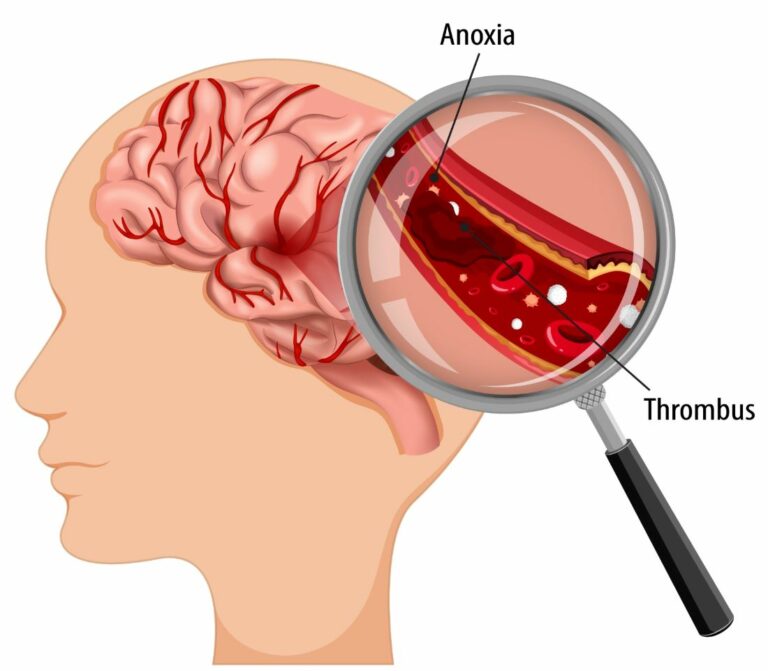
A brain tumour occurs when neurons are mutated and thus abnormal cells are formed. Glioma and meningioma are the two most common types or brain tumours, comprising approximately 75% of all brain tumours.

Who are Likely to Suffer a Brain Tumour?

The subject of diseases and their cure is not easy. It is never possible to say: “Don’t do this and you will never suffer that disease”. Currently only ‘risk factors’ can be presented. Risk factors are something which need to be avoided, if possible.

  • People residing or working in areas concurrently exposed to chemical factories or radiations being released are more prone to develop brain tumour.
  • People with sedentary lifestyle and with more stress in life are generally more seen to develop brain tumours.
  • Lifestyle and dietary factors play a major role in the development of brain tumours
  • People consuming food products such as cured meat, fruits, and vegetables, which contain large amounts of dietary N-nitroso compounds (NOCs) and their precursors, have an association with the development of brain tumours. Hence, they are more prone to develop tumours
  • Males are more prone to develop brain tumours compared to females.
  • The incidence of central nervous system tumours in India ranges from 5 to 10 per 100,000 population with an increasing trend and accounts for 2% of malignancies.
  • A recent study showed an increase in gliomas, but not meningiomas, in workers concurrently exposed to chemical solvents, pesticides or lead, and low-frequency electromagnetic fields.
  • Nitrite exposure has been hypothesized as an explanation for the association of both cured meat and fruits/vegetables. Cured meats are the primary source of dietary N-nitroso compounds (NOCs) and their precursors.
  • NOCs, contained in processed meat, have been long noticed to be associated with higher risk of brain tumour. A recent meta-analysis also indicated that processed meat consumption was associated with higher risk of brain tumour, while intake of vegetables, fruits, and Vitamin A might reduce its risk.
  • Intake of fish is associated with lower risk of brain tumours in children.

Research Findings

Since brain tumours cannot be prevented at this time, it is but natural that much research will continue to be done on the subject. Rather than prevention, currently the focus is on which are the likely people who might get a brain tumour. One major research showed that:

  • People with stressful, sedentary lifestyle and wrong diet and those addicted to alcohol consumption and the habit of cigarette smoking have higher risk of brain tumours.
  • Males are more prone to brain tumours.
  • Among subtypes, majority had glioblastoma and the least had meningioma.
  • According to location, majority had cerebellopontine angle tumour and the least had left thalamic glioma and multicentric glioma.

The other deductions from the above research was that once a brain tumour was detected, to slow down its rate of growth, immediate care should be:

  • Preventing exposure to radiations
  • Avoiding cigarette smoking
  • Providing healthy diet
  • Avoiding chronic stress
  • Avoiding environmental pollution

Even after a successful brain tumour surgery, and postoperative patients should be encouraged in avoiding infections by maintaining proper hygiene and eating a healthy diet for their speedy recovery.

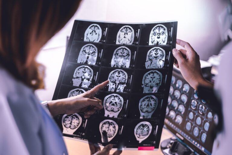
Early detection, better cure

Again, screening isn’t prevention, but it might help find a brain tumour early and when the tumour is small. And detection of the tumour when it is small greatly improves chances of a successful treatment.

Tobacco

Human beings have been using tobacco in various forms since over nine hundred years. Christopher Columbus was given dried tobacco leaves as a gift by the American Indians in 1492. The tobacco plant and smoking was introduced to Europeans. In 1531 Europeans start cultivation of the tobacco plant in Central America. Tobacco may be providing relief and pleasure to its users, but it’s use also lead to many diseases, cancer being one of them. Brain tumours are also attributed to the use of tobacco. Tobacco in every form is bad for health, whether it is chewed, snuffed or smoked.

Cigarette smoking is a major source of multiple systemically absorbed chemical carcinogens including, among others, polycyclic aromatic hydrocarbons and NOCs. Among smokers, tobacco smoke is by far the greatest source of exposure to NOCs.

Although nicotine may increase the permeability of the blood–brain barrier, it is unknown if NOCs penetrate the human brain tissue.

Lifestyle changes

Lifestyle changes, especially dietary habits, are at the basis of chronic systemic low-grade inflammation, insulin resistance, and majority of diseases. An inflammatory reaction jeopardizes the high glucose needs of our brain, causing various adaptations, including insulin resistance, functional reallocation of energy-rich nutrients, and changing serum lipoprotein composition.

With the advent of the agricultural and industrial revolutions, we have introduced numerous false inflammatory triggers in our lifestyle, driving us to a state of chronic systemic low-grade inflammation that eventually leads to typical Western diseases via an evolutionary conserved interaction between our immune system and metabolism. The underlying triggers are an abnormal dietary composition and microbial flora, insufficient physical activity and sleep, chronic stress, and environmental pollution. The disturbance of our inflammatory/anti-inflammatory balance is illustrated by dietary fatty acids and antioxidants. Association between long term exposure to pm2.5 absorbance and malignant brain tumours is seen.

Treatment for Brain Tumours

Treatment options for brain tumours include: Radiation therapy, Surgery, Chemotherapy. Many a times, a combination of two or all three methods is used.

Radiation therapy uses strong beams of energy to kill brain cancer cells. Special equipment ensures that the strong beams of radiation are focussed only on the tumour cells. Radiation is often used along with surgery or chemotherapy to treat brain tumours.

Conclusion

To conclude, brain tumours are rare, treatment options are available. Living a healthy lifestyle is the key point. People with stressful life condition, wrong diet, and sedentary lifestyle and those addicted to alcohol, with the habit of cigarette smoking, have higher risk of brain tumours. Males are more prone to brain tumours.

How to Stop Headache Immediately at Home

How to Stop Headache Immediately at Home

How to Stop Headache Immediately at Home

By Dr.Ravindra Patil

Nearly everyone experiences head pain once in a while. Headache is considered to be the second most common type of pain in human beings after backache. Luckily, most headaches are due to minor conditions and can be cured not with medicines but with home remedies. Your mother and grandmother may have more treatment suggestions for headache. And the surprising part is, most of them will be effective most of the times.

But alas, all headaches are not so benign. Some headaches need specialist doctors for treatment. We list out the special types of headaches which need a doctor’s treatment. All other types of headaches are likely to be relieved with the treatments suggested in the subsequent paragraphs. But first let us see the headaches for which you need to consult a doctor.

Table of Contents

Special types of Headaches

These headaches need a doctor:

  • Tension headache
  • Cluster headache
  • Migraine headache
  • Hemicrania continua
  • Thunderclap headache
  • Allergy or sinus headache
  • Hormone headache (also known as menstrual migraine)
  • Caffeine headache
  • Exertion headache
  • Hypertension headache
  • Rebound headache
  • Post-traumatic headache
  • Spinal headache

If you have headache which is severe and recurs again and again, it may be one of the above and you must consult a doctor.

Now let us see home remedies for ordinary non serious Headaches

Ease the Pressure on your Scalp or Head

This can be done by any of the following methods. Choose the method which suits you and which you can easily implement.

Use a heating pad

Electrical heating pads or a hot water rubber bag applied to the forehead or temples may often relieve your headache. Keep the temperature setting to ‘mild’, not ‘hot’.

Apply a cold compress

Paradoxically, a cold compress on the head or neck can also with pain relief! You may also take a cold shower or a warm water shower. Or take a bath.

Massage the back of your neck

You can do it yourself. Pressing the back of your neck with your index finger can help relieve a headache. This works because the muscles in this area tighten when you experience stress. A few minutes massaging your forehead, neck, and temples can help ease a tension headache, which may result from stress. Or apply gentle, rotating pressure to the painful area.

Acupressure

Pressing certain the pressure points on the back of your hand can help with headaches. This point is located between the base of your thumb and index finger. However, take advice from a trained person.

Hydrate

Drinking water or herbal tea can help with headaches.

Dim the lights

Turning down the lights can help reduce headaches.

Take a nap

After you take a nap, you may awaken refreshed!

Other home remedies include…

Try not to chew

Chewing sometimes increases certain types of headaches.

Get some caffeine

The age old remedy of tea or coffee cannot be forgotten.

Practice Relaxation

Whether it’s stretches, yoga, meditation, or progressive muscle relaxation, learning how to chill out when you’re in the middle of a headache can help with the pain. You might talk to your doctor about physical therapy if you have muscle spasms in your neck.

Take Some Ginger

A small recent study found that taking ginger, in addition to regular over-the-counter pain meds, eased pain for people in the ER with migraines. Another found that it worked almost as well as prescription migraine meds. In many Indian families, ginger is always added to regular tea.

Try some herbal Tea

Many special herbal teas are available for curing a headache.

Use Meds in Moderation

There are plenty of medicines for headaches but use them in moderation. To get the most benefit with the least risk, follow the directions on the label and these guidelines:

  • Avoid ibuprofen and other nonsteroidal anti-inflammatory drugs (NSAIDs) if you have heart failure or kidney failure.
  • Do not give aspirin to a child under age 18.
  • Take painkillers as soon as you start to hurt. You’ll probably beat it with a smaller dose than if you wait.
  • If you get sick to your stomach when you get a headache, ask your doctor what might help.
  • Ask your doctor what to take to avoid a rebound headache, which is pain that sets in after a few days of pain relievers.
  • And be sure to talk to your doctor about what headache symptoms you should not treat at home.

Types of Pain

Knowing the type of your pain greatly assists diagnosis. There are four types of pain that can be present individually or can be present at the same time which can cause a mixed pain pattern. With several types of pain, there are various unique treatment options to suit the intricacies of each type.The Four Major Types of Pain are…

Nociceptive Pain

Typically the result of tissue injury. Common types of nociceptive pain are arthritis pain, mechanical back pain, or post-surgical pain.

Inflammatory Pain

An abnormal inflammation caused by an inappropriate response by the body’s immune system. Conditions in this category include gout and rheumatoid arthritis.

Neuropathic Pain:

Pain caused by nerve irritation. This includes conditions such as neuropathy, radicular pain, and trigeminal neuralgia.

Functional Pain:

Pain without obvious origin, but can cause pain. Examples of such conditions are fibromyalgia and irritable bowel syndrome.

If you describe your pain precisely to your doctor, it will assist him/her in diagnosis and treatment of your condition. Also, when describing your pain to your doctor, keep these in mind:

Provocation and Palliation

What were you doing at the onset? What do you think provokes your pain? What in your opinion alleviates it?

Quality/Quantity

What does the pain feel like and how often? Is it sharp, dull, stabbing, crushing, throbbing, nauseating?

Region/Radiation

Where is the pain located? To be specific, point to the location of the pain or draw it on a face diagram. Does it radiate anywhere? If so, where and to what side? Is it equal if both sides are involved? The more specific you can be the easier it is for the doctor to make a diagnosis.

Severity Scale

How much does it hurt on a scale from 1-10?

Timing

Does the severity or character of the pain change based on time of day, activity, weather, time of year, or position?

When to Call Your Doctor

OK, you have tried all home remedies but your headache is not relieved, or is getting worse. It is time to call your doctor. Also, gGet medical care right away for:

  • A headache that follows a head injury
  • A headache along with dizziness, speech problems, confusion, or other neurological symptoms
  • A severe headache that comes on suddenly
  • A headache that gets worse even after you take pain medications

When is Emergency Medical Attention Needed

In some cases, a headache may require immediate medical attention. Seek immediate medical care if you’re experiencing any of the following symptoms alongside your headache:

  • Stiff neck
  • Rash
  • The worst headache you’ve ever had
  • Vomiting
  • Confusion
  • Slurred speech
  • Fever of 100.4°f (38°c) or higher
  • Paralysis in any part of your body
  • Visual loss

It is commonly thought that a simple symptom like a headache never requires the consultation of a neurosurgeon in a super specialty hospital like Samarth Neuro and Super Speciality Hospital of Miraj, District Sangli. Surprisingly some headaches may need the services of that hospital, and also the opinion of an expert neurosurgeon like Dr Ravindra Patil, who is the founder of that hospital.

How to Cure Sinusitis Permanently

How to Cure Sinusitis Permanently

How to Cure Sinusitis Permanently

By Dr.Ravindra Patil

Is it possible to cure sinus permanently?

We will try to answer this question first. Many people have allergies which lead to cold quite often. Cold is a viral infection. It may extend to inside the sinuses. Repeated attacks of cold may make sinusitis chronic, meaning a long-standing disease. In such situations it is necessary to get treatment for allergy by immunotherapy. There are injections which reduce the allergic reaction of the body to allergens, reduces episodes of cold and sinusitis.

Most sinus infections clear up without antibiotics.

Surgeries are thought to be cure for everything. However, they are the last resort.

Most cases of sinusitis can be cured but if they recur, it may require surgery. Fungal sinusitis is sometimes difficult to cure.

What Is Sinusitis?

Sinusitis or sinus infection is a disease in which the paranasal sinuses swell. The production of slimy mucus helps moisten the nasal pathways. However, when there is too much production, the sinuses can become irritated. Various Causes Can Lead to Sinusitis like Bacteria, allergies, fungi, and viruses.

There are many different types of sinusitis, each with its own symptoms. Some people have chronic sinusitis, acute sinusitis, recurrent sinusitis, or subacute sinusitis. Though the condition causes unpleasant sensations and pain, it usually goes away without medical help. However, if the symptoms become more severe, you may need to see a doctor.

Table of Contents

What Causes Sinus Problems?

Sinusitis can be caused by a virus, bacteria, or fungus that enlarges and clogs the sinuses There are many possible causes for the common cold, including cold viruses. Nasal allergies and allergies to mould are common.

Who Is at Risk for Sinusitis?

A sinus infection can affect anyone. People with allergies to pollen, mucus, or other substances in the air, asthma, or an abnormal nose structure are more likely to get sinusitis. In addition, smoking can increase your chances of getting a sinus infection.

Diagnosis of sinusitis

Your doctor may diagnose chronic sinusitis by:

  • Asking about your symptoms
  • Feeling for tenderness in your nose and face
  • Looking inside your nose through a fibreoptic endoscope
  • Using imaging tests like CT or MRI

Untreated sinus infections can lead to life-threatening infections if bacteria or fungi spread to your brain, eyes, or nearby bone.

Symptoms Of Acute Sinusitis

  • Facial pain or pressure
  • Runny nose
  • Stuffy nose
  • Loss of sense of smell
  • Cough or congestion
  • Fever
  • Bad breath
  • Fatigue
  • Toothache

If you have two or more symptoms, or a thick, green, or yellow nasal discharge, then you may have acute sinusitis.

Symptoms Of Chronic Sinusitis

You may have these symptoms if your sinusitis lasts for 12 weeks or longer:

  • A feeling of congestion or fullness in your face
  • Stuffy nose
  • Pus in the nasal cavity
  • Fever
  • Runny nose, discoloration of nasal discharge.

You may also suffer from headaches, bad breath, and toothaches. You may feel tired a lot because of your illness. There are many possible causes of these symptoms.

Can sinusitis be passed from person to person?

Bacterial sinusitis is not contagious, but viruses that can cause this condition are. Follow good hand washing practices to avoid transmitting infection by contaminated hands to other people. If you must sneeze or cough, face away from others or cover your face with your handkerchief.

Treatment For Sinusitis

There are many different ways to treat sinusitis. Some of the conditions can be cured with simple medications, while others may require surgery. There are various home remedies that you can use to get rid of your sinus infection. There are several treatments available for sinusitis, including medications. Take a look at some of the treatments listed here.

1. Oral corticosteroids

Oral corticosteroids are used to relieve severe symptoms when they occur. Patients who have nasal polyps can also take them. If they are used for a longer period of time, they may have side effects. Corticosteroids are only prescribed when other treatments such as nasal corticosteroids fail.

2. Nasal corticosteroids

Nasal corticosteroids are sprays that help to reduce inflammation. Doctors can prescribe various corticosteroids, such as budesonide, fluticasone, etc. If the medical expert thinks that the nasal rinse is not helping, they may suggest other methods of treatment. The doctor may prescribe a solution that has saline and medication.

3. Nasal sprays

These sprays are available in every pharmacy, and are used to relieve congestion from the nose. They can help reduce drainage and help flush away irritants.

4. Antibiotics

If the cause of your infection is a bacterium, then antibiotics may help. Sometimes, doctors prescribe other medicines along with this one. This is why so many diagnostic tests are performed so that a correct evaluation is assessed.

5. Immunotherapy

Immunotherapy is a possible treatment for sinusitis if the cause is due to allergies. With allergy shots, you might see a reduction in your symptoms as your body no longer reacts as strongly to allergens.

Home Remedies to Cure Sinusitis

1. Rest

You need to rest so your body can fight the infection. When you go to bed, use a pillow to raise your head. Breathing is important for healthy lungs.

2. Steam

Steam inhalation is a great way to cleanse your sinuses. You can add some essential oils to the steam bowl to make your home cleaning experience more pleasurable. Another option is to take a hot shower. You can use a warm towel to help cool your face.

3. Nasal Irrigation

Nasal irrigation with a saline solution is a good way to clean out the nasal passages. For respiratory problems such as congestion, you can use the neti pot, saline canister, or humidifier. Make sure to clean the water and check if the equipment is sterile.

4. Spicy foods

Adding spicy foods to your diet can help increase your appetite and satisfaction. Eating spicy foods can help clear your nasal passages However, if you have a medical condition that forbids eating spicy food items, you should be careful.

5. Warm compress

Warm compresses can be used to help reduce swelling in the area. It will provide relief from the discomfort and pain.

6. Fluids

You need to drink a lot of fluids. Drink more juice and water to stay hydrated. It will help to weaken the mucus layer. However, you should avoid drinking alcoholic beverages.

How Can I Cure My Sinuses Fast?

  • Drink plenty of water
  • Eat foods with antibacterial properties
  • Add moisture
  • Clean the sinuses with oils
  • Use a neti pot
  • Relieve facial pain with warm compresses
  • Use over-the-counter (OTC) medications

How to Treat Sinusitis at Home?

  • Use warm heat.
  • Try a nasal saline solution. While they don’t contain medicine (saline is salt water), they can help keep your nasal passages moist.
  • Flush out your sinuses.
  • Drink lots of fluids.
  • Rest.

When to see a doctor

Most people with acute sinusitis don’t need to see a doctor, but contact your doctor if you have any of the following:

  • Symptoms that last more than a week.
  • Symptoms that get worse after seeming to get better.
  • A fever that lasts.
  • A history of repeated or chronic sinusitis.

See a health care provider immediately if you have symptoms that might mean a serious infection:

  • Pain, swelling or redness around the eyes.
  • High fever.
  • Confusion
  • Double vision or other vision changes.
  • Stiff neck.

Thrombolysis

Thrombolysis

Thrombolysis

By Dr.Ravindra Patil

Blood is a magical organ of our body which keeps flowing throughout our body through arteries and veins all our life. It carries oxygen from the lungs to all the tissues needing it and carries carbon dioxide back to the lungs. It carries nutrition to every cell of the body and carries waste products from all tissues and organs to the kidney to be excreted. This is what we learn in high school science class.

While in school, we all injure ourselves. Our skin may be torn and we bleed. But our magical blood clots and stops further bleeding. We all have experienced this.

The magic of healthy people’s blood is, it clots only when there is an injury and we bleed and further blood loss is prevented by clotting. In healthy people, the blood never clots inside arteries, veins or the heart. It always stays in a liquid state. And as has been mentioned our blood does the supply chain work of providing O2 and nutrients and flushing out waste products and CO2. If blood suddenly stops flowing because of a clot, the organs affected will first be starved of oxygen, and may die in a very short while.

If the organs so affected are the brain or the heart, the person will die!

So, the blood must keep flowing at any cost.

Table of Contents

Thrombosis and Embolism

However, on some occasions because of different pre disease factors, blood clots inside a blood vessel. This is known as thrombosis.
These clots sometimes break, dislodge, and are carried to distant portions of the circulatory system, where they get stuck, and again block the blood supply beyond. This is known as embolism.
Sometimes both thrombosis and embolism occur together, and then it is known as a thrombo-embolic event.
If a clot forms in a vein and breaks loose, it will be carried to the arteries of the lungs and block them. This is known as pulmonary embolism and this is also a very serious life threatening condition.
In all the above cases, a blood clot has occurred inside the arteries of the human body, which is starving the organs being supplied by that artery, and the person is very seriously ill if the arteries with the blood clot supply blood to the heart or the brain.
All other organs are equally affected if their blood supply is blocked, but a block in the blood supply of the heart or the brain may cause immediate death.
Thrombolysis in stroke is one of the best treatments for brain stroke.
Clot in a brain artery results in an acute disease known as Cerebrovascular Stroke or Brain Stroke.
Clot in a heart artery results in an acute disease known as Myocardial Infarction or Heart Attack.
Clot in the pulmonary arteries is known as Pulmonary Embolism.

Thrombolysis

In simple words, thrombolysis [also known as thrombolytic therapy] is a treatment to dissolve dangerous clots in blood vessels, improve blood flow, and prevent damage to tissues and organs. Thrombolysis is done by the injection of clot-dissolving drugs through an intravenous (IV) line or through a long catheter that delivers drugs directly to the site of the blockage. It also may also be done by using a long catheter with a mechanical device attached to the tip that either removes the clot or physically breaks it up.

Thrombolysis procedure is often used as an emergency treatment to dissolve blood clots that form in arteries feeding the heart and brain — the main cause of heart attacks and ischemic strokes — and in the arteries of the lungs (acute pulmonary embolism).

Thrombolysis procedure is also used to treat blood clots in the following areas:

  • Inside veins: it is known as deep vein thrombosis (DVT). These are clots in the veins of legs, pelvic area, and upper extremities; if left untreated, pieces of the clot can break off and travel to an artery in the lungs, resulting in an acute pulmonary embolism.
  • Inside Bypass Grafts: these are clots formed in veins used in heart bypass surgeries, done for the treatment of a heart attack. These veins are harvested from the patient’s legs. This may lead to a second heart attack.
  • Inside Dialysis Catheters: these are lifesaving tubes inserted inside the neck veins of patients who need dialysis very often. A clot inside such a tube means the patient cannot be dialysed and this is a very serious condition.

If a blood clot is determined to be life threatening, thrombolysis may be an option if initiated as soon as possible — ideally within one to two hours — after the onset of symptoms of a heart attack, stroke, or pulmonary embolism (once a diagnosis has been made).

Different Thrombolysis Medications

The most commonly used clot-busting drugs — also known as thrombolytic agents or thrombolysis injections — include:

  • Eminase (anistreplase)
  • Retavase (reteplase)
  • Streptase (streptokinase, kabikinase)
  • t-PA (class of drugs that includes Activase)
  • TNKase (tenecteplase)
  • Abbokinase, Kinlytic (rokinase)

Depending on the circumstances, a doctor may choose to inject clot-busting drugs into the access site through a catheter. More often, however, doctors insert a longer catheter into the blood vessel and guide it near the blood clot to deliver medications directly to the clot.

During both types of thrombolysis, doctors use radiologic imaging to see if the blood clot is dissolving. If the clot is relatively small, the process may take several hours. But treatment for a severe blockage may be necessary for several days.

Thrombolysis Contraindications

Although thrombolysis can safely and effectively improve blood flow and relieve or eliminate symptoms in many patients without the need for more invasive surgery, it can’t be done in the following cases:

  • High blood pressure
  • Active bleeding or severe blood loss
  • Haemorrhagic stroke from bleeding in the brain
  • Severe kidney disease
  • Recent surgery
  • In pregnancy
  • In advanced age patients

Besides risk of serious internal bleeding, other possible risks of thrombolysis include:

  • Bruising or bleeding at the access site
  • Damage to the blood vessel
  • Migration of the blood clot to another part of vascular system
  • Kidney damage

The most serious possible complication is intracranial bleeding, but it is rare.

Prognosis after Thrombolysis

Although thrombolysis is usually successful, the treatment is not able to dissolve the blood clot in up to 25% of patients. Another 12% of patients subsequently redevelop the clot or blockage in the blood vessel.

In addition, thrombolysis alone — even when successful — cannot treat tissue that has already been damaged by compromised blood circulation. So, further treatment may be needed.

Thrombolysis in stroke

In advanced brain treatment hospitals like Samarth Neuro and Superspeciality Hospital of Miraj, thrombolysis is done in stroke patients in a very special manner. As soon as the hospital receives information about an incoming stroke patient, the Emergency Room and the CT Scan room are kept ready. The patient is transferred on arrival to the CT scan room and brain CT is done. If a blood clot in brain is detected, immediate treatment with Recombinant Tissue Plasminogen Activator [rtPA] is immediately begun and the patient is transferred to the ICU for continuous close monitoring.

If the patient has arrived in the “GOLDEN HOUR” or the first 60 minutes after a stroke, it is likely that the patient will recover fully from the stroke in a very short time.

Lacunar Infarcts

Diagnosis and Treatment of Lacunar Infarcts

Diagnosis and Treatment of Lacunar Infarcts

By Dr.Ravindra Patil

Introduction

Stroke is one of the most common illnesses causing paralysis and loss of sensation. Often one side of the body is paralysed when a major part of the brain is deprived of blood either by a blood clot or cerebral haemorrhage.
However there is another type of stroke on a much smaller scale. It is called Lacunar strokes, a type of stroke caused by the occlusion of small deep penetrating branches of the main brain arteries. They include branches from all the cerebral arteries.
Most lacunar strokes involve the non-cortical areas and remain asymptomatic because only small parts of the brain are affected. However, if many small parts of the brain suffer ischaemia [a condition where a body part suffers lack of blood supply and dies, which is known as an infarct], the multiple small lacunar infarcts can lead to significant movement, sensory and functional disabilities.

Table of Contents

Causes of Lacunar Infarcts

In lacunar infarction, the small penetrating cerebral vessels supplying the subcortical areas are occluded due to various vascular disease conditions.

In some cases, small pieces of blood clots called embolic fragments from a distant part can travel the small arteries of the brain and cause deep penetrating artery occlusion, ischemia and infarction.

Lacunar infarcts and strokes are in turn caused by hypertension, diabetes, smoking, high LDL levels, carotid artery atherosclerosis, peripheral artery disease, TIA, and hyperhomocysteinemia.


Certain genetic factors also increase the risk of developing small vessel disease. 

Pathophysiology

Occlusion or blockage of the small penetrating arteries causes small lacunar strokes. Their size varies from 3 mm to 20 mm in dimension. However, only 17% of lacunar strokes are less than 10 mm in size.

Small arterial occlusions are caused by lipohyalinosis and micro-atheroma formation.

History and Physical Examination

As with ischemic strokes, lacunar infarcts usually present with sudden onset of neurological deficits. However, some lacunar infarctions may present in a stepwise pattern and are known to worsen in a short time.

Lacunar infarcts are common in the thalamus, basal ganglia, pons, and white matter of the internal capsule.

Lacunar infarcts can be asymptomatic.

Clinical presentation depends on the area of brain involvement.

Certain lacunar infarcts, like in the posterior limb of the internal capsule or the pons, can present with severe hemiplegia.

Lacunar strokes seldom affect memory, language, and judgment.

Types of lacunar Infarcts

Pure motor hemiparesis – contralateral hemiparesis of the face, arm, and leg. [45%].

Ataxic hemiparesis comprises 10 to 18% of cases. It causes hemiparesis of the contralateral face and leg and ataxia of the contralateral limb. Lack of coordination is the prominent feature of this stroke.

Pure sensory stroke involves the thalamus or corona radiata; it presents with the absent or abnormal sensation of the contralateral of the face, arm, and leg. It accounts for 7% of cases of lacunar strokes.

Dysarthria: clumsy hand syndrome and problems pronouncing words due to voice box muscle weakness

A sensory: motor stroke is the second most common type of lacunar strokes. They account for 20% of cases.

Evaluation

Sudden onset of neurological deficit requires a plain head CT scan and CT angiogram of the head and neck. MRI is a superior imaging method. Carotid ultrasound also helps diagnose an atherosclerotic narrowing of the extracranial carotid artery.

Treatment

The initial goal of acute-stage treatment is ensuring medical stability and determining if the patient is fit for thrombolysis. Treatment with ‘tissue plasminogen activator’ (TPA) improves outcomes for ischemic stroke patients if TPA is administered within 4.5 hours of the first symptoms. But cerebral haemorrhage must be ruled out before TPA treatment. An acute lacunar infarct is efficiently treated with TPA.

After 4.5 hours, dual antiplatelet therapy [DAPT] with aspirin and clopidogrel within 24 hours of symptom onset is begun and continued for 21 days. DAPT in the acute phase effectively lowers recurrent ischemic stroke for 90 days from symptom onset.

Management of blood pressure if present, is done allowing for permissive hypertension. This means, if the BP is lowered to normal levels, the brain may be starved of oxygen. Lowering the blood pressure below185/110 mmHg is ideal. Blood sugar management is done to maintain normal blood glucose levels at 60–180 mg/dL. Blood volume is corrected by giving isotonic saline and oxygen saturation must be above 90%. Statin therapy to reduce LDL is begun if LDL is high.

Stroke Prevention

A lacunar infarct patient is likely to suffer stroke. Hence primary and secondary stroke prevention remains an essential part of the treatment plan. Primary prevention includes the prevention of the first episode of stroke, and secondary prevention includes the prevention of recurrence.

Antihypertensive medications, diabetes control, cholesterol-lowering agents, smoking cessation, dietary intervention, weight loss, and exercise as appropriate are done for primary prevention.

Secondary prevention is done to prevent recurrence of stroke. It includes antithrombotic agents like aspirin, clopidogrel, extended-release dipyridamole, and ticlopidine and managing underlying risk factors. Antiplatelet agents reduce the risk of recurrence in patients with lacunar strokes.

Differential Diagnosis

Differential diagnoses of lacunar infarcts include the following:

  • Ischemic stroke in the middle cerebral artery territory
  • Intracranial haemorrhages
  • Seizures
  • Complicated migraine
  • Brain tumours
  • Multiple sclerosis (MS)

Prognosis

Earlier studies suggested that lacunar stroke has a better prognosis compared to other strokes. It has a high survival rate, a low recurrence rate, and a relatively good functional recovery. But there is an increased risk of death, mainly from cardiovascular causes.

Complications

Lacunar strokes are thought to be the leading cause of vascular dementia and cognitive impairment. Accumulated lacunar infarcts can lead to other disease-related complications due to physical disability, including, but not limited to, aspiration pneumonia, deep vein thrombosis, pulmonary embolism, urinary tract infection, depression, and decubitus ulcers.

Rehabilitation

Physical therapy and rehabilitation are essential steps for lacunar stroke patients with physical disabilities. Speech therapy, occupational therapy may be needed after hospital care to regain strength and functions. The goal of rehabilitation is to optimize functional recovery.

How to prevent Stroke

People need to be aware of risk factors of stroke. Take medicines regularly for diabetes, BP, antithrombotic agents etc… Maintain a healthy diet, exercise regularly, avoid smoking, and avoid excessive alcohol use. Correct lipid disorders with medicines.

The timeline of recovery from a lacunar stroke is different for everyone. Home safety is essential. Fall risk due to physical disability is common. Depression is common in people who have experienced a stroke and should be addressed if present. Cognitive impairment due to multiple subcortical strokes can progress to vascular dementia and should be monitored.

Enhancing Healthcare Team Outcomes

Patients with lacunar infarctions must be managed by a neurologist, family clinicians, nursing staff, and physical, occupational, and social therapists, all operating as an interprofessional team, to receive comprehensive care. Samarth Neuro and Super Speciality Hospital has all the facilities and specialist doctors to mange acute Lacunar Stroke.

Rehabilitation therapy must be continued to maximize the patient’s neurologic function to bring them close to their baseline before the infarction.

Long-term care coordination is the responsibility of the Family Physicians. Emphasis on managing stroke risk factors includes intense antihypertensive therapy, lipid management, and strict control of blood sugars are essential after the lacunar ischemic event.

Is Bowel Leakage a Sign of Cancer?

Is Bowel Leakage a Sign of Cancer?

By Dr.Ravindra Patil

Talking about diarrhoeal disease is considered shameful. But this article describes about “is frequent diarrhoea a sign of cancer”. We hope to clarify myths and make people more aware about what is otherwise and embarrassing topic.

Another question which worries people is: “Is bowel leakage a sign of cancer?” In this context, anal leakage and bowel leakage are the same.

Cancer has many symptoms and blood in stool is one of them. Hence ‘Colon Cancer Poop” can often be bloody.

Many conditions can cause bowel leakage, and most of them are treatable, but if you experience bowel leakage that doesn’t seem to be connected to a temporary stomach illness or something you ate, you should see a doctor, as it can be a sign of cancer and other serious illnesses.

Table of Contents

What is bowel leakage?

Bowel leakage, or faecal incontinence, is characterized by difficulty controlling the release of faecal matter. It is the involuntary release of solid or liquid faeces.

  • Urge incontinence, where you know you need to defecate but can’t make it to the toilet in time
  • Passive incontinence, where you pass stool and are unaware

Faecal incontinence is not uncommon but is it likely that it is underreported. Minor illnesses, such as diarrhoea from many causes can lead to incontinence that typically resolves within a few hours to a couple of weeks. However, faecal incontinence may be cause for concern if it’s long-lasting or happens frequently.

Bowel leakage can range in severity. Some people may experience occasional leakage while passing gas, while others may lose control completely.

Expert Care at Samarth Neuro & Super Speciality Hospital

At Samarth Neuro & Super Speciality Hospital in Miraj, we understand that discussing bowel leakage and cancer can be uncomfortable. However, our dedicated medical teams are here to provide expert care, ensuring your well-being and peace of mind. Our experienced professionals are trained to address your concerns, provide accurate information, and offer appropriate guidance.

Causes of lasting bowel leakage

Numerous factors can trigger long-term bowel leakage:

  • Conditions associated with structural abnormalities in the muscles, nerves, tissue, or other structures
  • Inflammatory bowel disease
  • Previous injury or surgery (obstetrical injury or haemorrhoidectomy, for example)
  • Rectal prolapse, spinal cord injuries, and multiple sclerosis
  • Conditions associated with functional abnormalities, including diabetes mellitus and irritable bowel syndrome
  • Factors affecting stool characteristics include infections, certain medications, irritants (such as laxatives), and faecal impaction
  • Other factors and conditions include advancing age, dementia, antidepressant use, caffeine, and food intolerances

While it’s not among the most common causes, research indicates there are connections between bowel leakage and certain types of cancer.

Can neurological problems cause bowel problems?

Neurogenic bowel is the loss of normal bowel function due to a nerve problem. It causes constipation and bowel accidents. Nerve damage may be due to an injury or a health condition such as multiple sclerosis. Also spine tumours and injuries may in some cases cause faecal incontinence.

Relationships between bowel leakage and cancer

Fortunately, most cases of bowel leakage aren’t caused by cancer. However, there’s evidence of relationships between the two.

Among people with bowel leakage, the risk of developing cancer was still low

Compared to those without bowel incontinence, those with the condition were more likely to be diagnosed with one of the types of cancer within one year

Patients with bowel leakage went on to develop other types of cancer, including larynx, lung, and kidney cancer

Signs of colorectal cancer

Colorectal cancer often starts without symptoms, so screening is vital, but people with colorectal cancer may experience:

  • Blood in or on the stool
  • Changes in bowel habits
  • Diarrhoea
  • Constipation
  • A feeling that the bowel is not fully emptying
  • Abdominal pain, aches, or cramps
  • Unexplained weight loss
  • Signs of other gastrointestinal cancers

Gastrointestinal cancers can affect the oesophagus, liver, pancreas, and many other parts of the gastrointestinal system. The symptoms will depend on which part of the body is affected but may include:

  • Difficulty swallowing
  • Heartburn or indigestion
  • Abdominal pain or swelling
  • Loss of appetite
  • Vomiting blood or blood in the stool
  • Weakness and fatigue
  • Unexplained weight loss
  • Yellowing of the eyes and skin

Risk factors for colorectal cancer

About 5% of people with colorectal cancer have gene mutations passed down through families. But most people who develop colorectal cancer will have no family history of the condition. Other factors  include:

  • Crohn’s disease, ulcerative colitis
  • Sedentary lifestyle
  • Not eating enough fruits, vegetables, and fibre
  • Eating too much fat or too many processed meats
  • Overweight
  • Too much alcohol
  • Smoking
  • Advanced age, 50 or over

Causes of bowel cancer

  • A diet low in fibre.
  • High red meat consumption, especially processed meats
  • Being overweight or obese
  • Alcohol consumption
  • Smoking tobacco
  • Inherited genetic risk and family history
  • Inflammatory bowel disease such as Crohn’s disease.
  • Polyps

Risk factors for other gastrointestinal cancers

Like the associated symptoms, the risk factors for gastrointestinal cancers depend on the location. Risk factors may include:

  • Being 55 or older
  • Being a man
  • Pylori infection (the leading cause of GERD)
  • Having pernicious anaemia (when the body lacks B12 and can’t make enough red blood cells)
  • Having undergone stomach surgery
  • Having a hepatitis B or hepatitis C infection
  • Having alcohol-related liver disease
  • Having hemochromatosis (a condition in which the body stores too much iron)
  • Smoking
  • Drinking too much alcohol
  • Having a family history of gastrointestinal cancers
  • Low socioeconomic status
  • High-temperature beverages & foods

Treatment options for bowel leakage

To effectively treat your bowel leakage, you’ll need to determine the cause. If your incontinence is occasional and doesn’t last long, the solution may be as simple as avoiding certain foods. However, bowel leakage linked to an underlying illness may be more difficult to treat and will require gaining control of the underlying cause.

Bowel training

This sounds like you’re going back to being a toddler, but bowel training or retraining is used to treat both faecal incontinence and severe constipation. Encouraging bowel movement after meals using the gastro-colic reflex and encouraging defecating in a squatting position can be tried.

Pelvic floor exercises can be valuable if leakage is linked to muscle weakness.

Over-the-counter medicines

For diarrhoea, doctors usually recommend loperamide, codeine sulfate, or amitriptyline. Loperamide It slows down bowel movements and improves anal sphincter tone.

For constipation, your doctor may recommend a laxative, a stool softener, or a concentrated fibre supplement such as psyllium.

Consult your doctor before taking medications to help with your bowel leakage, even over-the-counter ones. Some may worsen your symptoms, and some may mask symptoms of a serious underlying condition that demands medical attention.

Dietary changes

Digestive troubles, including incontinence, may be caused or aggravated by your diet. A nutritionist is of help in such cases. You should keep a food diary, as disruptive foods can vary from person to person. Keep track of what you eat and how it makes you feel.

For constipation or haemorrhoids, the doctor may recommend increasing your fibre and fluid consumption. If you have diarrhoea, you should be cautious with alcohol, caffeine, dairy, spicy foods, sugar alcohols and foods with high levels of fructose.

Wearing absorbent pads

Wear absorbent pads in the worst cases. It will drastically improve quality of life.

When to see a doctor? Is Bowel Leakage a Sign of Cancer?​

You should always see a doctor if you have symptoms of bowel incontinence, because in some cases, it can be a sign of something more serious, including bowel cancer. Or, it is more likely to be related to a less severe, treatable condition.

You may be hesitant to see your doctor about bowel leakage, but remember, doctors and medical professionals have heard and seen it all. Seeing a doctor is essential to rule out serious conditions that may get worse with time or lead to severe complications.

How to Cure Cervical Spondylosis Permanently

मानेचा (म्हणजे सर्व्हायकल) स्पॉन्डिलायसिस कायमचा कसा बरा करावा स्पॉन्डिलायसिस उपचार सर्व्हायकल स्पॉन्डिलायसिस म्हणजे सकारात्मक आणि ताणशील गोंधळलेली असलेली समस्या

How to Cure Cervical Spondylosis Permanently

By Dr.Ravindra Patil

What is Cervical Spondylosis

Cervical spondylosis is a common, age-related disorder of the cervical spine that is located in the neck region. This condition is also known as neck arthritis or cervical osteoarthritis. With age, the cervical discs gradually face wear and tear resulting in the breakdown of the structure. The fluid present between the cervical discs is lost which makes the spine stiffer, leading to cervical pain. This results in changes in the bones, discs, and joints at the spine of the neck.

As a result of the fluid dehydration and degeneration of the discs and cartilage in the surrounding area, abnormal growth or spurs of bones called osteophytes can occur leading to the narrowing of the passage from where spinal nerves exit, causing a related condition called cervical spinal stenosis.

Table of Contents

What is Spondylosis?

Spondylosis is an umbrella term that is used to describe any degeneration of the spinal cord. Spondylosis symptoms involve acute pain in the neck and back in old-age. Cervical spondylosis is prevalent among middle-aged and elderly people.

Spondylitis vs Spondylosis

Spondylitis and spondylosis both affect the spinal cord but there is a basic difference between the two. The meaning of spondylitis is that it is the inflammation of one or more typical cervical vertebrae leading to pain in the spine. Cervical spondylosis mostly refers to any kind of degeneration of the spine. So, even though the effects of both the diseases might be similar, their origin and cause are completely different.

Symptoms of Cervical Spondylosis: What is the difference?

Cervical spondylosis symptoms can vary from person to person and the extent of cervical spine pain depends on the age of the affected person. Some of the common spondylosis symptoms are:

  • Excessive stiffness at the neck leads to pain in neck
  • Cervical spondylosis headache that may originate at the main cervical spondylosis pain areas
  • Pain in the shoulder and arms
  • Failure to properly turn the head around
  • Grinding noise or sensation when trying to turn the neck
  • Cervical pain is severe in the morning and at night before bedtime
  • If cervical spondylosis leads to spondylotic myelopathy, the following symptoms may occur:
    • Abnormal tingling, numbness, and weakness in the arms, hands, feet, and legs
    • Lack of balance and coordination
    • Abnormal reflexes, random muscle spasms
    • Loss of control over urinary bladder and bowels

Causes of Cervical Spondylosis

Wear and tear of the vertebrae in the spinal cord causes degenerative spondylosis. Other causes are:

  • Bone spurs or abnormal outgrowths of bones called osteophytes
  • Dehydrated Spinal Discs
  • Herniated Discs:
  • Injury of the neck
  • Stiffness of the Ligament
  • Excessive Stress and Overuse

Risk Factors of Cervical Spondylosis

The greatest risk factor of cervical spondylosis is aging. Other risk factors are:

  • Heavy physical actions like heavy lifting
  • Poor posture in people who work at desk jobs
  • Genetic factors and family history
  • Smoking
  • Being overweight and inactive

How to prevent cervical spondylosis?

  • Avoid loading heavy objects on the head or neck. Do not sleep on a high-pillow.
  • For people with desk jobs, maintain a proper posture
  • Lean back your head once in a while.
  • Swimming is considered to be a relaxed exercise for cervical pain
  • Perform general physical activities.
  • Do not be inactive for a long period of time, take exercise

Diagnosis of Cervical Spondylosis

1. Physical Examination

  • Testing the reflexes of the patient and checking for the presence of muscle weakness and deficits in the sensory nerve function is done.
  • Tests are done to determine the range of neck movement
  • Overall walking movement is checked

2. Imaging Tests

  • X-Rays
  • CT Scan
  • MRI Scan
  • Myelogram

3. Nerve Function Tests

  • Electromyogram (EMG) records the passage of nerve signals in the body by measuring the electrical activity of the nerves.
  • Nerve Conduction Study – the speed and strength of the signals sent by the nerve endings in the body are checked.

Treatment of Cervical Spondylosis

Spondylosis treatments mainly focus on how to cure cervical spondylosis permanently, give pain relief and lower the risk of extensive damage to your vertebrae.

Physiotherapy

  • Physiotherapy for cervical spondylosis is a great way for pain relief. A trained physical therapist can teach all the exercises for cervical spondylosis.
  • Neck traction: it uses weights to help increase the space between the cervical joints and reduce the pressure forming on the cervical discs and nerve roots.

Medications

Commonly administered medications for the treatment of Cervical Spondylosis are:

  • Muscle relaxants
  • Narcotics are used for severe neck pain
  • Anti-seizure medications help to reduce pain caused due to damage of nerves
  • Steroids: Used to reduce the occurrence of tissue inflammation and reduce pain
  • Non-Steroidal Anti Inflammatory Drugs [NSAIDS]
  • Antidepressants help in relieving severe neck and shoulder pain
  • Certain homeopathic medicines also help

Home Remedies for Cervical Spondylosis

  • Take enough rest
  • Maintain a proper posture
  • People who acquire spondylosis at an early age normally do so because of bad study habits
  • Heat and cold treatment help provide relief and also works in relaxing the sore muscles of the neck and shoulders. Using a heating pack or a cold pack can be a good home remedy for cervical spondylosis
  • Chiropractic manipulation that can help in order to control events of more severe pain due to cervical spondylosis. However the chiropractor must have the requisite skills of manipulating the neck
  • Wearing a neck collar for cervical spondylosis helps limit movement and provides support for the neck and shoulders. However, these neck collars should not be worn for a longer period of time as it can cause muscle weakness.
  • Several people opt for ayurvedic medicine for cervical spondylosis and Patanjali medicine for cervical spondylosis to cure their ailment

The Need for Cervical Spondylosis Surgery

Cervical spondylosis is not progressive even though patients might have to live with severe pain for long periods of time all their life as it is a chronic condition. Cervical Spondylosis Surgery is only advised by doctors in rare cases when other forms of treatment do not help to ease the pain. For people who suffer from a progressive loss of function in the arms, hands, and legs, surgery may be required to fix the defect at the source point. This is because any form of compression at the cord may result in permanent functional disability.

In cases of the surgery, the main goal is to remove the areas or sources that have been applying extra pressure to the spinal cord and nerve endings. Some extensive forms of surgery may also involve the insertion of implants or bone grafts to add stabilization. Doctors also perform a fusion of the degenerated discs as a cure for cervical spondylosis.

Road to Recovery and Aftercare

Cervical spondylosis is not uncommon among people who have crossed their 60s. Early diagnosis of this disease can help doctors recommend a conservative line of treatment to help ease the pain. Though this condition is not reversible, with proper care, home remedies, and lifestyle practices, patients overcome the stiffness and discomfort.

How to cure cervical spondylosis permanently?

This is a very difficult question to answer as has been mentioned, the treatment of cervical spondylosis is primarily aimed at pain relief cervical exercises by physiotherapy and medicines. Surgery in this condition is done only when there is a physical problem like a bone spur pressing on the spinal cord, or a herniated disc pressing on the nerves or a compressed disk ‘pinches’ a nerve or the spinal canal is narrowed, due to spinal stenosis. Samarth Neuro and Super-speciality Hospital in Miraj offers medical and physiotherapy treatment for cervical spondylosis. If required, surgery for cervical spondylosis can also be done at Samarth Hospital in Miraj, Maharashtra.

Cranio-Vertebral Junction Anomalies

Cranio-Vertebral Junction Anomalies

Cranio-Vertebral Junction Anomalies

By Dr.Ravindra Patil

Anomaly

An anomaly is something that deviates from what is standard, normal, or expected. Cranio-vertebral junction anomalies mean any situation where there is some deviation from what is the ‘normal’ cranio-vertebral junction.

CVJ

The cranio-vertebral junction (CVJ) is present between the base of the skull and the upper region of the cervical spine. Any deviations or deformities in this region are known as cranio-vertebral junction anomalies. They can occur as congenital anomalies, developmental or secondary to other disease processes. Cranio-vertebral anomalies are classified into different types based on their causative factors.

The craniovertebral junction (CVJ) has unique anatomical structures that separate it from the cervical spine. In addition to housing vital nerves and blood vessels, the majority of head movements like flexion, extension, and looking sideways is accomplished at the CVJ.

The CVJ is a complex combination of osseous and ligamentous supports allow for stability despite a large degree of motion. To understand the anomalies of the CVJ, an understanding of anatomy and biomechanics in short is essential to understand the various anomalies and diseases that may affect this region.

The craniovertebral junction (CVJ) is the joint between the skull, atlas [first cervical vertebra], and axis [second cervical vertebra]. It is a complex area that houses vital nerves and blood vessels. This a joint has the most mobility as compared to any other joint of the vertebral column or the spine. The CVJ represents the transition between the brain and cervical spine. The majority of the spine’s rotation, flexion, and extension occur between the occiput, the atlas, and axis.

Table of Contents

Causes and risk factors of Cranio-vertebral Junction Anomalies

There are various factors associated with CVJ anomalies. Some of them include:

  • Skeletal structure abnormalities
  • Congenital systemic disorders such as Achondroplasia, Down syndrome, etc.
  • Infections
  • Metastatic Tumours effecting bones
  • Slow growing tumours in the CVJ
  • Rheumatoid Arthritis
  • Traumatic injuries by an accident, a fall etc.

Signs and symptoms of Cranio-vertebral Junction Anomalies

The signs and symptoms of CVJ anomalies vary based on the cause, severity and specific structures affected. However, some common signs and symptoms may include:

  • Neck pain
  • Headache in the back region of the head
  • Worsening head and neck pain with movement.
  • Decreased range of motion in the CVJ
  • Increased pain intensity while coughing or bending

Symptoms associated with spinal cord compression in the CVJ may include:

  • Weakness in arms, legs or both.
  • Numbness and tingling sensation on bending the neck.
  • Decreased sensations of heat, cold and pain in the legs/hands/feet.
  • Confusion and dizziness

Symptoms associated with brain stem dysfunction may include:

  • Sleep apnoea
  • Impaired balance and coordination (Ataxia)
  • Involuntary eye movements (Nystagmus)
  • Facial paralysis

Symptoms associated with lower cranial nerve dysfunction may include:

  • Difficulty in swallowing
  • Muscle weakness
  • Impaired hearing
  • Paralysis of soft palate and tongue atrophy
  • Diagnosis of Cranio-vertebral Junction Anomalies

CVJ anomalies are diagnosed based on medical history, physical examination and diagnostic imaging scans. Lateral view X-rays, CT scan and MRI are used to visualise the structural abnormalities in the CVJ. CT scan is the most common diagnostic tool used for CVJ anomalies. However, if further investigations are required, MRI and CT myelography are considered.

Treatment of Cranio-vertebral Junction Anomalies

Surgery is the treatment of choice for CVJ anomalies. The type of surgery and approach is decided based on some factors which include: traumatic causes, non-traumatic causes, reducible or irreducible lesions. Considering these factors, the basic surgical procedures involved are reduction, immobilization, decompression and fixation. Treatment may include any one of the procedures or a combination which is determined based on the nature, severity and risk factors of the disease.

Treatments

  • Full endoscopic cervical canal stenosis decompression
  • Spinal fixation
  • Craniovertebral junction surgeries
  •  

CVJ Fixation

Due to the complex anatomical nature as well as the significant mobility of the CVJ, fixation of this region remains at times a challenging decision and a difficult execution. A wide variety of fixation methods exist and may include a combination of the following: screws, rods, wires/cables, bone grafts, hooks, or plates. Furthermore, arthrodesis [surgical immobilization of a joint by fusion of the bones] is also a challenge as there is little space and bone surface for sufficient bone grafting.

Fixation of the CVJ to the skull can be accomplished via small burr holes and wire or a combination of screws and plates, which allows for fixation to the cervical spine through connection of rods or additional plates in the spine. Bur holes with wire are not currently used as often, as the screw/rod/plate constructs have shown to be bio-mechanically superior in terms of screw pullout strength and stiffness.

The screws used in the CVJ are generally a larger diameter than those used in the cervical spine. They have a smaller pitch and blunt tips to prevent piercing the dura mater [a thick tissue layer covering the brain and the spinal cord].

Fixation in the cervical spine can be a combination of a number of methods as well, usually with the intention of connecting to the occiput fixation. The screws used in the cervical spine are generally polyaxial and placed into the lateral masses of the vertebrae.

Generally, the skull and the first cervical vertebra [atlas] segment is not fixated alone due to the large moment arm created by the cranium on C-1. However, there are situations in which the occiput and atlas can be fixated using the above listed devices, like screws, rods, wires and so on.  Sometimes a bone graft is placed in between the posterior arch of atlas and the base of the skull. Successful fusion rates of such surgeries have been reported up to 89%, but the patient’s head is not immediately stable after surgery. Such patients are required to remain in a halo neck fixation for 12 weeks.

Another option involves the use of fixing screws across joints. Similar to the above construct, this is usually reserved for isolated Skull-Atlas instability and is not easily incorporated into multisegment constructs. This technique increases stiffness in rotation but is poor in flexion-extension. Thus, this technique is used in conjunction with supplemental fixation to allow for sufficient stability.

Skull to Atlantoaxial Segment fixation

The occiput to atlantoaxial region is classically difficult to fixate as both the occipitoatlantal and atlantoaxial segments are highly mobile in flexion and extension, and additionally the atlantoaxial segment is very mobile in axial rotation. Any number of combinations of the previously mentioned techniques can be used. Occipitocervical fixation constructs consist of points of fixation along the skull, atlas vertebra and axis vertebra, with connection to some type of longitudinal element. These longitudinal elements span the segments in the CVJ and allow them to be rigidly fixated.

The above description may seem to be very complex to the lay reader. However, all efforts have been made to removed complicated medical words and use as simple words as possible.

To develop a strong CVJ construct, the longitudinal elements should be able to have multiple points of fixation along the junction, interface with all of the fixation points, have the ability to interface with the thickest regions of bone in the suboccipital region, and have the ability to be crosslinked.  Various types of longitudinal elements are available and may include rods, structural bone grafts, reconstruction plates, and hybrid devices combining plates and rods in preformed shapes. As with all spinal instrumentation, it is critical to choose the type of longitudinal element that best suits the individual patient’s needs based on the goal of surgery and the patient’s anatomy.

Samarth Neuro and Multispeciality hospital in Miraj, Maharashtra, has the infrastructure and team of doctors headed by neurosurgeon Dr Ravindra Patil to accomplish such complex surgeries. Besides as the hospital is located in a tier 2 city, the overall cost of such surgeries is much less in Samarth Hospital of Miraj as compared to surgeries done in large metropolitan cities.

Spina Bifida Causes, Symptoms and Treatment

Spina Bifida - Samarth neuro and super speciality hospital

Spina Bifida Causes, Symptoms and Treatment

By Dr.Ravindra Patil

What is Spina Bifida?

Spina bifida is a birth defect in which an area of the spinal column doesn’t form properly, leaving a section of the spinal cord and spinal nerves exposed through an opening in the back. Spinabifida occurs in 1 per 2,000 live births.

Table of Contents

Myelomeningocele

The most common and serious form of spinabifida is called myelomeningocele, in which a part of the spinal cord and surrounding nerves push through the open bones (vertebrae) in the spine and protrude from the foetus’s back. In myelomeningocele, spinal cord and nerves protrude from the back in a sac that is exposed to the amniotic fluid. Continuous bathing of the fragile developing spinal cord in amniotic fluid over the course of gestation is thought to result in progressive neurologic injury.

Other types of spina bifida

Other types of spinabifida include myeloschisis, lipomeningocele and myelocystocele. Open neural tube defects such as myelomeningocele and myeloschisis are treatable by foetal repair. Closed neural tube defects such as lipomeningocele and myelocystocele are not treatable by foetal repair.

Causes of spina bifida

Myelomeningocele has genetic and micronutrient causes. Mothers who have had a baby with spinabifida have up to a 4 percent risk of recurrence in subsequent pregnancies.

Spina bifida symptoms

The myelomeningocele lesion can occur at any level on the developing spine, but most are found in the lumbo-sacral region. Depending on the lesion’s location, myelomeningocele may cause:

  • Bladder and bowel problems (incontinence)
  • Sexual dysfunction
  • Weakness and loss of sensation below the defect
  • Inability to move the lower legs (paralysis) and other cognitive impairments
  • Orthopaedic malformations such as club feet or problems of the knees or hips
  • Generally, the higher the defect is located on the spine, the more severe the complications.

In many cases, the brain develops an Arnold-Chiari II malformation, in which the hindbrain herniates or descends into the upper portion of the spinal canal in the neck. This herniation of the hindbrain blocks the circulation of cerebrospinal fluid, causing hydrocephalus (accumulation of fluid in the brain), which can injure the developing brain. Ventricular shunting (placement of a thin tube into the ventricles of the brain) is used to drain fluid and relieve hydrocephalus.

Hindbrain Herniation and Ventricular Shunting

Ventricular shunting is done to treat hydrocephalus. It involves placement of a thin tube into the ventricles of the brain to drain fluid and relieve hydrocephalus. The other end of the tube is usually put in the peritoneum [abdomen] of the patient for continuous drain.

Evaluation and diagnosis of spina bifida

As with other birth defects, most myelomeningoceles are discovered by routine ultrasound evaluation between 16 and 18 weeks into the pregnancy. Sonographic features suggestive of myelomeningocele include a cystic mass anywhere on the spine. Abnormally high alpha-fetoprotein (AFP) will support the diagnosis.

Other evaluation methods for suspected spinabifida are:

  • High-resolution level II ultrasound — to confirm the diagnosis and determine the location of the lesion, and to assess for any other birth defects such as club feet
  • Ultrafast foetal MRI — to confirm presence of the Chiari II malformation and to screen for evidence of any other neurologic abnormalities
  • Foetal echocardiogram — to determine any problems with the heart
  • Amniocentesis — to confirm the presence of elevated amniotic fluid alpha-fetoprotein (AFAFP) levels and acetylcholinesterase (AChE), which indicate open neural tube defects
  • Maternal serum alpha-fetoprotein (MSAFP) test — to confirm elevated AFP levels in the mother’s blood

After evaluation, a multidisciplinary team works together to review test results, confirm the diagnosis, explain spina bifida treatment options and potential outcomes, and answer all the patient’s questions.

Treatment

The two main spina bifida treatment options are foetal surgery during pregnancy or surgery on the baby right after birth.

Deciding whether prenatal or postnatal spinabifida repair is appropriate is influenced by several considerations, including gestational age, the level of the myelomeningocele lesion on the spine, presence of the Chiari II malformation, and a number of important maternal health factors.

Spina bifida surgery after birth

If postnatal spina bifida surgery is recommended or selected, your pregnancy is monitored and a caesarean delivery is planned at 37 weeks.

Traditional spina Bifida treatment takes the form of surgical repair 24 to 48 hours after birth. Your child will undergo general anaesthesia. A paediatric neurosurgeon removes the MMC sac, if one is present, and closes the surrounding tissue and skin over the defect to protect the spinal cord. After surgery, your baby will receive care in the Neonatal Intensive Care Unit (NICU).

Spina bifida surgery before birth

Because spinal cord damage is progressive during gestation, prenatal repair of myelomeningocele may prevent further damage. Scientific studies have demonstrated that prenatal repair can offer significantly better results than traditional postnatal repair.

  • It reduced the need to divert fluid from the brain using ventricular shunting
  • Reduced the incidence or severity of neurological effects such as impaired motor and sensory function of the legs
  • Improved mobility and improved the chances that a child will be able to walk independently
  • Reversed the hindbrain herniation component of the Arnold-Chiari II malformation

Foetal surgery for spinabifida is a complex and challenging procedure, requiring the most expert, comprehensive care for both mother and foetus. The surgical team’s level of experience and all aspects of care surrounding the operation are of paramount importance.

If your unborn baby has been diagnosed with myelomeningocele, the surgery is performed between 23 weeks and 25 weeks of gestation.

Spina bifida prognosis

Rarely, children can die due to complications associated with Chiari II malformation or hydrocephalus. Many patients require shunts (85 percent), and 45 percent of shunts develop complications within one year.

Follow-up care

Children operated for spina Bifida require comprehensive long-term follow-up, from the time of repair through adolescence. It is team work of a paediatrician, nurse, social worker, physical therapist, neurosurgeon, orthopaedic specialists, urologist and genetic counsellor.

Depending on the location of the lesion and outcome after surgery, follow-up care may also include:

  • Clean intermittent catheterization
  • Bowel management
  • Lower extremity bracing
  • Physical therapy evaluation and guidance to outside physical therapists and early intervention
  • Pressure sore management
  • Referral to appropriate psychosocial and financial resources
  • Consultation with other subspecialties including ophthalmology, nephrology, nutrition and feeding specialists, and plastic surgery

The Spina Bifida team works closely with each child’s primary care paediatrician and school, and helps families access appropriate community services. Ongoing follow-up is critical when it comes to improving outcomes and advancing treatment for patients with myelomeningocele.

Spina Bifida can lead to a variety of social and emotional challenges and lifelong quality-of-life issues. Hence proper diagnosis during pregnancy is essential.

Spina bifida occulta

Spina bifida occulta is the mildest type of spinabifida. It is sometimes called “hidden” spinabifida. In this birth defect, there is a small gap in the spine, but no sac comes out of the back. The spinal cord and the nerves usually are normal.

At Samarth Neuro & Super Speciality Hospital in Miraj, we are dedicated to providing expert care, ensuring the well-being and quality of life for individuals facing this condition.

Atlantoaxial Dislocation

Atlantoaxial dislocation

Atlantoaxial Dislocation

By Dr.Ravindra Patil

This condition occurs in the neck when the first two cervical vertebrae move away from their positions. Luckily this is a very rare condition. But if ever it occurs, it may have serious implications. The patient may suffer quadriplegia, that is, paralysis of all the body below the neck. But before we understand what is atlantoaxial dislocation, we must understand the anatomy of the cervical spine, and especially the first vertebra of the spinal column, called the ‘atlas’, and the second vertebra, which is called the ‘axis’. The two vertebrae, the atlas and axis, are joined at what is called the atlantoaxial joint.

Table of Contents

Anatomy of the Cervical Spine

The cervical spine is the upper portion of the vertebral column, lying between the skull and the thoracic or chest vertebrae. It has seven vertebrae, two of which are given unique names:

  • The first cervical vertebrae (C1) is known as the atlas.
  • The second cervical vertebrae (C2) is known as the axis.

The cervical vertebrae, seven in all, are different from the other vertebrae and are a bit more delicate. We will focus on the first two cervical vertebrae.

Atlas vertebra

The atlas is the first cervical vertebra and lies between the skull and the axis vertebra. It differs from the other cervical vertebrae considerably.

Axis vertebra

The axis lies between the atlas and the third cervical vertebra. It articulates with the atlas through the medial atlanto-axial joint. This joint allows for rotation of the head independently of the torso.

The axis also articulates with the atlas to form the two lateral atlanto-axial joints.

Ligaments of the cervical spine

There are six major ligaments in the cervical spine. The ligaments are strong tensile connective tissue which hold the vertebrae together.

Injuries of the cervical spine

Injury due to vertical fall
A vertical fall onto an extended neck e.g. diving into excessively shallow water can compress the lateral masses of the atlas between the occipital condyles and the axis. This causes them to be driven apart, fracturing one or both anterior/posterior arches.
If the fall occurs with enough force, the transverse ligament of the atlas may also be ruptured.
Hyperextension (Whiplash) Injury
A rear-end traffic collision or a poorly performed rugby football tackle can both result in the head being whipped back on the shoulders, causing whiplash. In minor cases, the anterior longitudinal ligament of the spine is damaged which is acutely painful for the patient.
Atlantoaxial Subluxation
The worst-case scenario for these injuries is that dislocation or subluxation of the atlas and axis occurs. Subluxation means a slight misalignment of two vertebrae in relation to each other. It is the cause of many health problems. This often happens at the axis vertebra level, where its body of moves forward with respect to the third cervical vertebra. Such an injury may well lead to spinal cord damage. Quadriplegia or death may occur. More commonly, subluxation occurs at the lower levels of the cervical spine.
Hangman’s Fracture
Hangman’s fracture refers to a fracture of the bony column of the axis vertebra. It typically occurs as a result of high velocity hyperextension and distraction of the head. Such an injury is likely to be lethal as rupture the spinal cord may occur, causing deep unconsciousness, respiratory failure and cardiac failure.
Fracture of the Dens
Occurs in the axis vertebra. These fractures are often unstable and are at high risk of avascular necrosis. As with any fracture of the vertebral column, there is a risk of spinal cord involvement.
Dislocation of the Atlanto-Axial Joint
Atlanto-axial Dislocation or Fracture can cause severe spinal cord compression resulting in significant neurological injuries of upper spinal cord and lower medulla oblongata. Patient may suffer paralysis of all four extremities (quadriplegia). Severe neck pain is spread over back of the neck and arms. Patient will also have continuous chronic pain.
Atlanto-axial dislocations (AADs) may be classified into four varieties depending upon the direction and plane of the dislocation i.e. anterior-posterior, rotatory, central, and mixed dislocations. However, from the surgical point of view these are divided into two categories i.e. reducible [RAADs or Reducible Atlanto Axial Dislocations] and irreducible [IAADs or Irreducible Atlanto Axial Dislocations].

Surgical Treatment

For RAAD, Posterior fusion is done. Screws are fixed across joints and vertebral bodies and wiring is done. Often, IAAD is due to inadequate extension in dynamic X-ray study which may also be due to spasm of muscles.

The above is a very simplistic explanation of a very complex surgical process. Various different methods are there to treat AAD [atlanto-axial dislocation]. However, it will be too complicated for non-medical people to understand, hence it is not described in detail.

The surgeries require high precision. That is why hospitals like Samarth Neuro and Multispeciality Hospital located in Miraj are able to conduct such complex surgeries. Because Samarth Hospital has surgical navigation system which is the only way to manage such complex injuries.

While it may seem to the reader that the complexity of atlanto axial subluxation or dislocation is being exaggerated, it should be remembered that if the surgery goes wrong even slightly, the patient may suffer quadriplegia or even death. There is only one chance for the perfect surgery. Otherwise the patient may suffer with failed surgery.

Hence the use of surgical navigation, which pinpoints the exact locations where the initial incisions are to be taken and the exact spots where screws and wires are to be fitted so as to prevent injury to the spinal cord and the spinal nerves coming out between vertebrae.

Medical Care

There are no are medicines for atlantoaxial instability (AAI), subluxation or dislocation. Because of the chronicity of the instability at the time of presentation in most cases, corticosteroids have little, if any benefit. Neurologic problems do not get better with corticosteroids. In fact they may cause side effects. In the acute traumatic setting, corticosteroids remain controversial. Guidelines provided by the American Academy of Neurological Surgeons (AANS) include level I evidence against the use of corticosteroids or gangliosides in the acute trauma patient.

Unless symptoms of spinal cord compression occur, AAI requires no treatment. Once symptoms arise, cervical spine stabilization is indicated until surgical stabilization is performed.

What are the symptoms of rotatory atlantoaxial dislocation?

Main symptom is torticollis. It is painless, and often, there are no neurological symptoms. Rotatory atlantoaxial dislocation can be subdivided into reducible or irreducible types. Reducible type rotatory atlantoaxial dislocation is classified as one where the dislocation reduces on dynamic images or after institution of cervical traction. The torticollis is painless, and often, there are no neurological symptoms. Rotatory atlantoaxial dislocation can be subdivided into reducible or irreducible types. Reducible type rotatory atlantoaxial dislocation is classified as one where the dislocation reduces on dynamic images or after institution of cervical traction.

What is the treatment for occipitoatlantal dislocation?

For Type II lesions associated with occipito-atlantal dislocation and/or when the diagnosis was delayed for more than 14 days, a posterior atlantoaxial or occipito atlantoaxial arthrodesis [fusing the two bones of the joint for ever] was recommended. Other lesions are all treated with posterior arthrodesis after reduction.

How to treat rotatory atlantoaxial instability?

Holding and manipulation of the spinous process of axis, posterior arch of atlas, and the facets of atlas and axis can result in reduction of the rotatory dislocation.

To summarise

As has been mentioned, Atlantoaxial Dislocation or subluxation is very rare. It is not so easy to understand the complex disease condition for most people. Samarth Neuro and Multispeciality Hospital has the infrastructure and surgical teams to treat Atlantoaxial Dislocations.