ट्रायजेमिनल मज्जातंतुवेदना (न्युरालजिया)

trigeminal neuralgia

ट्रायजेमिनल मज्जातंतुवेदना (न्युरालजिया)

By Dr. Ravindra Patil

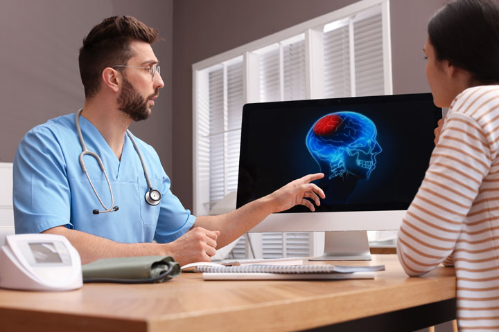
ट्रायजेमिनल न्यूराल्जिया (टीएन), कधीकधी मानव जातीला होत असलेल्या वेदनांमध्ये ‘सर्वाधीक वेदनादायक’ असे वर्णन केले जाते. वेदनांमध्ये सामान्यत: खालचा चेहरा आणि जबडा समाविष्ट असतो. काहीवेळा नाकाच्या आसपास आणि डोळ्यांच्या वरच्या भागावर देखील दुखते. ही तीव्र, सळी घुसवल्यासारखी, विजेच्या धक्क्यासारखी वेदना ट्रायजेमिनल नर्व्हला सतत होणार्‍या बारीक इजांमुळे होते. ट्रायजेमिनल नर्व्हच्या कपाळ, गाल आणि खालच्या जबड्यात फांद्या असतात व तिथे अतिशय तीव्रतेने दुखते. हे दुखणे सहसा चेहऱ्याच्या एका बाजूलाच मर्यादित असते. दात घासणे, खाणे किंवा अचानक वार्‍याची झुळूक यासारख्या नित्य आणि किरकोळ गोष्टींमुळे वेदना सुरू होऊ शकतात. उपचार न केल्यास, ट्रायजेमिनल मज्जातंतुवेदना हळूहळू अधीक वाढत जाऊ शकतात. फेसियल न्युराल्जिया म्हणजे चेहर्याचा मज्जातंतुवेदना, म्हणजेच चेहऱ्यावर मज्जातंतूशी संबंधित वेदना.

ट्रायजेमिनल न्यूराल्जियाचा संपूर्ण उपचार होऊ शकत नाही पण अति तीव्र वेदना कमी करण्यासाठी उपचार उपलब्ध आहेत. सामान्यतः, अँटीकॉन्व्हल्सिव्ह औषधे ही उपचारांची पहिली पायरी असते. पण ज्या रूग्णांना औषधांनी बरे वाटत नाही किंवा ज्यांना औषधांचे गंभीर दुष्परिणाम होतात त्यांच्यासाठी शस्त्रक्रिया हा एक प्रभावी पर्याय असू शकतो.

Table of Contents

ट्रायजेमिनल नर्व्ह

ट्रायजेमिनल नर्व्ह हा क्रॅनियल नर्व्हचा एक संच आहे, याचा अर्थ ट्रायजेमिनल नर्व्ह मेंदूतून निघते. हा मज्जातंतू चेहऱ्याला संवेदना देण्यासाठी जबाबदार आहे. आपल्या चेहऱ्याच्या दोन्ही बाजूला दोन ट्रायजेमिनल नसा असतात. ट्रायजेमिनल नर्व मेंदूतून बाहेर निघते आणि कवटीच्या आत जाते, जिथे ती तीन लहान शाखांमध्ये विभागते, व शेवटी संपूर्ण चेहऱ्यावरील संवेदना नियंत्रित करते. तिच्या तीन शाखा खालील प्रमाणे असतातः

  • ऑप्थाल्मिक नर्व्ह- आपल्या डोळ्यांत आणि कपाळावर संवेदना नियंत्रित करते.
  • मॅक्सिलरी नर्व्ह- खालच्या पापण्या, गाल, नाकपुडी, वरचे ओठ आणि वरच्या हिरड्यांमध्ये संवेदना नियंत्रित करते.
  • मॅंडिब्युलर नर्व- जबडा, खालचा ओठ, खालची हिरडी आणि चघळण्यासाठी वापरल्या जाणार्‍या काही स्नायूंमधील संवेदना मंडिब्युलर नर्व्ह नियंत्रित करते.

ट्रायजेमिनल

असे नोंदवले जाते की दरवर्षी 150,000 लोकांना ट्रायजेमिनल न्यूराल्जियाचे निदान होते. हा विकार कोणत्याही वयात उद्भवू शकतो, परंतु 50 वर्षांपेक्षा जास्त वयाच्या लोकांमध्ये हा विकार जास्त प्रमाणात असतो. पुरुषांपेक्षा स्त्रियांमध्ये हा रोग दुप्पट वेळा आढळतो.

ट्रायजेमिनल न्यूराल्जियाचे प्रकार

ट्रायजेमिनल न्यूराल्जियाचे दोन मुख्य प्रकार आहेत:

  • टिपिकल (प्रकार १) ट्रायजेमिनल मज्जातंतुवेदना – रुग्णाला तीक्ष्ण, तीव्र आणि तुरळक वेदना होतात.
  • एटिपिकल (प्रकार २) ट्रायजेमिनल न्यूराल्जिया – हे कमी वेदनादायक आणि तीव्र परंतु अधिक व्यापक असते.

कारणे

TN चे नेमके कारण अद्याप अज्ञात आहे, परंतु त्याच्याशी संबंधित वेदना मज्जातंतूची जळजळ दर्शवते. प्राथमिक ट्रायजेमिनल मज्जातंतुवेदना मज्जातंतूच्या संकुचनामुळे होते, विशेषत: डोक्याच्या तळाशी जेथे मेंदू पाठीच्या कण्याला भेटतो. हे सहसा निरोगी धमनी किंवा शिरा आणि मेंदूच्या पायथ्याशी असलेल्या ट्रायजेमिनल नर्व्ह यांच्यातील संपर्कामुळे होते. हे मेंदूमध्ये प्रवेश करताना मज्जातंतूवर दबाव आणते आणि मज्जातंतूंमध्ये तीव्र वेदना उत्पन्न होतात. ट्यूमर, सिस्ट, चेहऱ्यावरील दुखापत किंवा मायलिन आवरणांना हानी पोहोचवणाऱ्या कोणत्याही वैद्यकीय स्थितीसारख्या मेंदूच्या रोगातून देखील मज्जातंतूवर दाब पडल्यामुळे दुय्यम TN होतो.

ट्रायजेमिनल न्यूराल्जियाची लक्षणे

दर वेळी चेहऱ्याच्या एका बाजूचे दुखणे म्हणजे ट्रायजेमिनल न्यूराल्जियाच असतो असे नही. बहुतेक रुग्ण म्हणतात की त्यांच्या वेदना आपोआप सुरू होतात. इतर रुग्ण म्हणतात की त्यांच्या मज्जातंतूचा वेदनांची सुरुवात कार अपघातानंतर, चेहऱ्यावर आघात किंवा डेन्टिस्टच्या ट्रीटमेन्ट नंतर झाली.

टीएन चे चक्र असते. रुग्णांवर अनेकदा वारंवार हल्ले होतात, त्यानंतर आठवडे, महिने किंवा कधी कधी अनेक वर्षे कमी वेदना होतात किंवा अजिबात वेदना होत नाहीत. वेदना सामान्यत: विजेच्या शॉक बसल्यासारखे वाटून सुरू होतात व त्यांचा शेवट 20 सेकंदांपेक्षा कमी वेळात तीव्र वेदना होऊन होतो.

वेदना एका ठिकाणी केंद्रित केली जाऊ शकते किंवा ती संपूर्ण चेहऱ्यावर पसरू शकते. सामान्यतः, हे फक्त एका बाजूने चेहऱ्याचे दुखणे असते, उदा.… चेहऱ्याच्या उजव्या बाजूला, जबडा आणि कानात दुखणे; तथापि, क्वचित प्रसंगी आणि कधीकधी मल्टिपल स्क्लेरोसिसशी संबंधित दुखणे. रुग्णांना त्यांच्या चेहऱ्याच्या दोन्ही बाजूंनाही वेदना जाणवू शकतात. वेदना क्षेत्रांमध्ये गाल, जबडा, दात, हिरड्या, ओठ, डोळे आणि कपाळ यांचा समावेश होतो.

TN चे हल्ले खालील कारणांमुळे होऊ शकतात:

  • त्वचेला हलके स्पर्श करणे
  • धुणे
  • दाढी करणे
  • दात घासणे
  • नाक शिंकरणे
  • गरम किंवा थंड पेये पिणे
  • हलक्या वाऱ्याची झुळूक अनुभवणे
  • मेकअप लावणे
  • हंसणे
  • बोलणे

TN प्रमाणेच इतर अनेक रोगांमध्ये अशा प्रकारेच दुखते. मेंदूचे विशेषज्ञ अचूक निदान करण्यासाठी विविध चाचण्या वापरतात.

ट्रायजेमिनल न्यूराल्जियाचे निदान

TN चे निदान करणे खूप कठीण आहे, कारण या साठी कोणत्याही विशिष्ट निदान चाचण्या नाहीत आणि लक्षणे चेहर्यावरील वेदनांच्या इतर विकारांसारखीच असतात. म्हणूनच, डोळे, ओठ, नाक, जबडा, कपाळ आणि टाळूभोवती असामान्य, तीक्ष्ण वेदना जाणवत असल्यास वैद्यकीय मदत घेणे महत्वाचे आहे. खास करून जर तुमची अलीकडे दात किंवा चेहऱ्याची कोणतीही शस्त्रक्रिया करवून घेतली असेल आणि दुखणे सुरु झाले तर टीएन साठी डॉक्टरांना दाखवणे आवश्यक आहे.

TN चे निदान सामान्यत: रुग्णाने दिलेल्या लक्षणांचे वर्णन, रुग्णाचा तपशीलवार इतिहास आणि क्लिनिकल मूल्यमापनाच्या आधारे केले जाते.

चाचण्या

TN साठी कोणत्याही विशिष्ट निदान चाचण्या नाहीत, म्हणून डॉक्टरांनी लक्षणे आणि इतिहास, वेदना प्रकार, वेदनांचे स्थान आणि वेदना सुरू करणाऱ्या गोष्टींवर खूप अवलंबून असते. एमआरआय स्कॅन ट्यूमर (मेंदूची गाठ) किंवा मल्टिपल स्क्लेरोसिस शोधू शकते. जर रक्तवाहिनीच्या नर्व वरील दाबामुळे दुखत असते तर तर नवीन स्कॅनिंग तंत्रे तंत्रिका वर दाबत आहे की नाही हे दर्शवू शकतात आणि नर्व संकुचित होण्याची डिग्री देखील दर्शवू शकतात.

ट्रायजेमिनल न्यूराल्जियाचा शस्त्रक्रिये शिवाय उपचार

वेदना कमी करण्याचे अनेक प्रभावी मार्ग आहेत, ज्यात विविध औषधे वापरतात. औषधे सामान्यत: कमी डोसमध्ये सुरू केली जातात आणि रुग्णाच्या प्रतिसादावर हळूहळू डोस वाढवतात.

  • कार्बामाझेपाइन
  • गॅबापेंटिन
  • ऑक्सकार्बाझेपाइन

इतर औषधांमध्ये बॅक्लोफेन, अॅमिट्रिप्टाइलीन, प्रीगाबालिन, फेनिटोइन, व्हॅल्प्रोइक ऍसिड इ.

औषधांचे साइड इफेक्ट्स असू शकतात आणि दीर्घकाळ वापर केल्यानंतर ते निकामी होऊ शकतात.

ट्रायजेमिनल न्यूराल्जियाचा शस्त्रक्रियाने उपचार

TN वर उपचार करण्यासाठी औषधे निकामी ठरली तर अनेक प्रकारच्या शस्त्रक्रिया वेदना नियंत्रित करण्यात मदत करू शकतात.

ओपन सर्जरी

मायक्रोव्हस्कुलर डीकंप्रेशन शस्त्रक्रियेमध्ये ट्रायजेमिनल नर्व्ह रूटचे मायक्रोसर्जरी करून उघडतात आणि मज्जातंतूला दाबत असलेली रक्तवाहिनी दूर करतात. ही शस्त्रक्रिया जरी हे सर्वात उत्तम असली तरी तरी ही सर्वात आक्रमक देखील आहे.

जखम प्रक्रिया

पर्क्यूटेनियस रेडिओफ्रिक्वेंसी राइझोटॉमी इलेक्ट्रोकोएग्युलेशन (उष्णता) च्या वापराद्वारे टीएनवर उपचार करतात. हे मज्जातंतूचा वेदना कारणीभूत भाग नष्ट करून आणि मेंदूला वेदना सिग्नल दाबून मज्जातंतूच्या वेदना कमी करू शकते.

पर्क्यूटेनियस बलून कॉम्प्रेशन सुईचा वापर करते जी गालातून ट्रायजेमिनल नर्व्हकडे जाते. सुई द्वारा एक बारीक फुगा मज्जातंतू संकुचित करतो, वेदना निर्माण करणार्‍या तंतूंना इजा करतो आणि नंतर काढला जातो. त्यामुळे सतत होणारे दुखणे थांबते.

पर्क्यूटेनियस ग्लिसरॉल राइझोटॉमीमध्ये सुईद्वारे ग्लिसरॉलचे इंजेक्शन ट्रायजेमिनल मज्जातंतूं ज्या ठिकाणी तीन मुख्य शाखांमध्ये विभाजीत होतो त्या ठिकाणी देण्यात येते. यामुळे मेंदूला वेदना सिग्नल प्रसारित करण्यात व्यत्यय आणण्यासाठी मज्जातंतूवर निवडकपणे नुकसान करणे हे ध्येय असते. यामुळे देखील वेदना कमी होतात.

गामा चाकू, सायबरनाइफ, लिनियर एक्सीलरेटर (लिनाक) यांसारख्या प्रक्रियांद्वारे स्टिरिओटॅक्टिक रेडिओसर्जरी करून ट्रायजेमिनल मज्जातंतूच्या मुळाशी लहान, अचूक आयनीकरण रेडिएशनचा एकच उच्च केंद्रित डोस देतात. हे उपचार अजिबात आक्रमक नाहीत आणि म्हणून यात अनेक धोके टळतात. ओपन शस्त्रक्रिये मुळे होणारे सर्व दुष्परिणाम देखील होत नाही.

एकंदरीत, शस्त्रक्रिया किंवा ट्रायजेमिनल नर्वला मुद्दाम केलेल्या इजेच्या तंत्राचे फायदे व तोटे काळजीपूर्वक तोलले पाहिजेत. मिरज येथील समर्थ न्यूरो आणि सूपरस्पेश्यालिटी हॉस्पिटलमध्ये न्यूरोसर्जन डॉ. रवींद्र पाटील यांच्या तज्ञ देखरेखीखाली ट्रायजेमिनल न्युरोल्जिया किंवा एका बाजूच्या चेहऱ्याचे दुखणे असलेले अनेक रुग्ण वैद्यकीय किंवा शस्त्रक्रियेनंतर बरे झाले आहेत.
Trigeminal Neuralgia

trigeminal neuralgia

Trigeminal Neuralgia

By Dr. Ravindra Patil

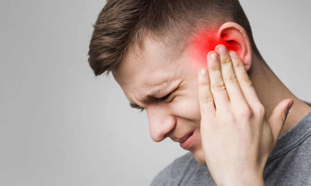
Trigeminal neuralgia (TN), is sometimes described as the most excruciating pain known to humanity. The pain typically involves the lower face and jaw, although sometimes it affects the area around the nose and above the eye. This intense, stabbing, electric shock-like pain is caused by irritation of the trigeminal nerve, a nerve in face, which sends branches to the forehead, cheek and lower jaw. It usually is limited to one side of the face. The pain can be triggered by an action as routine and minor as brushing your teeth, eating or a sudden sharp breeze. If left untreated, trigeminal neuralgia can progressively worsen. Facial neuralgia means pain on the face related to a nerve.

Trigeminal neuralgia cannot always be cured, there are treatments available to alleviate the debilitating pain. Normally, anticonvulsive medications are the first treatment choice. Surgery can be an effective option for those who become unresponsive to medications or for those who suffer serious side effects from the medications.

Table of Contents

The Trigeminal Nerve

The trigeminal nerve is one set of the cranial nerves, which means it originates from the brain. It is the nerve responsible for providing sensation to the face. There are two trigeminal nerves, one on either side of our face. The trigeminal nerve leaves the brain and travels inside the skull, where it divides into three smaller branches, controlling sensations throughout the face:

  • Ophthalmic Nerve controls sensation in our person’s eyes and forehead.
  • Maxillary Nerve controls sensation in the lower eyelid, cheek, nostril, upper lip and upper gum.
  • Mandibular Nerve controls sensations in the jaw, lower lip, lower gum and some of the muscles used for chewing.

Prevalence

It is reported that 150,000 people are diagnosed with trigeminal neuralgia every year. While the disorder can occur at any age, it is most common in people over the age of 50. TN is twice as common in women than in men.

Types of Trigeminal Neuralgia

There are two main forms of trigeminal neuralgia:

  • Typical (Type 1) trigeminal neuralgia. Patient is likely to experience painful episodes that are sharp, intense and sporadic.
  • Atypical (Type 2) trigeminal neuralgia. This will be less painful and intense but more widespread.

Causes

The exact cause of TN is still unknown, but the pain associated with it represents an irritation of the nerve. Primary trigeminal neuralgia has been linked to the compression of the nerve, typically in the base of the head where the brain meets the spinal cord. This is usually due to contact between a healthy artery or vein and the trigeminal nerve at the base of the brain. This places pressure on the nerve as it enters the brain and causes the nerve to misfire. Secondary TN is caused by pressure on the nerve from a brain lesion like a tumour, cyst, facial injury or any medical condition that damages the myelin sheaths.

Trigeminal Neuralgia Symptoms

Any one side face pain cannot be trigeminal neuralgia. Most patients report that their pain begins spontaneously. Other patients say their neuralgia pain came after a car accident, a blow to the face or dental work.

TN tends to run in cycles. Patients often suffer long stretches of frequent attacks, followed by weeks, months or even years of little or no pain. The pain typically begins with a sensation of electrical shocks that culminates in an excruciating stabbing pain within less than 20 seconds.

Pain can be focused in one spot or it can spread throughout the face. Typically, it is only one side face pain, eg… pain in right side of face and jaw and ear; however, in rare occasions and sometimes when associated with multiple sclerosis, patients may feel pain in both sides of their face. Pain areas include the cheeks, jaw, teeth, gums, lips, eyes and forehead.

Attacks of TN may be triggered by the following:

  • Touching the skin lightly
  • Washing
  • Shaving
  • Brushing teeth
  • Blowing the nose
  • Drinking hot or cold beverages
  • Encountering a light breeze
  • Applying makeup
  • Smiling
  • Talking

There are many other conditions which are similar to TN. Brain specialists use various tests to pinpoint the exact diagnosis.

Trigeminal Neuralgia Diagnosis

TN can be very difficult to diagnose, because there are no specific diagnostic tests and symptoms are very similar to other facial pain disorders. Therefore, it is important to seek medical care when feeling unusual, sharp pain around the eyes, lips, nose, jaw, forehead and scalp, especially if you have not had dental or other facial surgery recently.

TN usually is diagnosed based on the description of the symptoms provided by the patient, detailed patient history and clinical evaluation.

Testing

There are no specific diagnostic tests for TN, so physicians must rely heavily on symptoms and history, type of pain (sudden, quick and shock-like), the location of the pain and things that trigger the pain. MRI can detect tumour/s or Multiple Sclerosis and if there is compression caused by a blood vessel. Newer scanning techniques can show if a vessel is pressing on the nerve and may even show the degree of compression.

Treatment of Trigeminal Neuralgia – Non-Surgical

There are several effective ways to alleviate the pain, including a variety of medications. Medications are generally started at low doses and increased gradually based on patient’s response to the drug.

  • Carbamazepine
  • Gabapentin
  • Oxcarbazepine

Other medications include baclofen, amitriptyline, pregabalin, phenytoin, valproic acid etc.

Medicines may have side effect and may become ineffective after long usage.

Trigeminal Neuralgia Treatment: Surgery

If medications have proven ineffective in treating TN, several surgical procedures may help control the pain.

Open Surgery

Microvascular decompression surgery involves microsurgical exposure of the trigeminal nerve root, identification of a blood vessel that may be compressing the nerve and gentle movement of the blood vessel away from the point of compression. Although most effective, it is also most invasive.

Lesioning Procedures

Percutaneous radiofrequency rhizotomy treats TN through the use of electrocoagulation (heat). It can relieve nerve pain by destroying the part of the nerve that causes pain and suppressing the pain signal to the brain.

Percutaneous balloon compression utilizes a needle that is passed through the cheek to the trigeminal nerve. The balloon compresses the nerve, injuring the pain-causing fibres, and is then removed.

Percutaneous glycerol rhizotomy utilizes glycerol injected through a needle into the area where the nerve divides into three main branches. The goal is to damage the nerve selectively in order to interfere with the transmission of the pain signals to the brain.

Stereotactic radiosurgery (through such procedures as Gamma Knife, Cyberknife, Linear Accelerator (LINAC) delivers a single highly concentrated dose of ionizing radiation to a small, precise target at the trigeminal nerve root. This treatment is non-invasive and avoids many of the risks and complications of open surgery and other treatments.

Overall, the benefits of surgery or lesioning techniques should always be weighed carefully against its risks. At Samarth Neuro and Trauma Hospital in Miraj, many patients with Trigeminal neuralgia or one side face pain have recovered after medical or surgical treatment, under the specialist care of Neurosurgeon Dr Ravindra Patil.

Pain in Ears and Jaw

Pain in Ears and Jaw

By Dr. Ravindra Patil

Pain in the area near our ear, our jaw or the muscles on the side of our face, sometimes accompanied by a clicking or popping sound and/or restricted jaw movement is called Temporo-Mandibular Disorders or TMD in short. There is another abbreviation related to the joints of the jaws. It is TMJ for Temporo-Mandibular Joint. We have two TMJs on either side of our face.

Jaw pain reasons are many and may be due to TMJ disease or even mental stress!

TMD pain describes a group of conditions characterized by pain and dysfunction of the TMJ and/or the muscles surrounding it. It’s not always easy to figure out the cause of these symptoms. However, most TMD cases can be treated with conservative remedies. Only if these remedies are not useful should major procedures like dental treatment or surgery should be done.

The two TMJs that connect your lower jaw, the mandible, to the temporal bones of the skull on either side, are actually very complex joints that allow movement in three dimensions. The TMJ anatomy is that the lower jaw and temporal bone fit together as a ball and socket, with a cushioning cartilage disk in between. Large pairs of muscles in the cheeks and temples move the lower jaw. Any of these parts — the disk, the muscles or the joint itself — can become the source of a TMD problem.

For people suffering from TMD pain or for people having difficulty opening or closing their jaw, a thorough investigation workup is necessary to pinpoint the cause of the pain or difficulty in opening and closing the jaw.

Table of Contents

What causes TMD?

The temporo-mandibular joint or TMJ can suffer orthopaedic problems like any other joint. These problems include inflammation, sore muscles, strained tendons and ligaments, and cartilage problems.

Heredity also plays a role in causing TMD. Compared to men, women appear to be more prone to it. As we age, the chances of getting TMD are higher. Physical and psychological stresses can also cause TMD. In some cases, jaw pain may be related to a more widespread, pain-inducing medical condition such as fibromyalgia. Fibromyalgia is a pain of muscles and connective tissue around the TMJ [or any other joint].

Because we have two Temporo Mandibular Joints, one on each side our the face, pain in left side of head, jaw pain left side, pain on left side of head above ear, pain in right side of head, jaw pain left side near ear or jaw pain one side, all permutations and combinations of pain are possible.

Signs and Symptoms

Clicking Sounds: some people with TMD hear a clicking, popping or grating sound coming from the TMJ when opening or closing the mouth. This is usually caused by a shifting of the cartilage disk inside the joint. This clicking or popping sound can sometimes be heard by anyone standing next to the patient. Clicking by itself is actually not a significant symptom because it has been found that over one third of all the population has jaw joints which click. However, if the clicking is accompanied by pain or limited jaw movement and function, or the jaw is getting ‘stuck’ in an open or closed position often, this definitely is TMD.

TMD Muscle Pain Cycle

Every joint is operated by muscles. The jaw can be clenched by two pairs of very powerful muscles, the temporalis muscles and the masseter muscles. They are located on either side of our face. Pain in these muscles is significant and should be taken care of. Jaw pain is not so uncommon.

Pain on right side of the head or left side head pain, the side depends on which side TMJ is affected. Often both sides jaw pain may occur.

The reason for jaw pain may be muscle spasm of temporalis and masseter muscles.

The muscle pain may be felt in the cheeks (masseter muscles) and temples (temporalis muscles), where the two big pairs of jaw-closing muscles are located. If a person feels soreness and stiffness upon waking up in the morning, it’s often related to habits of clenching and/or grinding the teeth at night. If you have this type of nocturnal habit, it can be treated with a custom-made guard which is very helpful in decreasing the force applied to your teeth. This in turn will allow your muscles to relax and be painless. This will relieve pressure on your jaw joints. Some of the other self-care remedies are discussed below.

TMJ headache means headache or jaw pain caused because of TMD.

Joint Pain

Pain that originates one or both jaw joints is called arthritis of the TMJs. When we look at X-Ray images of the TM joints, we may or may not find arthritic changes. The funny part is, some people have arthritic-looking TMJs but no symptoms of pain or dysfunction; while others have significant symptoms of pain and dysfunction but their joints look normal in X-Ray images. Alas, there is no permanent cure for arthritis anywhere in the body, but medicines definitely help in relieving arthritic pain and swelling.

Pain Relief

As has been mentioned, a thorough investigative work up on a TMD patient will help in finding the cause of the pain and then treat or reduce the same. Sometimes an intervention as simple as switching to a soft diet which does not need thorough chewing can reduce stress on the muscles and joints and reduce pain.

Application of Ice and/or moist heat can help relieve swelling, soreness and inflammation. If the pain is caused because of muscle spasm, it can be relieved with gentle stretching exercises. Non-steroidal anti-inflammatory drugs [NSAIDs] and muscle relaxants can also provide relief. NSAIDs apart from reducing inflammation, also reduce pain.

Other Treatments

Severe TMD cases may require more complex forms of treatment, which might include orthodontics, dental restorations like bridgework, or minor procedures inside the joint such as cortisone injections or lavage (flushing) of the joint. It’s rare for major surgery ever to be necessary in a case of TMD. Again, it’s important to try the wide range of conservative, reversible treatments available, and give them enough time to work as they almost always prove effective.

Chewing tobacco and TMD

It is well known that chewing tobacco causes cancers of the mouth. But chewing tobacco also causes difficulty with chewing, swallowing, speaking or moving the tongue and jaw. Tobacco users find it very difficult to open their mouth and so are unable to eat properly. While the risk of cancer with tobacco chewing is great, the stiffening of the jaw and inability to open the jaw fully is also a major symptom. There may be cheek swelling too.

Many people who had been chewing tobacco and Gutkha for a few years, experience inability to open their mouth. The mouth barely opens more than 30 mm and the opening is further reduced over a period of time. The jaws become stiff. Such patient are firstly unable to eat because of small mouth opening. Besides, they are unable to eat spicy food. This condition is known as Oral Sub Mucous Fibrosis.

Stopping tobacco chewing is the first step in treating Oral Sub Mucous Fibrosis. Surgical treatment also helps open the jaw wide.

To summarise…

TMD, or Temporomandibular Disorders, is an umbrella term for various painful conditions that affect the jaw joints. There are different treatment approaches to TMD problems, but not all are based on science. It’s important to be up on the latest information and to be an educated patient. We have tried to provide useful information in this article about TMD.

क्रॅनिओटॉमी शस्त्रक्रिया

Craniotomy surgery

क्रॅनिओटॉमी शस्त्रक्रिया

By Dr. Ravindra Patil

क्रॅनियोटॉमी ही एक शस्त्रक्रिया आहे, परंतु कोणत्याही एका विशिष्ट रोगावर उपचार करण्यासाठी नाही. क्रॅनिओटॉमी म्हणजे मेंदूच्या गाठीची शस्त्रक्रिया किंवा एपिलेप्सी शस्त्रक्रिया करण्यासाठी कवटी उघडणे. क्रॅनियोटॉमी या शब्दाचे दोन भाग आहेत: “क्रॅनिअम” आणि “ओटोमी”.

“क्रॅनिअम” हा शब्द लॅटिन भाषेतून आला आहे. याचा अर्थ कवटी. आणि “ओटोमी” हा शब्द ग्रीक भाषेतून आला आहे आणि त्याचा अर्थ शरीराचा एक भाग कापून टाकणे असा होतो. बहुतेक एलोपथी वैद्यकीय संज्ञा लॅटिन आणि ग्रीक भाषेतील आहेत. अशाप्रकारे, गॅस्ट्रोटॉमी म्हणजे पोटा काढणे, फॅरिंगोटॉमी म्हणजे घशाची पोकळी काढणे. त्याच प्रकारे, क्रॅनिओटॉमी म्हणजे कपाळ किंवा कवटी काढणे.

मेंदूच्या या शस्त्रक्रियेमध्ये मेंदूमध्ये प्रवेश करण्यासाठी कवटीचा एक भाग काढून टाकला जातो. शस्त्रक्रिया झाल्यावर हाड परत लावले जाते. क्रॅनिओटॉमी मेंदूतील गाठ काढून टाकण्यासाठी, मेंदूतील एन्युरिझमवर उपचार करण्यासाठी, एपिलेप्सीवर उपचार करण्यासाठी, सेरेब्रल हॅमरेजवर उपचार करण्यासाठी, इंट्राक्रॅनियल प्रेशर कमी करण्यासाठी केले जाते. अशा शस्त्रक्रिया न्यूरोसर्जन करतात.

भारतात मेंदूच्या शस्त्रक्रियेची किंमत

जेव्हा अशा मोठ्या शस्त्रक्रियेचा उल्लेख केला जातो तेव्हा खर्चाचा विषय येतोच. मेंदूच्या शस्त्रक्रियांसाठी एक लाख ते पाच लाख किंवा त्याहून अधिक खर्च येऊ शकतो. पण हा खर्च आयुष्मान भारत योजना, आरोग्य विमा किंवा नियोक्त्याने (नोकरी देणार्यांनी) दिलेल्या सुविधा असे पर्याय आहेत. तुम्हाला वरीलपैकी कोणतेही फायदे नसल्यास, वैद्यकीय विमा खरेदी करणे अत्यंत उचित आहे.

क्रॅनिओटॉमीच्या आधीची तयारी

रुग्ण सुरक्षितपणे प्रक्रिया पार पाडू शकतो याची पुष्टी करण्यासाठी विविध चाचण्या केल्या जातात. त्या म्हणजे:

  • शारीरिक परीक्षा
  • रक्त चाचण्या
  • न्यूरोलॉजिकल परीक्षा
  • मेंदूचे इमेजिंग (सीटी स्कॅन किंवा एमआरआय)
  • वैद्यकीय स्थितीवर आधारित डोक्यावर नक्की कुठे ऑपरेशन करायचे ते ठरवणे

शस्त्रक्रियेच्या आदल्या रात्री, रुग्णाला उपाशी ठेवतात. डोक्याच्या केसांचे मुंडन केले जाते.

प्रक्रियेदरम्यान, रुग्ण ऑपरेटिंग टेबलवर झोपतो. मेंदूच्या कोणत्या भागावर ऑपरेशन केले जात आहे यावर रूग्णाची स्थिती अवलंबून असते. तुमच्या हातामध्ये इंट्राव्हेनस लाइन घातली जाते. रुग्णाच्या मूत्राशयात मूत्र कॅथेटर लावतात. आणि नंतर सामान्य भूल (जनरल एनेस्थेशिया) दिली जाते. कवटी उघडण्याची शस्त्रक्रिया म्हणजे क्रेनियोटॉमी सर्जरीचा चाकू, मेडिकल ड्रिल आणि करवतीने केली जाते. मग ब्रेन ट्यूमरचे ऑपरेशन, एपिलेप्सी सर्जरी किंवा ब्रेन हॅमरेज असा जो काही रोग असेल त्याची शस्त्रक्रिया करतात. मग सर्जन ताबडतोब किंवा काही महिन्यांनंतर वायर, टाके किंवा प्लेट्सच्या मदतीने काढलेला हाडाचा तुकडा परत कवटीला जोडतात. शेवटी त्वचेवर टाके घेऊन किंवा सर्जीकल स्टेपल्स लावून जखम बंद करतात. जखमेवर स्टराईल (निर्जंतुकीकरण केलेले) ड्रेसिंग ठेवण्यात येते आणि निर्जंतुकीकरण केलेली मलमपट्टी किंवा बॅंडेज बांधले जाते. क्रॅनियोटॉमी शस्त्रक्रियेसाठी सुमारे तीन ते चार तास लागू शकतात.

क्रॅनियोटॉमीचे प्रकार

क्रॅनियोटॉमीचे अनेक प्रकार आहेत. प्रत्येक प्रकाराला शस्त्रक्रियेमध्ये वापरल्या जाणार्‍या तंत्र किंवा स्थानासाठी नाव दिले जाते.

स्टिरिओटॅक्टिक क्रॅनिओटॉमी

क्रॅनिओटॉमीमध्ये कवटीला स्टिरिओटॅक्टिक फ्रेमने फिक्स केले असल्यास आणि एमआरआय किंवा सीटी स्कॅन वापरल्यास, त्याला स्टिरिओटॅक्टिक क्रॅनिओटॉमी म्हणतात.

एंडोस्कोपिक क्रॅनिओटॉमी

एंडोस्कोपद्वारे कवटीच्या एका लहान चीराद्वारे केले जाते.

Table of Contents

जागृत क्रॅनियोटॉमी

रुग्ण जागृत असताना जागृत क्रॅनिओटॉमी केली जाते.

की-होल क्रॅनिओटॉमी

ब्रेन ट्यूमर काढून टाकण्यासाठी की-होल (शक्य तेवढ्या लहान छीद्रातून) क्रॅनिओटॉमी केली जाते. या शस्त्रक्रियेत कमीत कमी आकराची सर्जीकल जखम होते.

सुप्रा-ऑर्बिटल ‘आयब्रो’ क्रॅनिओटॉमी

सुप्रा-ऑर्बिटल म्हणजे डोळ्याच्या खाचांच्या वरची शस्त्रक्रिया. ही शस्त्रक्रिया मेंदूच्या पुढच्या भागात असलेला ब्रेन ट्यूमर काढण्यासाठी केली जाते.

टेरोनिअल (फ्रंटो टेम्पोरल) क्रॅनिओटॉमी

कवटीत टेरिऑन म्हणजे कपाळाचे हाड आणि टेम्पोरल, स्फेनोइड आणि पॅरायटल हाडे एकत्र होतात ते ठिकाण. यात टेरिऑनचा भाग काढून टाकतात.

ऑर्बिटो-झायगोमॅटिक क्रॅनिओटॉमी

काठण्यास अवघड असलेले ट्यूमर आणि एन्युरिझम्सचा उपचार ऑर्बिटो-झायगोमॅटिक क्रॅनिओटॉमीद्वारे केला जाऊ शकतो.

शल्यचिकित्सक कवटीच्या ज्या हाडांमुळे चेहेर्‍याचा गोलाकार आकार येतो त्या हाडांचा काही भाग तात्पुरता काढून टाकतो. नंतर ती हाडे परत जोडली जातात.

पोस्टरियर फोसा क्रॅनिओटॉमी

हे ऑपरेशन कवटीच्या पायथ्याशी एका चीराद्वारे केले जाते.

ट्रान्सलेबिरिन्थाइन क्रॅनिओटॉमी

ट्रान्सलेबिरिन्थाइन क्रॅनिओटॉमीमध्ये, सर्जन कानाच्या मागे चीरा करून ऑपरेशन करतात.

बायफ्रंटल क्रॅनिओटॉमी

बायफ्रंटल क्रॅनिओटॉमी, किंवा विस्तारित बायफ्रंटल क्रॅनिओटॉमी, पुढच्या मेंदूतील काढण्यास कठीण ट्यूमर काढण्यासाठी केली जाते.

क्रॅनिओटॉमी का केली जाते?

भारतात ब्रेन ट्यूमर शस्त्रक्रियेसाठी लागणारा खर्च विविध योजनांद्वारे सर्वांना परवडणारा होत असल्याने, या शस्त्रक्रिया अनेकदा केल्या जातात. मेंदूच्या खालील रोगांवर उपचार करण्यासाठी क्रॅनिओटॉमी केली जाते:

  • ट्यूमर
  • एन्युरिझम
  • संसर्ग
  • सूज (सेरेब्रल इडिमा)
  • कवटीच्या आत रक्तस्त्राव
  • रक्ताची गुठळी
  • मेंदूत संक्रमण व पस होणे
  • कवटीचे फ्रॅक्चर
  • ड्युरा मॅटर फाटणे
  • धमनी विकृती
  • आर्टिरिओव्हेनस फिस्चुला
  • इंट्राक्रॅनियल दाब वाढणे
  • अपस्मार (एपिलेप्सी)
  • पार्किन्सन रोगासारख्या सतत होणार्‍या हालचालीच्या विकारांसाठी उपकरणे रोपण करणे.

ब्रेन ट्यूमर काढून टाकल्यानंतर सहसा बायोप्सीसाठी पाठवले जाते. भारतात ब्रेन ट्यूमर शस्त्रक्रियेचा खर्च बहुतेक लोकांना वाटतो तितका जास्त नसतो.

क्रॅनिओटॉमी जोखीम व दुष्परिणाम

रुग्णाची विशिष्ट मेंदूची शस्त्रक्रिया आणि वैद्यकीय स्थिती आणि अनेक इतर घटक यावर दुष्परिणाम होणे आवलंबून असते. संभाव्य दुष्परिणाम खालील प्रमाणे असतात:

  • डोक्यावर जखमांचे व्रण
  • कवटीची हाडा काढल्यामुळे दिसणारा खड्डा
  • डोक्यात उपकरणे लावल्यामुळे दुखापत
  • चेहऱ्याच्या मज्जातंतूचे नुकसान
  • सायनसचे नुकसान
  • हाडांच्या काढलेल्या भागात किंवा त्वचेत संसर्ग
  • दौरे (फेफरं येणे)
  • मेंदूला सूज
  • सेरेब्रोस्पाइनल फ्लुइडची गळती
  • स्नायू कमकुवत होणे
  • स्ट्रोक

क्वचितच, क्रॅनिओटॉमीमुळे असे ही होऊ शकते:

  • बोलण्यात समस्या
  • स्मृती समस्या
  • चालतांना तोल राखण्याची समस्या
  • लकवा
  • कोमा

क्रॅनियोटॉमीमुळे सर्वसामान्य शस्त्रक्रियेमुळे होणारे दुष्परिणाम देखील होऊ शकतात, जसे की:

  • रक्तस्त्राव
  • रक्ताच्या गुठळ्या
  • न्यूमोनिया
  • जनरल ऍनेस्थेसियाचे दुष्परिणाम
  • अस्थिर रक्तदाब

या सर्व दुष्परिणामांविषयी सर्जन स्वताःच जास्त माहिती देऊ शकतात.

क्रॅनियोटॉमी नंतर बरे होण्याची प्रक्रिया

शस्त्रक्रियेनंतर लगेच रुग्णाला रिकव्हरी युनिट किंवा अतिदक्षता विभागात [ICU] नेले जाते. नर्सेस सतत रुग्णाच्या जीवनावश्यक गोष्टींचे बारकाईने निरीक्षण करतात कारण रूग्ण हळूहळू एनेस्थेशियाच्या परिणामातून बाहेर पडतो. त्यानंतर रुग्णाला वेगळ्या खोलीत नेले जाते. रुग्णालयात मुक्काम शस्त्रक्रियेच्या प्रकारावर अवलंबून असतो. परंतु सहसा एक आठवडा तरी रहावेच लागते.

या महत्त्वपूर्ण बरे होण्याच्या कालावधीत:

  • सूज टाळण्यासाठी डोके उंचावले जाते
  • ऑक्सिजन दिला जातो
  • न्युमोनिया टाळण्यासाठी खोल श्वास घेण्याचे व्यायाम आणि स्पायरोमेट्री शिकवली जाते
  • पायांभोवती विशेष टूर्निकेट्स बांधतात. त्या मध्ये हवा आलटून पालटून भरली जाते व बाहेर टाकली जाते. यामुळे पायांच्या स्नायुंना मसाज मिळतो व हे शिरामध्ये रक्ताच्या गुठळ्या होण्यास प्रतिबंध करते.
  • युरिनरी कॅथेटर अनेक दिवस मूत्राशयात ठेवतात.
  • मेंदू आणि शरीराची कार्ये तपासण्यासाठी वारंवार न्यूरोलॉजिकल तपासणी केली जाते.

रूग्णाला डिस्चार्ज मिळून घरी जाणे

क्रॅनियोटॉमीनंतर, डोक्याच्या जखमेची काळजी घेणे आवश्यक आहे. वेदना बरे होईपर्यंत आणि संसर्ग प्रतिबंधासाठी औषधे घेणे आवश्यक आहे. बरे व्हायला किमान सहा आठवडे लागू शकतात. रुग्णाने दैनंदिन नियमित क्रियाकलाप करताना अत्यंत काळजी घेणे आवश्यक आहे जसे की:

  • चालणे
  • बोलणे
  • ज्या क्रियाकलापांसाठी शक्ती आवश्यक असते
  • ज्या क्रियाकलापांसाठी समतोलता आवश्यक असते

फिझिओथेरपी, ऑक्युपेशनल थेरपी आणि स्पीच थेरपीचा घेणे अनेकदा आवश्याक असते. आणि अर्थातच, विश्रांती आवश्यक आहे. जखमेच्या ड्रेसिंगसाठी आणि मेंदूच्या कार्यात्मक मूल्यांकनासाठी रुग्णाला हॉस्पिटलमध्ये न्यावे लागते. मेंदूच्या शस्त्रक्रियेनंतर दीर्घ विश्रांतीची आवश्यकता असते. विश्रांतीच्या काळात रूग्ण काही काम धंधा करू शकत नाही आणि यामुळे भारतात मेंदूच्या शस्त्रक्रियेचा खर्च वाढतो.

क्रॅनिओटॉमी नंतरचे जीवन

रुग्ण पूर्णपणे बरा झाल्यानंतर, काळजी घेणे अजूनही महत्त्वाचे आहे. असे करणे हे भविष्यातील दुष्परिणाम टाळण्यासाठी आणि दीर्घकालीन आयुष्य सुधारण्यासाठी मदत करते. अशा मोठ्या शस्त्रक्रियेतून बरे झाल्यानंतर खालील गोष्टी करणे अत्यंत आवश्यक असते:

  • नियमित व्यायाम
  • सकस आहार
  • अल्कोहोल (दारू) मर्यादित किंवा टाळा
  • धूम्रपान सोडा
  • पुरेशी झोप घ्या

कॉम्प्लिकेशन्स – दुष्परिणाम

सर्व शस्त्रक्रियांप्रमाणे, क्रॅनिओटॉमीमुळे वेदना, रक्तस्त्राव, संसर्ग इत्यादी गुंतागुंत होऊ शकतात. परंतु असे झाल्यास सर्जन त्यांवर उपचार करतात. रुग्णांना गंभीर डोकेदुखी, फेफरे किंवा जखमेच्या संसर्गाची चिन्हे असल्यास त्यांनी ताबडतोब हॉस्पिटलशी संपर्क साधावा.

थोडक्यात…

क्रॅनिओटॉमी म्हणजे फक्त कवटी उघडणे. ही कोणती विशिष्ट शस्त्रक्रिया नाही. हे ऑपरेशन भारतातील मोठ्या आणि मध्यम आकाराच्या शहरांमध्ये केले जाऊ शकते. “आयुष्यमान भारत – प्रधान मंत्री जन आरोग्य योजनेद्वारे ” किंवा इतरांना वैद्यकीय विम्याद्वारे भारतात ब्रेन ट्यूमर शस्त्रक्रियेचा खर्च अगदी गरीबांनादेखील परवडणारा आहे.

Craniotomy surgery

Craniotomy surgery

Craniotomy surgery

By Dr. Ravindra Patil

Craniotomy is a surgery, but not to treat one specific disease. Craniotomy means opening the skull to do brain tumour surgery or epilepsy surgery. The word craniotomy has two parts: Cranium and Otomy.

The word Cranium originates from the Latin language. It means the skull. And ‘Otomy’ comes from the Greek language and it means cutting into a part of the body. Most medical terminology is from Latin and Greek. Thus, a gastrotomy would be cutting into the stomach, a pharyngotomy means cutting into the pharynx. In the same way, craniotomy means cutting into the cranium or skull.

In this surgery for the brain, a part of the skull is removed to access the brain. The bone is replaced when the surgery is done. Craniotomy is done to remove brain tumours, treat brain aneurysms, treat epilepsy, treat cerebral haemorrhage, to decrease intracranial pressure and so on. Neurosurgeons performs the procedure.

Cost of brain surgery in India

When such a major surgery is mentioned, the subject of cost must be cleared. Brain surgeries may cost between one lakh to five lakhs or more. But there are options like Ayushman Bharat Yojana, health insurance or employer given benefits to cover the costs. If you have none of the above benefits, it is highly advisable to buy a medical insurance.

Before a craniotomy procedure

Various tests are done to confirm that the patient can safely undergo the procedure. They are:

  • Physical exam
  • Blood tests
  • Neurological exam
  • Imaging of the brain (ct or mri)
  • Determination of the surgical site based on the medical condition

On the night before surgery, the patient will be NBM [nil by mouth]. Scalp will be shaven.

During the procedure the patient lies down on the operating table. The position depends on the part of the brain being operated on. Intravenous line is inserted into your arm or hand. A urinary catheter into the patient’s bladder. And then general anaesthesia is given. Surgery is done with a knife, a medical drill and a saw. Then the brain tumour operation, epilepsy surgery or brain haemorrhage is done. Then the surgeons fix the bone with wires, stitches, or plates either immediately or a few months later. Finally the skin will be stitched or stapled. A sterile dressing and a sterile bandage are applied. Craniotomy surgery may take about three to four hours.

Types of Craniotomy

There are many types of craniotomies. Each type is named for the technique or location used in the surgery.

Stereotactic craniotomy

If in a craniotomy the skull is fixated with a stereotactic frame and uses an MRI or CT scan, it’s called a stereotactic craniotomy.

Endoscopic craniotomy

Done through a tiny incision in the skull through an endoscope.

Awake craniotomy

Table of Contents

An awake craniotomy is done while the patient is awake.

Keyhole craniotomy

A keyhole craniotomy is used to remove brain tumours. It’s a minimally invasive surgery.

Supra-orbital ‘eyebrow’ craniotomy

A supra-orbital means surgery above the eye sockets, done to remove brain tumours in the front of the brain.

Pteronial (frontotemporal) craniotomy

In the skull, the pterion is where the frontal, temporal, sphenoid, and parietal bones meet. It involves removing part of the pterion.

Orbitozygomatic craniotomy

Difficult tumours and aneurysms may be treated with an orbitozygomatic craniotomy. The surgeon temporarily removes part of the bone that creates the curve of your orbit, or eye socket, and cheek.

Posterior fossa craniotomy

This is done through an incision at the base of the skull.

Translabyrinthine craniotomy

In a translabyrinthine craniotomy, the surgeon makes a cut behind a ear.

Bifrontal craniotomy

A bifrontal craniotomy, or extended bifrontal craniotomy, is done to remove difficult tumours in front of the brain.

Why is a craniotomy done?

As the cost for brain tumour surgery in India is becoming affordable through various schemes to everybody, these surgeries are done often. A craniotomy is done to treat the following conditions of the brain:

  • Tumour
  • Aneurysm
  • Infection
  • Swelling (cerebral oedema)
  • Bleeding inside the skull
  • Blood clot
  • Brain abscess
  • Skull fracture
  • Dura mater tear
  • Arteriovenous malformation
  • Arteriovenous fistula
  • Raised Intracranial pressure
  • Epilepsy
  • To implant devices for movement disorders like Parkinson’s disease.

A brain tumour after removal is usually sent for a biopsy. Brain tumour surgery cost in India is not as high as most people think.

Craniotomy risks, side effects, and complications

The risk of complications depends on many factors, including the patient’s specific brain surgery and medical condition. Possible complications include:

  • Head scarring
  • Dent where bone flap was removed
  • Injury from the head device
  • Facial nerve damage
  • Damage to the sinuses
  • Infection of the bone flap or skin
  • Seizures
  • Brain swelling
  • Leaking of cerebrospinal fluid
  • Muscle weakness
  • Stroke

Rarely, a craniotomy may lead to:

  • speech problems
  • memory problems
  • balance issues
  • paralysis
  • coma

A craniotomy can also cause general surgical side effects like:

  • Bleeding
  • Blood clots
  • Pneumonia
  • Reaction to general anesthesia
  • Unstable blood pressure

However, the surgeon is the best person who can explain about these complications.

Recovery following craniotomy

Immediately after surgery the patient is taken to the recovery unit or intensive care unit [ICU]. Nurses closely monitor the patient’s vitals as he/she gradually come out of anaesthesia. Then the patient is shifted to a room. Hospital stay will depend on type of surgery but may be as long as a week.

During this crucial recovery period:

  • Head is elevated to prevent swelling
  • oxygen is given
  • Deep-breathing exercises and spirometry are taught to prevent pneumonia
  • Special tourniquets are wound around legs into which air is pumped in and out alternately. This gives massage to the muscles of the calves and thighs. This prevents blood clots in veins.
  • The urinary catheter stays in the urinary bladder for several days.
  • Frequent neurological check-ups are done to check brain and body functions.

At home

After a craniotomy, the head wound care is critical. Medicines for pain and infection prevention are to be taken till the would heals. Recovery may take at least 6 weeks. The patient must take extreme care while attempting routine activities like:

  • Walking
  • Talking
  • Activities needing strength
  • Activities needing Balance

Physiotherapy, occupational therapy and speech therapy is usually advised. And of course, rest is necessary. The patient needs to visit his/her hospital for wound dressing and functional brain assessment. A long rest is required after brain surgery and during that time the patient cannot do any productive earning work, and this increases the brain surgery cost in India.

Life after craniotomy

After the patient has fully recovered, it’s still important to take care. This will help manage future complications and improve the long-term outlook. After recovering from such a major surgery, it is highly advisable to:

  • Exercise regularly
  • Eat a healthy diet
  • Limit or avoid alcohol
  • Quit smoking
  • Get enough sleep

Complications

Like in all surgeries, a craniotomy can cause complications like pain, haemorrhage, infection and so on. But surgeons will treat them if the occur. Patients must contact the hospital immediately if he/she has severe headaches, seizures, or signs of a wound infection.

To summarise…

Craniotomy means opening the bone, not a specific surgery. It can be done in large and medium sized cities in India. Cost of brain tumour surgery in India is affordable even to the poorest through AB-PMJAY yojana or through medical insurance to others.

Epilepsy surgery

Epilepsy surgery

Epilepsy surgery

By Dr. Ravindra Patil

Overview

Epilepsy is a brain disorder that causes seizures or unusual sensations and behaviour. Medicines can control seizures. But sometimes medicines do not work and brain surgery for seizures or brain surgery for epilepsy is done.

Epilepsy brain surgery is not the first line of treatment, because it is a super major surgery. But surgery for epilepsy is most effective when seizures always occur in a single location in the brain. So, every epileptic patient does not get relief from seizures after brain surgery.

Special tests can determine if a patient is eligible for epilepsy surgery.

In epilepsy brain surgery a part area of the brain where seizures occur is removed.

Table of Contents

Goal of Epilepsy Surgery

Is to limit seizures after brain surgery, limit their severity with or without the use of medications.

How Do Seizures Kill

Poorly controlled epilepsy can have complications like:

  • Physical injuries during a seizure – a head injury can cause permanent disability or death
  • Drowning, if the seizure occurs during a bath or swimming
  • Depression and anxiety
  • It delays growth in children
  • Sudden death, a rare complication of epilepsy
  • Memory and other thinking skills loss

Types of epilepsy surgery

The type of brain surgery for seizures depends on the location of the electrical disturbance in the brain and the age of the patient:

  • Resective surgery, the most common epilepsy surgery, is the removal of a small portion of the temporal lobes of the brain.
  • MRI guided Laser interstitial thermal therapy uses a laser to pinpoint and destroy a small portion of brain tissue.
  • Just as a pacemaker controls heart rhythms, an implantable device for the brain detects abnormal activity and corrects it – before a person with epilepsy experiences a seizure. This innovative device is called a neurostimulator or a seizure pacemaker. It is a proven treatment for adults who have disabling seizures not controlled by medication.
  • MRI guided Deep Brain Stimulation is a permanently implanted seizure pacemaker in patient’s chest, which stimulates deep inside the brain. It releases regularly timed electrical signals that disrupt abnormal seizure-inducing activity.
  • Corpus callosotomy is a surgery to completely or partially remove part of the brain that connects nerves on the right and left sides of the brain.
  • Hemispherectomy removes one side of the gray matter of the brain.
  • Functional hemispherectomy is used in children that removes the connection nerves without removing actual brain.

Risks of brain surgery for epilepsy

Different areas of the brain control different functions. Therefore, risks vary depending on the surgical site and the type of surgery. The surgical team will help patients or their relatives understand the specific risks of your procedure, as well as the strategies the team will use to reduce the risk of complications. Risks may include the following:

  • Memory and language problems
  • Visual impairment
  • Depression, mood changes
  • Headache
  • Stroke
  • Seizure after brain surgery

Before operation

Special tests will detect exactly which brain part is to be operated upon.

  • Baseline electroencephalogram (EEG).
  • Continuous EEG with video monitoring records seizures as they occur. Evaluating EEG with during a seizure helps pinpoint the area of your brain to be operated.
  • MRI imaging to identify damaged cells, tumours or other abnormalities that can cause seizures.
  • Invasive EEG monitoring.
  • Video Invasive EEG monitoring.
  • PET scan: measures brain function.
  • Single-photon emission computerized tomography (SPECT) measures blood flow in the brain during a seizure.

Evaluations to understand brain function

Depending on the surgical site, your team may recommend tests to determine the precise areas of the brain that control language, sensory functions, motor skills or other critical functions. This information helps surgeons preserve function to the greatest extent possible when removing or altering a site in your brain. They are:

  • Functional MRI identifies regions of brain activity when you’re doing a particular task, such as listening or reading. This helps the surgeon know the precise locations in your brain that control a particular function.
  • Wada test. With this test, an injected medication temporarily puts one side of brain to sleep at a time. You’re then administered a test for language and memory function. This test can help determine which side of your brain is dominant for your language usage.
  • Brain mapping. Small electrodes match tasks with measurements of brain’s electrical activity.

Neuropsychological tests

Additionally, testing is usually recommended to measure verbal and nonverbal learning skills and memory function. These tests may provide additional insight into the area of the brain affected by seizures, as well as a baseline for measuring function after surgery.

How is surgery done

Hair will be shaved over the section of your skull that before the operation. IV tubes will be placed in veins and pre-operative medicines will be given

General anaesthesia is given. In some cases, awake surgery is done, more details are given at the end. Awake surgery helps save parts brain which control language and movement. But the patient feels no pain.

The surgeon operated through a small window in the skull after surgery the window of bone is replaced and fastened to the remaining skull for healing. Pain is reduced with appropriate medicine. Antibiotics are given to prevent infection. Total hospital stay for epilepsy surgeries will be about three to seven days.

Returning to work or school may take one to three months. Normal activity must started gradually.

Results

The outcomes of epilepsy surgery vary depending on the type of surgery performed. The best expected outcome is seizure control without medication.

The most common and best-understood procedure — resection of tissue in the temporal lobe — results in seizure-free outcomes for about two-thirds of people. Studies suggest that if you do not have a seizure in the first year after temporal lobe surgery — with medication — the likelihood of being seizure-free at two years is 87% to 90%. If you have not had a seizure in two years, the likelihood of being seizure-free is 95% at five years and 82% at 10 years.

If a patient remains seizure-free for at least one year, medicines may be discontinued. If seizures occur, they are controlled by medicines.

Awake brain surgery

Awake brain surgery is a type of brain surgery for seizure performed on the brain while the patient is awake and alert. Awake brain surgery is used to treat some brain (neurological) conditions, including some brain tumours or epileptic seizures.

To summarise, surgery on the brain for epilepsy is useful for those people whose seizures originate from one part of the brain. It is a procedure for seizure which is useful when medicines don’t work.

एपिलेप्सी (अपस्मार) शस्त्रक्रिया

एपिलेप्सी (अपस्मार) शस्त्रक्रिया

एपिलेप्सी (अपस्मार) शस्त्रक्रिया

By Dr. Ravindra Patil

आढावा

एपिलेप्सी हा मेंदूचा विकार आहे की ज्यामुळे दौरे (फेफरं) किंवा असामान्य संवेदना आणि वर्तन होते. औषधे फेफरे येणे वगैरेचा त्रास कमी करू करू शकतात. परंतु काहीवेळा औषधे काम करत नाहीत आणि फेफरेसाठी मेंदूची शस्त्रक्रिया किंवा अपस्मारासाठी मेंदूची शस्त्रक्रिया केली जाते.

एपिलेप्सी च्या उपचारासाठी मेंदूची शस्त्रक्रिया हा सर्वप्रथम उपचार नाही, कारण ती एक मोठी शस्त्रक्रिया आहे. त्या शिवाय अपस्मारासाठी शस्त्रक्रिया सर्वात प्रभावी तेव्हा असते जेव्हा मेंदूच्या एकाच ठिकाणातील रोगामुळे फेफरे येतात. त्यामुळे, प्रत्येक अपस्माराच्या रुग्णाला मेंदूच्या शस्त्रक्रियेनंतर फेफरे येण्यापासून आराम मिळत नाही.

कुठलाही एपिलेप्सीचा रुग्ण शस्त्रक्रियेसाठी पात्र आहे की नाही हे विशेष चाचण्या करून ठरवतात.

मेंदूच्या एपिलेप्सी शस्त्रक्रियेमध्ये मेंदूचा तो भाग काढून टाकला जातो की ज्यामुळे फेफरे येतात.

Table of Contents

एपिलेप्सी शस्त्रक्रियेचे ध्येय

मेंदूच्या शस्त्रक्रियेनंतर झटके मर्यादित करणे, औषधे देऊन किंवा न देऊन फेफरांची तीव्रता मर्यादित करणे.

फेफरांचे दौरे कसे घातक होऊ शकतात

असमाधानकारकपणे नियंत्रित केलेल्या एपिलेप्सीमध्ये असे दुष्परिणाम होऊ शकतात:

  • जप्ती दरम्यान शारीरिक इजा – डोक्याला दुखापत झाल्यास कायमचे अपंगत्व किंवा मृत्यू होऊ शकतो
  • आंघोळ करताना किंवा पोहताना जप्ती आली तर बुडणे
  • नैराश्य आणि चिंता
  • यामुळे लहान मुलांच्या शारीरिक व मानसिक वाढीत विलंब होतो
  • आकस्मिक मृत्यू (हे अपस्मारचे दुर्मिळ कॉम्प्लिकेशन आहे)
  • स्मरणशक्ती आणि इतर विचार कौशल्य कमी होणे

एपिलेप्सी शस्त्रक्रियेचे प्रकार

दौरे कमी करण्यासाठी कोणती शस्त्रक्रिया करायची हे प्रकार मेंदूतील विद्युत गडबडीचे स्थान कुठे आहे आणि रुग्णाचे वय किती आहे यावर अवलंबून असते:

  • रेसेक्टिव्ह सर्जरी, सर्वात सामान्य एपिलेप्सी शस्त्रक्रिया म्हणजे मेंदूच्या टेम्पोरल लोबचा एक छोटासा भाग काढून टाकणे.
  • MRI मार्गदर्शित लेझर इंटरस्टिशियल थर्मल थेरपी – यात मेंदूच्या ऊतींचा एक लहान भाग लेसरने शोधतात आणि नष्ट करतात.
  • हृदयात जसा पेसमेकर हृदयाच्या तालांवर नियंत्रण ठेवतो, त्याचप्रमाणे मेंदूसाठी पण एक प्रत्यारोपण करण्यायोग्य उपकरण असते. अपस्मार असलेल्या व्यक्तीला दौरा येण्यापूर्वी हे उपकरण मेंदूतील असामान्य क्रियाकलाप ओळखते आणि लगेच ते दुरुस्त करते. ज्या प्रौढ रूग्णांना जप्तीचे दौरे औषधांद्वारे नियंत्रित केले जाऊ शकत नाहीत त्याच्या साठी हे अभिनव उपकरण (याला न्यूरोस्टिम्युलेटर किंवा सिझ्यर (जप्ती) पेसमेकर म्हणतात) एक सिद्ध उपचार आहे.
  • एमआरआय मार्गदर्शित डीप ब्रेन स्टिम्युलेशन हे रुग्णाच्या छातीत कायमचे प्रत्यारोपित केलेले जप्ती पेसमेकर आहे, जे मेंदूच्या आत खोलवरची जागा उत्तेजित करते. हे नियमितपणे कालबद्ध विद्युत सिग्नल सोडते. हे सिग्नल असामान्य जप्ती-प्रेरित क्रियाकलापात व्यत्यय आणतात आणि जप्तीचे दौरे कमी करतात.
  • कॉर्पस कॅलोसोटॉमी – ही मेंदूच्या उजव्या आणि डाव्या बाजूंच्या मज्जातंतूंना जोडणारा मेंदूचा भाग पूर्णपणे किंवा अंशतः काढून टाकण्याची शस्त्रक्रिया आहे.
  • हेमिस्फेरेक्टॉमी मेंदूच्या ग्रे मॅटरची एक बाजू काढून टाकते.
  • मुलांमध्ये फंक्शनल हेमिस्फेरेक्टॉमी वापरली जाते जी वास्तविक मेंदू न काढता कनेक्शनच्या नसा काढून टाकते.

एपिलेप्सीसाठी केलेल्या मेंदूच्या शस्त्रक्रियेचे धोके

मेंदूचे वेगवेगळे क्षेत्र वेगवेगळे कार्य नियंत्रित करतात. त्यामुळे, शस्त्रक्रियेची जागा आणि शस्त्रक्रियेच्या प्रकारानुसार जोखीम बदलू शकतात. सर्जिकल टीम रूग्णाला व त्याच्या नातेवाईकांना प्रक्रियेचे विशिष्ट धोके समजून घेण्यास मदत करेल, तसेच गुंतागुंत होण्याचा धोका कमी करण्यासाठी काय करण्यात येईल ते समजावेल. ऑपरेशनच्या जोखमींमध्ये खालील गोष्टींचा होउ शकतात:

  • स्मृती आणि भाषा समस्या
  • दृष्टीदोष
  • नैराश्य, मूड बदलणे
  • डोकेदुखी
  • स्ट्रोक
  • मेंदूच्या शस्त्रक्रियेनंतर जप्ती

ऑपरेशन करण्यापूर्वी

विशेष चाचण्यांद्वारे मेंदूच्या नेमक्या कोणत्या भागावर शस्त्रक्रिया करायची आहे हे कळेल. खालील तपासण्या करतात…

  • बेसलाइन इलेक्ट्रोएन्सेफॅलोग्राम (EEG).
  • जप्ती आल्यावर सतत व्हिडिओ मॉनिटरिंग रेकॉर्ड करतात. जप्तीच्या वेळी ईईजीचे मूल्यांकन केल्याने रोग्याच्या मेंदूचे ऑपरेशन केले जाणारे क्षेत्र निश्चित करण्यात मदत होते.
  • ज्यामुळे दौरे होऊ शकतात अशा खराब झालेले पेशी, ट्यूमर किंवा इतर विकृती MRI इमेजिंगने ओळखता येतात..
  • आक्रमक ईईजी निरीक्षण.
  • व्हिडिओ इनवेसिव्ह ईईजी मॉनिटरिंग.
  • पीईटी स्कॅन: मेंदूचे कार्य मोजते.
  • सिंगल-फोटॉन उत्सर्जन संगणकीकृत टोमोग्राफी (SPECT) जप्तीच्या वेळी मेंदूतील रक्त प्रवाह मोजते.

मेंदूचे कार्य समजून घेण्यासाठी मूल्यांकन

भाषा, संवेदी कार्ये, मोटर कौशल्ये किंवा इतर गंभीर कार्ये नियंत्रित करणारे मेंदूचे अचूक क्षेत्र निर्धारित करण्यासाठी सर्जन्सची टीम अनेक तपासण्या करते. हे सर्जिकल साइटवर अवलंबून असते. ही माहिती सर्जनला मेंदूतील काही भाग काढून टाकताना कोणता भाग कार्य जतन करण्यासाठी जपून ठेवायचा ते कळते.

  • जेव्हा तुम्ही ऐकणे किंवा वाचणे यासारखे विशिष्ट कार्य करत असता तेव्हा कार्यात्मक MRI मेंदूच्या क्रियाकलापांचे क्षेत्र ओळखते. हे सर्जनला तुमच्या मेंदूतील विशिष्ट कार्य नियंत्रित करणारी नेमकी ठिकाणे जाणून घेण्यास मदत करते.
  • वाडा चाचणी (Wada test). या चाचणीत इंजेक्ट केलेले औषध मेंदूच्या एका बाजूला तात्पुरते झोपवते. त्यानंतर भाषा आणि मेमरी कार्यांसाठी चाचणी केली जाते. ही चाचणी तुमच्या मेंदूची कोणती बाजू तुमच्या भाषेच्या वापरासाठी प्रबळ आहे हे निर्धारित करण्यात मदत करू शकते.
  • मेंदूचे मॅपिंग. लहान इलेक्ट्रोड्स मेंदूच्या विद्युत क्रियाकलाप व कार्य यांना जुळवतात.

न्यूरोसायकोलॉजिकल चाचण्या

याव्यतिरिक्त, सामान्यतः मौखिक आणि गैर-मौखिक शिक्षण कौशल्ये आणि मेमरी फंक्शन मोजण्यासाठी चाचणीची शिफारस केली जाते. या चाचण्या जप्तीमुळे प्रभावित मेंदूच्या क्षेत्रामध्ये होणार्‍या बदलांची अतिरिक्त माहिती देतात. तसेच शस्त्रक्रियेनंतर कार्य मोजण्यासाठी आधारभूत माहिती प्रदान करू शकतात.

शस्त्रक्रिया कशी केली जाते

ऑपरेशनपूर्वी कवटीच्या भागावर केस मुंडले जातील. नसेत सुई द्वारा नळ्या लावल्या जातील आणि शस्त्रक्रियापूर्व औषधे दिली जातील.

सामान्य भूल (जनरल एनेस्थेशिया) दिली जाते. काही प्रकरणांमध्ये, जागृत शस्त्रक्रिया केली जाते, अधिक माहिती या लेखाच्या शेवटी दिली आहे. जागृत शस्त्रक्रिया मेंदूचे असे भाग वाचवण्यास मदत करते की जे भाषा आणि हालचाल नियंत्रित करतात. रूग्ण जरी जागा असला तरी जागृत शस्त्रक्रियेत रुग्णाला वेदना होत नाहीत.

सर्जन कवटीच्या एका लहान छीद्रातून शस्त्रक्रिया करतात. शस्त्रक्रियेनंतर सर्जन कवटी मध्ये झालेले छीद्र कवटीच्या काढलेल्या हाडांनी बंद करतात. योग्य औषधांनी वेदना कमी करण्यात येतात. संसर्ग टाळण्यासाठी प्रतिजैविके औषधे (एन्टीबायोटीक्स) दिली जातात. एपिलेप्सी शस्त्रक्रियेसाठी एकूण रुग्णालयात मुक्काम तीन ते सात दिवसांचा असु शकतो.

ऑपरेशन नंतर कामावर किंवा शाळेत परत येण्यासाठी एक ते तीन महिने लागू शकतात. सामान्य क्रियाकलाप हळूहळू सुरू करणे आवश्यक आहे.

परिणाम

एपिलेप्सी शस्त्रक्रियेचे परिणाम शस्त्रक्रियेच्या प्रकारानुसार बदलतात. अपेक्षित परिणाम म्हणजे औषधोपचारा शिवाय जप्ती बंद होणे.

सर्वात सामान्य आणि सर्वोत्कृष्ट समजली जाणारी प्रक्रिया म्हणजे मेंदूच्या टेम्पोरल लोबमधील ऊतींचे विच्छेदन — याने सुमारे दोन-तृतीयांश लोकांना जप्ती पासून मुक्ती मिळते. शास्त्रीय अभ्यास असे सूचित करतात की जर टेम्पोरल लोब सर्जरीनंतर औषधे घेऊन पहिल्या वर्षात जप्ती आली नाही तर दोन वर्षात जप्तीमुक्त होण्याची शक्यता 87% ते 90% आहे. जर तुम्हाला दोन वर्षात जप्ती आली नसेल, तर पाच वर्षात 95% आणि 10 वर्षात 82% जप्तीमुक्त होण्याची शक्यता आहे.

जर एखादा रुग्ण कमीत कमी एक वर्ष जप्तीमुक्त राहिला तर औषधे बंद केली जाऊ शकतात. फेफरे आल्यास ते औषधांद्वारे नियंत्रित केले जातात.

जागृत मेंदू शस्त्रक्रिया

जागृत मेंदूची शस्त्रक्रिया ही एक प्रकारची मेंदूची शस्त्रक्रिया आहे जी रुग्ण जागृत आणि सतर्क असताना मेंदूवर केली जाते. जागृत मेंदूच्या शस्त्रक्रियेचा उपयोग काही मेंदूच्या (न्यूरोलॉजिकल) स्थितींवर उपचार करण्यासाठी केला जातो, ज्यामध्ये काही मेंदूच्या गाठी किंवा अपस्माराच्या झटक्यांचा समावेश होतो.

सारांश, अपस्मारासाठी मेंदूवरील शस्त्रक्रिया अशा लोकांसाठी उपयुक्त आहे ज्यांना मेंदूच्या एकाच भागातून फेफरे येतात. हा जप्तीचा उपचार जेव्हा औषधे काम करत नाहीत तेव्हा उपयोगी होतो.

लकवा, अर्धांगवायू, पक्षघात वगैरे

Paralysis

लकवा, अर्धांगवायू, पक्षघात वगैरे

By Dr. Ravindra Patil

हे तिन्ही शब्द थोडेफार सारखे आहेत. लकवा म्हणजे आपल्या शरीराच्या काही भागामध्ये स्नायूंचे कार्य कमी होणे. जेव्हा तुमचा मेंदू आणि स्नायूंमधून संदेश जातो तेव्हा काहीतरी चूक होते. लकवा पूर्ण किंवा आंशिक असू शकतो. हा रोग तुमच्या शरीराच्या एका किंवा दोन्ही बाजूंना होऊ शकतो. हा फक्त एका क्षेत्रात देखील होऊ शकतो किंवा ते व्यापक असू शकतो. अर्धांगवायू नेहमी मेंदू किंवा मज्जातंतूंच्या नुकसानाशी संबंधित असतो. लक्षात ठेवा, आपण जे काही करतो, म्हणजे विचार करणे, बोलणे, बघणे, कोणतीही कृती करणे, हे सर्व शेकडो मज्जातंतूंद्वारे व आपल्या मेंदूद्वारे नियंत्रित केले जाते. मेंदूला काही इजा झाली तर अर्धांगवायू होतो, तर मज्जातंतू किंवा पाठीच्या कण्याला झालेल्या नुकसानामुळे अर्धांगवायू तुलनेने लहान भागात होतो.

मेंदूला इजा होण्याची कारणे म्हणजे प्राणवायु न मिळणे, इजा होणे, कॅन्सरमुळे मेंदूवर दाब येणे, रक्तस्त्राव झाल्यामुळे दाब येणे वगैरे.

अर्धांगवायू म्हणजे खरे तर हेमिप्लेजिया, म्हणजे अर्ध्या अंगाचा लकवा. पण लकवा हा शब्द कमी वापरात आहे. पक्षघात म्हणजे डाव्या अथवा उजव्या अर्ध्या शरीराचा लकवा.

अर्धांगवायू ही एक गंभीर स्थिती आहे. अर्धांगवायूवर इलाज नाही. परंतु अर्धांगवायू रोखणे आणि पक्षाघातानंतर पुनर्वसन घडवून आणणे हे अनेक केस मध्ये शक्य असते.

शरीराला मोठ्या प्रमाणावर अर्धांगवायूचा म्हणजेच पक्षघाताचा झटका आला, म्हणजे शरीराच्या डाव्या किंवा उजव्या अर्ध्या भागात लकवा झाला, तरी रुग्ण बरे होतात आणि स्वतःचे जीवन एकटे जगू शकतात. लकव्यातून वाचलेले त्यांचे स्नायू नवीन गोष्टी शिकू शकतात जेणेकरुन रुग्णाला या निरोगी स्नायूंचा उपयोग त्याच्या दैनंदिन क्रियाकलापांसाठी कसा करायचा हे शिकता येते.

अर्धांगवायू, लकवा किंवा पक्षघात कसा होतो आणि त्याचे विविध प्रकार काय आहेत ते पाहू या. सर्वप्रथम अर्धांगवायू बरा होऊ शकतो का ते पाहू. हे प्रश्न आणि उत्तरे तुम्हाला अर्धांगवायूबद्दल बरीच महत्त्वाची माहिती देतील.

पक्षाघात बरा होऊ शकतो का?

सध्या पक्षाघातावर कोणताही इलाज नाही. परंतु वर नमूद केल्या प्रमाणे अनेक रूग्ण बरे होतात. रूग्ण बरा होईल की नाही हे अर्धांगवायूचे कारण आणि प्रकार यावर अवलंबून असते.

तात्पुरता लकवा, जसे की बेल्स पाल्सी किंवा स्ट्रोक उपचारांशिवाय देखील स्वतःहून बरो होऊ शकतो. हेमिप्लेजियाचे रुग्ण हळूहळू त्यांची कार्ये पुनर्प्राप्त करतात. पॅराप्लेजिया आणि क्वाड्रिप्लेजिया मात्र अधिक गंभीर आहेत.

लकव्याचे प्रकार

अर्धांगवायूचे वेगवेगळे प्रकार आहेत. क्वाड्रिप्लेजिया [सर्व चार अंगांचा लकवा], पॅराप्लेजिया [खालच्या अंगाचा लकवा], मोनोप्लेजिया [एका अवयवाचा लकवा], डायप्लेजिया [शरीराच्या सममितीय भागांचा लकवा] आणि अर्धांगवायू [शरीराच्या उजव्या बाजूस अर्धांगवायू किंवा शरीराच्या डाव्या बाजूस अर्धांगवायू].

पक्षाघाताचे सुरुवातीची लक्षणे कोणती?

हेमिप्लेजिया या साठी पक्षाघात हा शब्द वापरत आहोत. चेहऱ्याच्या एका बाजूला अचानक अशक्तपणा, अस्पष्ट बोलणे, चेहऱ्याची एक बाजू ओढली जाणे, शरीराचा अर्धा भाग कमकुवत किंवा हालवता येत नाही आणि हळू हळू हालचाल बंद होऊ शकते. खालचे अंग कमकुवत होऊ शकते किंवा पूर्णपणे हालचाल बंद होते. हा कमकुवतपणा हळूहळू स्नायूंच्या स्पॅस्टिकिटी किंवा कडकपणामध्ये विकसित होऊ शकते. नंतर स्नायू शिथिल होतात.

पक्षाघाताचे मुख्य कारण काय आहे?

बहुतेक वेळा सेरेब्रोव्हस्क्युलर स्ट्रोक हे होय. पाठीच्या कण्याला दुखापत झाली तर पॅराप्लेजिया होऊ शकतो. मानेला इजा झाली तर क्वाड्रिप्लेजिया होतो.

गुइलेन-बॅरे सिंड्रोम मध्ये ही लकवा होतो. हा एक दुर्मिळ ऑटोइम्यून रोग आहे व तो संसर्गामुळे होतो.

जेव्हा चेहऱ्याच्या मज्जातंतूला वायरस संसर्ग होतो किंवा फेसियल नर्वला सूज येते तेव्हा चेहर्याचा लकवा किंवा बेल्स पाल्सी होते. चेहेरा एक बाजू बाजूला खेचला जातो आणि रुग्ण सममितीयपणे हसू शकत नाही. चूळ थुंकण्यातही त्रास होतो.

सेरेब्रोव्हास्कुलर स्ट्रोक मेंदूतील धमन्यांमध्ये रक्तात गाठी होता, (म्हणजे थ्रोम्बोसिस) मुळे होतो, किंवा], एम्बोलिझम [हृदयातून मेंदूत रक्ताची गुठळी जाते] किंवा रक्तस्त्राव म्हणजेच हेमरेज [उच्च रक्तदाब किंवा कमकुवत धमनीच्या भिंती फाटल्यामुळे मेंदूच्या धमनी फुटणे] मुळे होतो.

विष बाधा झाल्यामुळे देखील अचानक पक्षाघात होऊ शकतो. हे साप किंवा कीटक चावल्यानंतर होते.

सेरेब्रोव्हस्कुलर स्ट्रोक कसे टाळायचे?

संतुलित आहार ठेवा, सक्रिय रहा, दिवसातून किमान 30 मिनिटे जमेल तेवढा व्यायाम करा, वर्षातून किमान एक किंवा दोनदा आरोग्य तपासणी (हेल्थ चेक अप) करा. तुमचे कोलेस्टेरॉल, साखर, रक्तदाब आणि शरीराचे वजन मर्यादेत ठेवण्याचा प्रयत्न करा. धूम्रपान सोडा.

हेमिप्लेजिया

हेमिप्लेजिया म्हणजे अर्धांगवायू. हेमिप्लेजिया शरीराच्या उजव्या किंवा डाव्या बाजूला प्रभावित करते. हे मेंदू किंवा स्पाईलन कॉर्डच्या दुखापतीमुळे होते. हेमिप्लेजिया तात्पुरता किंवा कायमचा असू शकतो.

अर्धांगवायूत शरीराच्या एका बाजूला शक्ती कमी होते किंवा हालचाल पूर्णपणे बंद होते. हे सहसा सेरेब्रल गोलार्धातील ज्या बाजूला मेंदूचे नुकसान होते त्याच्या चा विरुद्ध बाजूला लकवा होतो. हेमिप्लेजियामुळे मूत्राशयावर व गूदमार्गावर  नियंत्रण जाऊ शकते, गिळण्यात, श्वास घेण्यास आणि बोलण्यात त्रास होऊ शकतो.

हेमिप्लेजिया आणि हेमिपेरेसिसमध्ये काय फरक आहे?

हेमिपेरेसिस म्हणजे शरीराच्या एका बाजूला सौम्य किंवा आंशिक अशक्तपणा किंवा शक्ती कमी होणे. हेमिप्लेजिया म्हणजे शरीराच्या एका बाजूला शक्ती कमी होणे किंवा लकवा होणे. दोन्हीत फरक फक्त तीव्रतेमध्ये आहे

हेमिप्लेजिया हा स्ट्रोक सारखाच आहे का?

लोक अनेकदा वरील दोन गोष्टीत गोंधळात पडतात. स्ट्रोकमुळे हेमिप्लेजिया म्हणजे पक्षाघात होतो हे लक्षात ठेवणे.

हेमिप्लेजिया असलेली व्यक्ती चालू शकते का?

2015 मध्ये केलेल्या संशोधन अभ्यासात असे आढळून आले की हेमिप्लेजिक रुग्णावर जर अतिशय जलद उपचार केले गेले तर स्वतंत्र चाल साध्य करण्याची 93.8% शक्यता असते. याचाच अर्थ ‘गोल्डन अवर’ मध्ये उपचार मिळणे.

पायांची हालचाल लवकर परत येते पण वरच्या अंगाची हालचाल, संवेदना, शरीराची ढब, मानसिक क्षमता आणि बोलणे वगैरे काही प्रमाणात बरे होऊ शकतात. पहिल्या आठवड्यापासून सातव्या आठवड्यापर्यंत हळुहळु रूग्ण बरे होतात.

हेमिप्लेजियासाठी सर्वोत्तम उपचार कोणता आहे?

सर्वोत्तम हेमिप्लेजियाचे उपचार म्हणजे सतत फिझियोथेरपी द्वारा हलनचलन, व्यायाम वगैरे. लकवा झालेल्या स्नायूंना वारंवार हलवल्याने तुमच्या मेंदूला सिग्नल मिळतात आणि न्यूरोप्लास्टिसिटी सुरू होते. न्यूरोप्लास्टिसिटी वाढवण्यासाठी तुम्ही इलेक्ट्रिकल स्टिम्युलेशन, मानसिक सराव आणि इतर गोष्टी देखील वापरू शकता.

न्यूरोप्लास्टिसिटी म्हणजे काय

कोणत्याही प्रकारच्या अर्धांगवायूनंतर पुनर्प्राप्तीची ही गुरुकिल्ली आहे. ही मज्जासंस्थेची रचना, कार्ये किंवा कनेक्शन पुनर्रचना करून आंतरिक किंवा बाह्य उत्तेजनांना प्रतिसाद म्हणून त्याच्या क्रियाकलाप बदलण्याची क्षमता आहे. याचा अर्थ असा की एक स्नायू किंवा स्नायूंचा समूह पुरेशा पुनर्वसन थेरपीनंतर अर्धांगवायू झालेल्या स्नायूंची क्रिया करतो. हे सर्व केस मध्ये सफळ होत नाही, परंतु बर्याच रुग्णांमध्ये न्यूरोप्लास्टिसिटी किंवा न्यूरल प्लास्टिसिटी कार्यशीलता वाढवून बरे होण्यास मदत करते.

पॅराप्लेजिया

पॅराप्लेजिया म्हणजे कंबर आणि पाय या अवयवांची सर्व किंवा अंशिक कार्यशीलता गुमावणे. हे पाठीच्या कण्याला दुखापत झाल्यानंतर हे होऊ शकते. हे मणक्याचे अस्थिबंधन (फ्रॅक्चर) किंवा स्पाइनल कॉलमच्या गादीला (डिस्कला) झालेल्या इजेमुळे होते. फिझियोथेरपी, औषधोपचार आणि वैद्यकीय उपकरणे वगैरे वापरून पाठीच्या कण्याला दुखापत असलेल्या अनेक लोकांना पुन्हा स्वतंत्र व क्रियाशील जीवन जगता येते. भारतात दरवर्षी दहा लाखांहून कमी पॅराप्लेजियाच्या केसेस होतात. पॅराप्लेजियाबद्दल वाईट गोष्ट अशी आहे: उपचार मदत करू शकतात, परंतु ही स्थिती बरी होऊ शकत नाही.

पॅराप्लेजियाचे मुख्य कारण काय आहे?

पॅराप्लेजिया हा पाठीचा कणा किंवा मेंदूला झालेल्या दुखापतीमुळे होतो. त्यामुळे शरीराच्या खालच्या भागात सिग्नल पोहोचणे थांबते. जेव्हा मेंदू शरीराच्या खालच्या भागात सिग्नल पाठवू शकत नाही, तेव्हा त्याचा परिणाम पॅराप्लेजिया मध्ये होतो. अपघात हे पॅराप्लेजियाचे सर्वाधिक मोठे कारण आहे.

पॅराप्लेजिक चालतात का?

हे पाठीच्या कण्याला झालेल्या दुखापतीच्या प्रमाणात अवलंबून असते. जर स्पायनल कॉर्डला अपूर्ण दुखापत असली तर अंदाजे 80% रुग्ण पुनर्वसन कार्यक्रमानंतर चालण्याच्या उपकरणांच्या मदतीने चालू शकतात. तथापि, पाठीचा कणा पूर्णपणे जखमी असल्यास, चालण्याची शक्यता शून्य आहे.

क्वाड्रिप्लेजिया

यात दोन्ही हात आणि पाय निकामी होतात. पाठीच्या कण्याला दुखापत झाल्यानंतर हातपाय नियंत्रित करण्याची क्षमता दोन घटकांवर अवलंबून असते: तुमच्या पाठीच्या कण्याला कुठे दुखापत झाली आहे आणि दुसरे म्हणजे दुखापत किती तीव्र आहे. मानेच्या कण्याला झालेल्या दुखापतीमुळे क्वाड्रिप्लेजिया होतो, तर पाठीच्या कण्याला दुखापत झाल्यामुळे पॅराप्लेजिया होतो. दोन्ही मध्ये खालच्या अंगांचे, मूत्राशय आणि गूद मार्गावरी नियंत्रणा कमी किंवा बंद होऊ शकते. वर नमूद केल्याप्रमाणे, जर स्पायनल कॉर्डला संपूर्णपणे इजा झाली असेल असे रूग्ण बरे होण्याची शक्यता शून्य आहे.

एखादी व्यक्ती पॅराप्लेजियापासून बरे होऊ शकते का?

पॅराप्लेजियावर कोणताही उपचार नसला तरी थोडा बहूत फरक नक्कीच होऊ शकतो. पण ही एक लांब आणि कठीण प्रक्रिया असू शकते कारण पॅराप्लेजियाच्या रूग्णांना त्यांच्या पॅराप्लेजियाच्या लक्षणांचा सामना करण्यास शिकावे लागते.

समर्थ न्यूरो आणि सुपरस्पेशालिटी हॉस्पिटल मधील डॉक्टर रवींद्र पाटील न्यूरोसर्जन यांना या क्षेत्रात चांगले अनुभव आहेत.

Paralysis

Paralysis

Paralysis

By Dr. Ravindra Patil

Paralysis is the loss of muscle function in part of our body. It happens when something goes wrong with the way messages pass between your brain and muscles. Paralysis can be complete or partial. It can occur on one or both sides of your body. It can also occur in just one area, or it can be widespread. Paralysis is always associated with damage to brain or nerves. Remember, everything we do, from thoughts to speech to vision to action, is controlled by our brain via hundreds of nerves. Damage in the brain results in extensive paralysis, while damage to nerves or spinal cord results in relatively smaller area of paralysis.

Paralysis is a serious condition. There is no cure for paralysis. But preventing paralysis and rehabilitation after paralysis offer great hope in terms of functional recovery.

Even when our body suffers extensive paralysis, say in one half of our body, either left or right, patients do recover and can manage to live on their own. Their muscles which escaped paralysis learn new things so that the patient learns how to use these healthy muscles to do his or her daily activity.

Let us see how paralysis occurs and what are the various types or paralysis and so on. First of all, lets see if paralysis can be cured. These questions and answers will give you a lot of important information about paralysis.

Table of Contents

Can paralysis can be cured?

Currently there is no cure for paralysis. However, depending on the cause and type of the paralysis, patients do experience partial or complete recovery. Temporary paralysis, such as that caused by Bell’s palsy or stroke, may resolve on its own without medical treatment. While hemiplegia patient gradually recover their functions. Paraplegia and Quadriplegia are however, more serious.

Types of paralysis

The different types of paralysis are quadriplegia [all four limb functions affected], paraplegia [lower limb functions affected], monoplegia [one limb functions affected], diplegia [paralysis affecting symmetrical parts of the body] and hemiplegia [either the right side or the left side of the body is affected].

What are the early signs of paralysis?

Sudden weakness on one side of the face, arm/s, slurred speech, half smile on face, half part of the body may become weak and gradually stop being able to move, lower limbs become weak or are totally unable to move. This weakness may slowly develop into spasticity or rigidity of the muscles. Later the muscles become flaccid.

What is the main reason for paralysis?

Most cases of paralysis occur due to cerebrovascular stroke [hemiplegia] or injuries such as spinal cord injury [paraplegia] or a broken neck [quadriplegia].

Guillain-Barré syndrome, which is a rare autoimmune disorder possibly triggered by an infection.

When the facial nerve suffers a viral infection or a swelling, facial palsy or Bell’s palsy results. One side of the face is pulled on the side and the patient can’t smile symmetrically.

Other causes of paralysis include nerve diseases like amyotrophic lateral sclerosis. Autoimmune diseases such as Guillain-Barre syndrome.

Cerebrovascular stroke occurs due to thrombosis [blood clotting inside brain arteries], embolism [clot from the heart lodging in the brain] or haemorrhage [rupture of a brain artery due to high blood pressure or a weak arterial wall] in the brain.

Toxins such as venom or poison may also cause sudden paralysis, especially after snake or insect bites.

How to prevent cerebrovascular stroke?

Maintain a balanced diet, be active, take some exercise for at least 30 minutes a day, get a health check-up done at least once or twice a year. Try and keep your cholesterol, sugar, blood pressure and body weight levels within normal limits. Quit smoking.

Hemiplegia

Hemiplegia is a symptom that involves one-sided paralysis. Hemiplegia affects either the right or left side of your body. It happens because of brain or spinal cord injuries and conditions. Depending on the cause, hemiplegia can be temporary or permanent.

In a case of hemiplegia, there is partial or complete loss of strength leading to paralysis on one side of the body. It is usually the result of brain damage in the cerebral hemisphere, opposite the side of paralysis. Hemiplegia may be caused by several vascular conditions, injuries, infections, and congenital disorders. Hemiplegia may also lead to loss of bladder control, trouble swallowing, breathing, and speaking.

What is difference between hemiplegia and hemiparesis?

Hemiparesis is a mild or partial weakness or loss of strength on one side of the body. Hemiplegia is a severe or complete loss of strength or paralysis on one side of the body. The difference between the two conditions primarily lies in severity

Is hemiplegia the same as stroke?

People often confuse the above two. Hemiplegia [and also hemiparesis] occur because of a stroke.

Can a person with hemiplegia walk?

A research study in 2015 found that hemiplegic patients had a 93.8% chance of achieving independent gait within 6 months, provided the patient was treated very fast after the onset of stroke. This is a result of getting treatment in the ‘Golden Hour’. Recovery is usually best in the lower limb, but upper limb movement, sensation, body image, mental ability, and speech may also recover to some extent. Recovery may begin as early as the first week or as late as the seventh. ‘’

What is the best treatment for hemiplegia?

Overall, the best hemiplegia treatments involve repetitive, passive rehab exercise. Repetitively moving your affected muscles sends signals to your brain and sparks neuroplasticity. You can also use electrical stimulation, mental practice and so on, to boost neuroplasticity.

What is neuroplasticity?

This is the key to recovery after any type of paralysis. It is the ability of the nervous system to change its activity in response to intrinsic or extrinsic stimuli by reorganizing its structure, functions, or connections. It means that a muscle or group of muscles perform activities of paralysed muscles after adequate rehabilitation therapy. It does not always work, but in many patients neuroplasticity or neural plasticity helps in functional recovery.

Paraplegia

Paralysis that affects all or part of the torso, legs and pelvic organs is called paraplegia. It can occur after a spinal cord injury. It’s caused by damage to the vertebrae, ligaments or disks of the spinal column.

Rehabilitation, medication and medical devices allow many people with spinal cord injuries to lead productive, independent lives. There are less than than 1 million cases per year in India.

The bad thing about paraplegia is: treatment can help, but this condition can’t be cured.

What is the main cause of paraplegia?

Paraplegia is caused by injury to spinal cord or brain that stops signals from reaching the lower body. When the brain cannot send signals to the lower body, it results in paralysis. Road accidents are the most common cause of paraplegia.

Can paraplegics walk?

It depends on the amount of injury to the spinal cord. Approximately 80% of patients with incomplete spinal cord injury can walk after a rehabilitation program with a walking device. If however, the spinal cord is totally injured, possibility of walking is nil.

Quadriplegia

Paralysis below the neck, including both arms and legs, is called quadriplegia. The ability to control the limbs after a spinal cord injury depends on two factors: where the injury occurred on your spinal cord and the severity of injury.

Spinal cord Injuries in neck result in quadriplegia, while injuries lower down the spinal cord may result in paraplegia or partial loss of activities of the lower limbs, bladder and bowel control.

As has been mentioned, if the injury to the spinal cord is total, chances of recovery are nil.

Can a person recover from paraplegia?

While there is no known cure for paraplegia, partial recovery can be a long and difficult process as paraplegics learn to cope with their paraplegia symptoms.

Dr. Ravindra Patil Neurosurgeon at Samarth Neuro and Superspeciality Hospital has good experience in this field.

डोळ्यांच्या मागे डोकेदुखी

डोळ्यांच्या मागे डोकेदुखी

By Dr. Ravindra Patil

डोकेदुखी कधीच चांगली वाटत नाही, परंतु डोळ्यांच्या मागे डोकेदुखी तुम्हाला दयनीय बनवू शकते. डोळे दुखणे आणि डोके दुखणे एकत्र येऊ शकते. अशा डोकेदुखीची अनेक कारणे आहेत.

कृपया लक्षात घ्या की जर तुम्हाला तुमच्या डोळ्यांमागे डोकेदुखी वाटत असेल तर याचा अर्थ असा नाही की तुम्हाला डोळ्यांचा रोग आहे. लक्षात ठेवा, आपल्या डोळ्यांच्या मागे मेंदूचा एक भाग असतो आणि डोळ्यांच्या मागे डोकेदुखी ही मेंदूतील गाठीसारखी गंभीर गोष्ट असू शकते.

डोके आणि डोळ्याच्या उजव्या बाजूला डोकेदुखी आणि डोके आणि डोळ्याच्या डाव्या बाजूला डोकेदुखी हे विरुद्ध बाजूंनी समान रोगांचे कारण असू शकतात.

काही प्रकारचे डोकेदुखी अनेक दिवस सतत राहते, तर इतर प्रकारची डोकेदुखी येते आणि जाते. तर काही वेळा डोळ्यांच्या दुखण्यामुळे डोकेदुखी होते.

असेही होऊ शकते की डोकेदुखीमुळे दृष्टी समस्या होऊ शकतात.

डोकेदुखी हे कदाचित जगभरातील मानव जातीत सर्वात अधिक दिसणारे लक्षण आहे. पण चांगली बातमी अशी आहे की बहुतेक डोकेदुखी क्षुल्लक कारणांमुळे होतात आणि एक कप चहा किंवा कॉफी किंवा सामान्य औषधांनी माणूस बरा होऊ शकतो. पण, डोकेदुखी सतत राहिल्यास, तज्ञ न्यूरोसर्जनला भेटण्याची आणि स्वतःची तपासणी करण्याची वेळ आली आहे हे लक्षात ठेवा. कारण मेंदूचे आजार कितीही दुर्मिळ असले तरी ते असण्याची शक्यता असू शकते आणि त्याची तपासणी, निदान आणि सुरुवातीच्या टप्प्यावरच उपचार करणे फार आवश्यक असते.

डोळ्यांशी संबंधित डोकेदुखीची अनेक कारणे असू शकतात. तुम्हाला तुमच्या डोळ्यांच्या मागे वेदना जाणवण्याची काही कारणे येथे देत आहोत.

Table of Contents

सायनस किंवा दबावामुळे होणारी डोकेदुखी

सायनस म्हणजे कवटीच्या हाडांच्या आतील रिकाम्या जागा असतात. या पोकळ्यांमुळे कवटी खूप हलकी होते. त्याच वेळी ह्या सायनस पोकळ्या अशी ठिकाणे आहेत जिथे संसर्ग होतो आणि त्यामुळे खूप वेदना आणि अस्वस्थता येते. सायनस डोकेदुखीचे निदान या वस्तुस्थितीवरून केले जाते की ते सायनसच्या जागीच होतात, जे कपाळावर, डोळ्याभोवती, गालावर इत्यादी असू शकतात. सायनसमध्ये बाहेरच्या हवेशी संबंध ठेवणारी छिद्रे असतात. ही छीद्रे जर बंद झाली तर त्यामुळे सायनसमधील हवा शोषली जाते आणि आंशिक व्हॅक्यूम तयार होते. बाहेरील हवेचा दाब सायनसवर म्हणजेच आपल्या चेहेर्‍यावर दाबतो आणि सायनसच्या आतील व्हॅक्यूममुळे ‘प्रेशर पेन’ म्हणजे दबावामुळे डोक्याचा तो भाग दुखतो.

डोळ्यांच्या वरच्या कपाळात वेदना सामान्यतः फ्रंटल सायनुसायटिसमुळे होते, म्हणजे कपाळावर स्थित फ्रंटल सायनसच्या आत संक्रमण.

कशामुळे होते – सायनुसायटिस (क्रोनिक सायनुसायटिस), सर्दी किंवा ऍलर्जी.

उपचार: ह्युमिडिफायर वापरणे, उबदार शेक घेणे किंवा वेपोरायझर किंवा उकळत्या पाण्याच्या भांड्यातून वाफ घेणे. ओलसर हवेने मदत होऊ शकते. पाण्याची वाफ सायनसच्या आत जाते आणि गुदमरलेली सायनस उघडण्यास मदत करते. काहीं व्यक्तिंना औषधे घेतल्याने आराम मिळतो.

तणाव डोकेदुखी

म्हणजे टेन्शन डोकेदुखी. हा डोकेदुखीचा सर्वात अधिक होणारा प्रकार आहे आणि यामुळे तुमच्या डोळ्यांच्या मागे, तसेच तुमच्या डोक्याच्या एका किंवा दोन्ही बाजूंना आणि तुमच्या मानेपासून खालपर्यंत तुमच्या खांद्यापर्यंत वेदना होऊ शकतात. मानसिक ताण, बैठे काम, कोणत्याही प्रकारच्या स्क्रीनवर जास्त लक्ष केंद्रित केल्याने, मग तो मोबाईल फोन, कॉम्प्युटर किंवा टीव्ही असो, अशा प्रकारची डोकेदुखी होऊ शकते. फोन, कॉम्प्युटर आणि टीव्ही या तिन्ही गोष्टी केवळ अत्यावश्यक नसून जीवनातील गरजेच्या वस्तु आहेत यात शंका नाही. तथापि, ही उपकरणे वापरणे आणि तणावग्रस्त डोकेदुखी न होणे या दोन्हींचा सुवर्णमध्या साधणे हे केवळ आपल्यावर अवलंबून आहे.

कशामुळे होते – अयोग्य स्थितीत बसणे, चालणे, झोप न लागणे, उपाशी राहणे, डिहायड्रेट होणे किंवा संगणकाच्या स्क्रीनकडे जास्त वेळ टक लावून पाहण्याने डोळ्यांवर ताण येणे वगैरे.

उपचार: जर तुम्हाला अधूनमधून तणावाची डोकेदुखी होत असेल, तर डॉक्टर पॅरासिटामॉल किंवा एस्पिरिन सारख्या ओव्हर-द-काउंटर डोकेदुखीची औषधे देऊ शकतात. तुम्ही उबदार शेक, किंवा थंड शेक घ्या, अंधाऱ्या खोलीत बसून किंवा पडून राहवा, चमकते दिवे पाहू नका, किंवा फक्त डोळे मिटून आराम करा. जर तुम्हाला अशा प्रकारची डोकेदुखी वारंवार होत असेल, तर तुम्ही तपासणी आणि उपचारांसाठी वैद्यकीय तज्ञांशी संपर्क करावा.

मायग्रेन डोकेदुखी

या प्रकारची डोकेदुखीत वेदनांशिवाय दृश्य दिसणे, जसे की प्रभामंडल दिसणे किंवा चमकणारे दिवे दिसणे किंवा प्रकाश पाहतांना अतिसंवेदनशील होणे. त्याच बरोबर मळमळ आणि वाहणारे नाक देखील कधीकधी वेदनांसोबत असू शकते. मायग्रेन डोकेदुखीची आणखी बरीच लक्षणे आहेत, जसे की बद्धकोष्ठता, अचानक मूड बदलणे, अन्नाची लालसा, वारंवार तहान लागणे आणि लघवी लागणे, वारंवार जांभया येणे, आभा येणे आणि दृष्टीमध्ये बदल होणे.

कशामुळे होते – नीट झोप न लागणे, तणाव, तेजस्वी दिवे, विशिष्ट अन्न आणि पेय (जसे की अल्कोहोल किंवा चॉकलेट) किंवा विशिष्ट वास येणे वगैरे.

उपचार: मायग्रेनच्या उपचारांसाठी, कधीकधी एक कप चहा किंवा कॉफी पुरेसे असते. इतर लोकांना मायग्रेनच्या उपचारांसाठी डॉक्टरांनी लिहिलेली औषधे आवश्यक असू शकतात.

काचबिंदू (ग्लुकोमा)

डोळा दुखण्याचे एक कारण म्हणजे काचबिंदू (ग्लुकोमा). ६० वर्षांपेक्षा जास्त वयाच्या लोकांमध्ये दृष्टी कमी होण्याचे हे प्रमुख कारण आहे. या रोगात नेत्रगोलकाच्या आत दाब वाढतो आणि ऑप्टिक नर्व्हला नुकसान पोहोचते. डोकेदुखी शिवाय डोळ्यात तीव्र धडधडणारे दुखणे ही काचबिंदूची दोन मुख्य लक्षणे आहेत. अंधुक दृष्टी, डोळा लाल होणे, प्रभामंडल दिसणे आणि मळमळ आणि उलट्या देखील होऊ शकतात. तीव्र काचबिंदू ही वैद्यकीय आणीबाणी (इमरजन्सी) आहे, त्वरित उपचार घेणे आवश्यक असते.

क्लस्टर डोकेदुखी

तुम्हाला अशा प्रकारची डोकेदुखी असल्यास, तुम्हाला तुमच्या डोळ्याभोवती (आणि विशेषतः फक्त एका डोळ्याभोवती) तीव्र वेदना जाणवू शकतात आणि तुमच्या डोळ्यांत पाणीही येऊ शकते. याला “क्लस्टर” डोकेदुखी म्हणायचे कारण असे आहे की ही डोकेदुखी सामान्यतः अनेक दिवस किंवा अनेक आठवडे होत राहते. नंतर ती पुन्हा येण्यापूर्वी काही काळ अजिबात होत नाही. क्लस्टर डोकेदुखी चक्रीय पॅटर्न मध्ये होते. डोकेदुखीच्या सर्वात वेदनादायक प्रकारांपैकी एक म्हणजे क्लस्टर डोकेदुखी. ती साधारणपणे मध्यरात्री तुम्हाला जागी करते आणि तुमच्या डोक्याच्या एका बाजूला एका डोळ्याच्या आसपास तीव्र वेदना होत राहतात.

वारंवार होणारे डोकेदुखीच्या हल्ल्यांना क्लस्टर पीरियड्स म्हणतात. हे हल्ले काही आठवडे ते काही महिने टिकू शकतात. सामान्यतः जेव्हा डोकेदुखी थांबते तेव्हा माफीचा कालावधी येतो. जेव्हा ही डोकेदुखी कमी होते त्या दरम्यान, काही महिने आणि काहीवेळा काही वर्षांपर्यंत डोकेदुखी होतच नाही.

सुदैवाने, क्लस्टर डोकेदुखी दुर्मिळ आहे आणि जीवघेणी नाही. उपचारांमुळे क्लस्टर डोकेदुखीचे हल्ले कमी केले जाऊ शकतात आणि दुखण्याचे प्रमाण कमी होऊ शकते. औषधे क्लस्टर डोकेदुखीची संख्या कमी करू शकतात.

कशामुळे होते – धूम्रपान, दारू पिणे आणि विशिष्ट औषधे यांमुळे होऊ शकते.

उपचार: व्हेरापामिल किंवा प्रेडनिसोन यांसारखी औषधे किंवा ट्रिप्टन्स किंवा लिडोकेन नाकाच्या थेंबांच्या द्वारे . काही लोकांना शुद्ध ऑक्सिजन श्वासात घेतल्याने आराम मिळतो.

ऑप्टिक न्यूरिटिस

ही डोळ्यांच्या मज्जातंतूचा रोग आहे. यामुळे डाव्या डोळ्यात दुखणे आणि डोकेदुखी किंवा उजव्या डोळ्यात दुखणे आणि डोकेदुखी होऊ शकते. यात अंधुक दृष्टी, धूसर दिसणे आणि डोळ्यांच्या हालचाली वेदनादायक होणे असे होते.

डोक्याच्या मागे वेदना होणे

डोक्याच्या मागील बाजूचे क्षेत्र म्हणजे ओक्सीपीटल क्षेत्र. तेथील वेदनांना ओक्सीपीटल न्यूराल्जिया म्हणतात. ही एक अति वेदनादायक स्थिती असते. हे दुखणे अचानक होते व काही सेकंद ते काही मिनिटे टिकते. डॉक्टरांना या स्थितीचे अचूक निदान होणे आवश्यक असते. याच्या अनेक उपचार पद्धती आहेत, ज्यापैकी काही खूप प्रभावी आहेत. NSAIDs आणि antidepressants सारखी विशेष औषधे अशा वेदना कमी करण्यास मदत करू शकतात. वेदना कमी करण्यासाठी मज्जातंतू ब्लॉक (नर्व ब्लॉक) केले जाऊ शकतात.

समर्थ न्यूरो आणि सुपरस्पेशालिटी हॉस्पिटल मधील डॉक्टर रवींद्र पाटील न्यूरोसर्जन यांना या क्षेत्रात चांगले अनुभव आहेत.