Perioperative Management of Comatose Patients in Neuro-ICU

Perioperative Management of Comatose Patients in Neuro-ICU

Perioperative Management of Comatose Patients in Neuro-ICU

Many a times we are faced with situations where our relatives are being treated in the ICU. We are frightened, we are worried that our friend, relative, parent or child may die.

But half our worries will go away if we understand how an ICU works and how a neurology or neurosurgical ICU works. Let us understand how ICUs work and how patients after neurosurgery are taken care of in Neuro-ICU. Lets learn about ICUs and NICUs.

By Dr.Ravindra Patil

Table of Contents

What is an ICU

An Intensive Care Unit (ICU) is a specialized department of a hospital to provide intensive and specialized care for patients who are critically ill or have life-threatening conditions. Each bed has electric sockets nearby to power the ICU equipment. Piped gas outlets for oxygen, medical air and vacuum outlets are available nearby. The staff changes into scrub suits to prevent entry of outside dust in the ICU. Special footwear is always worn when entering the ICU. Caps and masks are worn by hospital staff as well as visitors. Visitors may have to wear gowns over their clothes. ICU work goes on 24×7.

ICU Staff

The ICU team consists of various healthcare professionals like intensivists (physicians or anaesthetists specialized in critical care medicine), critical care nurses, respiratory therapists, pharmacists, nutritionists, physical therapists and patient care officers [PROs]. Collaboration among team members is essential to ensure coordinated care and optimize patient outcomes.

Equipment

Some of the ICU equipment is: ventilators, monitors, syringe or infusion pumps, bedside ultrasound machines, trans-cranial Doppler, medical gas outlets, laryngoscopes, five-movement electrically operated beds and central air-conditioning. A patient transfer system for sliding the patient from the patient stretcher to the bed is often used to prevent pain to the patient and reduce difficulty of ICU staff. Defibrillators, Deep Vein Thrombosis [DVT] prevention pumps, air beds, built in weighing scales inpatient beds, glucometers, BP instruments, dialysis machines and nebulisers are other essential ICU biomedical items.

Goals of treatment in the ICU

The primary goal of an ICU is to stabilize patients who have undergone major surgeries, have critical illnesses or injuries need support for vital organ and to prevent further deterioration while treating the cause of the main illness or injury.
ICUs are organized into specialized units based on the type of care provided. Thus Medical icu is MICU, Surgical ICU is SICUs, Cardiac ICU is CICU and Neurological ICU is NICU.

Neuro-ICU or an NICU

The NICU will have advanced devices like electroencephalography (EEG), intracranial pressure (ICP) monitoring and trans-cranial Doppler ultrasound. CT-Scan and MRI facilities must be nearby an NICU.

NICU patients may have undergone a Craniotomy surgery, head injury, tumour surgery, stroke or cerebral haemorrhage may be admitted and treated in the NICU.

What is a comatose patient?

Comatose patient means one who is unresponsive to external stimuli such as light, sound, or touch, makes no purposeful movements, no signs of awareness. The patient cannot be awakened.

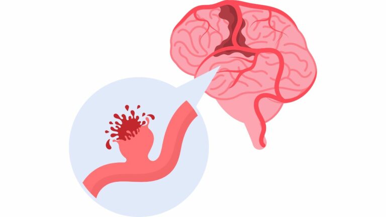
There are many causes for the above, like, head injury, stroke, brain haemorrhage, metabolic disorders, intoxication, or prolonged hypoxia.

Treating such comatose patients before, during and after surgery is the subject of this blog. It is known as perioperative management.

What is Perioperative management?

It is the comprehensive care of patients before, during, and after a surgery. Let us break it into three parts:

Preoperative:

It is first checked if the patient is ‘fit’ to be operated. It will mean a long list of blood tests, X-Ray, CT, MRI, Echo of the heart, give anaesthesia, checking for high BP, diabetes and existing infection. Then a physician gives his fitness. Many a times in brain surgery, the patient may not be ‘fit’ to be operated, yet surgery is done because the very condition which makes the patient ‘unfit for surgery’ will be cured with surgery. Being comatose is one such condition where the physician may say that the patient is ‘unfit for surgery’ but the neurosurgeon will treat that very condition with surgery. Patient is not given food, either orally or by a tube. This is called NBM or Nil By Mouth. Lastly, the patient is checked by the anaesthetist, ideally a day before and just before surgery. Surgery is undertaken only if the patient ‘passes’ all the above tests and criteria.

Intraoperative:

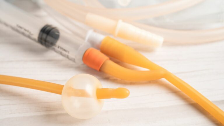
Means during surgery. Various lines [actually they are tubes] like intravenous, central venous, Foley’s catheter, ICP monitoring line, arterial line etc may be introduced to deliver medicines, remove urine, measure pressure or take blood samples will be inserted by the Anaesthesia team. Then the anaesthetist ‘induces’ the patient, meaning makes the patient unconscious and pain-free.  The patient’s vital signs like BP, Pulse, Respiratory rate per minute, oxygen saturation by SpO2 and temperature are monitored. Any adverse changes in the vital signs will be immediately treated by the anaesthetist in consultation with the neurosurgeon. If there is unexpected blood loss, blood transfusions may be given.

Postoperative:

This is the time to help the patient recover from his surgical wounds and also manage his/her general condition. Recovery from anaesthesia may take time in a comatose patient. Hence such patients need to be monitored very carefully. Their GCS [explained below] monitoring becomes crucial, along with their intake, output, their antibiotics, their blood coagulation, their intracranial pressure, their vital signs and their general conditions. A comatose patient needs to be moved to prevent pressure sores, but the neurosurgeon may refuse permission to move him, and such challenging situations are handled with frequent discussions amongst the super specialists.

All of the above together are called perioperative patient management.

GCS

The GCS score is an important tool for neurological monitoring. The Glasgow Coma Scale (GCS) is most commonly used in emergency rooms and ICUs by trained staff. The GCS is used to predict survival in hospitalized patients. It is the sum of the scores for three components: eye opening, verbal response, and motor response. The highest score is 15, and the lowest is 3. A score of 15 indicates that the patient is fully awake, responsive, and has no memory or thinking problems. The lower the score, the deeper the coma is.

The Glasgow Coma Scale (GCS) score of a comatose patient is usually 8 or less.

Critical Care in the NICU

Intensive care management in the perioperative period revolves around vigilant neurological monitoring and prompt intervention. Continuous assessment of neurological status, including pupil reactivity, motor response and GCS. The above guide in decision-making about sedation levels, ventilator settings, and pharmacological interventions to optimize cerebral perfusion and prevent secondary insults to the body.

In Summary

Perioperative management of comatose patients in the NICU demands a comprehensive and multidisciplinary approach which includes neurosurgical interventions, anaesthesia, critical care medicine, and Neuro-rehabilitation.

Many people are afraid of ICUs. Actually ICUs in good hospitals are the best places for curing patients. If a patient is intubated and is on ventilator, it doesn’t mean that the person is going to die. Intensive care specialists are stabilising the patient so that they can gradually reduce the air given by the ventilator, and the patient breathes on his/her own. This is done by ‘weaning’, where the oxygen supply to the patient is gradually reduced. This is how surgical comatose patients are treated in a good hospital.

Cerebral Vascular Aneurysms- Symptoms and Treatment Options

Cerebral Vascular Aneurysms- Symptoms and Treatment Options

Cerebral Vascular Aneurysms- Symptoms and Treatment Options

Cerebral vascular aneurysms, also known as brain aneurysms, are a serious medical condition characterized by a weakened area in the wall of a blood vessel within the brain. This weakening can lead to a bulging or ballooning of the blood vessel, which can potentially rupture and cause life-threatening complications. In this blog, we will explore the symptoms, treatment options, risk factors, diagnosis, prevention tips, and recovery process associated with cerebral vascular aneurysms.
The occurrence rate of cerebral vascular aneurysms can vary depending on several factors, including age, gender, genetics, and lifestyle factors. However, studies have estimated that cerebral aneurysms occur in approximately 2-3% of the population worldwide.
It’s important to note that not all cerebral aneurysms will cause symptoms or complications, and many may go undetected throughout a person’s life. Additionally, the risk of developing a cerebral aneurysm may increase with age, particularly after the age of 40.
Certain risk factors, such as a family history of cerebral aneurysms, smoking, high blood pressure, and certain genetic conditions, can also increase the likelihood of developing an aneurysm.
Overall, while cerebral aneurysms are relatively common, the majority of them do not rupture or cause significant health problems. However, for those individuals who do experience a ruptured aneurysm, it can be a life-threatening medical emergency requiring immediate treatment.
Let us see in detail the definition, symptoms and treatment of cerebral vascular aneurysms.
By the way, aneurysms occur only in arteries. So it is not really necessary to call cerebral aneurysms as cerebral vascular aneurysms. Vascular means relating to the blood vessels. Cerebral aneurysms are also called brain aneurysms.

By Dr.Ravindra Patil

Table of Contents

1. Cerebral Vascular Aneurysms:

Cerebral vascular aneurysms, often referred to simply as brain aneurysms, occur when a weakened area in the blood vessel wall within the brain leads to a bulging or ballooning of the vessel. These aneurysms can develop anywhere in the brain and vary in size and shape. The worst thing that can occur in an aneurysm is that it can rupture. That causes cerebral haemorrhage and may lead to serious medical condition called cerebrovascular stroke or brain stroke.

2. Brain Aneurysm Symptoms:

Brain aneurysms often do not cause symptoms until they rupture. However, when symptoms do occur, they may include severe headaches, nausea, vomiting, neck pain, blurred or double vision, sensitivity to light, and loss of consciousness. In the case of a ruptured aneurysm, symptoms can be severe and may include a sudden, severe headache, nausea, vomiting, stiff neck, confusion, seizures, and loss of consciousness.

3. Aneurysm Treatment Options:

Treatment options for cerebral vascular aneurysms depend on various factors, including the size and location of the aneurysm, as well as the individual’s overall health. Treatment options may include observation, medication to control symptoms and prevent complications, surgical clipping to prevent rupture, endovascular coiling to seal off the aneurysm, or a combination of these approaches. Remember, as cerebral aneurysms are inside the brain, any surgery or intervention to clip them or repair them may involve brain surgery, which itself is a very dangerous and risky procedure.

4. Ruptured Brain Aneurysm:

A ruptured brain aneurysm occurs when the weakened area in the blood vessel wall gives way, causing bleeding into the surrounding brain tissue. This is a medical emergency that requires immediate treatment to prevent further damage and complications. The intracranial pressure [ICP] of such patients will rise and this will lead to many symptoms like

  • Visual disturbances: blurred vision, double vision (diplopia), or temporary vision loss.
  • Changes in mental status: confusion, irritability, drowsiness, or difficulty concentrating.
  • Nausea and vomiting often occur in conjunction with headaches.
  • Raised ICP can sometimes trigger seizures, which are abnormal electrical discharges in the brain that can cause convulsions, loss of consciousness, and other neurological symptoms.
  • Papilledema: swelling of the optic disc due to increased pressure within the skull.
  • In some cases, there may be alterations in vital signs such as increased blood pressure, changes in heart rate, or irregular breathing patterns.

5. Cerebral Aneurysm Signs:

Signs of a cerebral aneurysm include sudden and severe headaches, nausea, vomiting, neck pain or stiffness, blurred or double vision, sensitivity to light, and loss of consciousness. These signs may indicate a ruptured aneurysm and require prompt medical attention.

6. Aneurysm Risk Factors:

Several factors can increase the risk of developing a cerebral vascular aneurysm, including a family history of aneurysms, high blood pressure, smoking, excessive alcohol consumption, drug abuse (particularly cocaine), and certain genetic conditions such as polycystic kidney disease and connective tissue disorders.

7. Cerebral Aneurysm Diagnosis:

Diagnosing a cerebral vascular aneurysm typically involves a combination of imaging tests, such as magnetic resonance imaging (MRI), computed tomography (CT) scans, cerebral angiography, and lumbar puncture. These tests help determine the size, location, and severity of the aneurysm.

8. Aneurysm Prevention Tips:

While it may not be possible to prevent all cerebral vascular aneurysms, certain lifestyle modifications can help reduce the risk. These include maintaining a healthy blood pressure, avoiding smoking and excessive alcohol consumption, managing stress, and staying physically active.

9. Brain Aneurysm Awareness:

Raising awareness about the signs, symptoms, and risk factors of cerebral vascular aneurysms is crucial for early detection and intervention. Education efforts can help individuals recognize the warning signs and seek prompt medical attention if necessary.

10. Aneurysm Surgery Recovery:

Recovery from aneurysm surgery depends on several factors, including the type of procedure performed, the size and location of the aneurysm, and the individual’s overall health. In general, recovery may involve a hospital stay, followed by a period of rest and rehabilitation to regain strength and function.

11. Cerebral Haemorrhage Symptoms:

Cerebral haemorrhage may result from a cerebral aneurysm. Symptoms of cerebral haemorrhage include sudden and severe headaches, nausea, vomiting, weakness or numbness on one side of the body, difficulty speaking or understanding speech, and loss of consciousness. Immediate medical attention is required in the event of a cerebral haemorrhage.

12. Intracranial Aneurysm Treatment:

Treatment for intracranial aneurysms, which occur within the skull, may include surgical clipping, endovascular coiling, or flow diversion techniques to prevent rupture and reduce the risk of complications.

13. Aneurysm Screening Guidelines:

Screening guidelines for cerebral vascular aneurysms may vary depending on individual risk factors and medical history. However, individuals with a family history of aneurysms or certain risk factors may benefit from regular screening and monitoring.

14. Aneurysm Rupture Complications:

Complications of aneurysm rupture can be severe and may include cerebral haemorrhage, brain stroke, brain damage, neurological deficits, and even death. Prompt diagnosis and treatment are essential to minimize the risk of complications and improve outcomes.

Aneurysm rupture is usually a life threatening event. The possibility of the survival of the patient depends only on whether the best medical or surgical treatment is available and that too in a short period of time.

15. Aneurysm Embolization Procedure:

Aneurysm embolization is a minimally invasive procedure that involves inserting a catheter into the blood vessel and using coils, glue, or other materials to seal off the aneurysm and prevent further bleeding. This procedure is often used as an alternative to surgical clipping for certain types of aneurysms.

To conclude, cerebral vascular aneurysms are a serious medical condition that requires prompt diagnosis and treatment to prevent complications such as rupture and cerebral haemorrhage.

Brain Tumour Diet and Nutrition

Brain Tumour Diet and Nutrition

Brain Tumour Diet and Nutrition

By Dr.Ravindra Patil

Table of Contents

Supporting health during treatment

During any illness, a proper diet is indeed necessary. What foods and beverages are considered ‘bad’ even during good health should obviously be avoided. This blog aims to search out for the best diet for a patient undergoing brain tumour treatment.

Is there a Brain Tumour Diet?

Are there nutritional guidelines for brain tumour patients?

That is the first question that comes to mind. Brain tumour is a serious condition and requires treatment. Is there a requirement of special nutrition during brain tumour treatment?

There isn’t a specific “brain tumour diet” that universally applies to all individuals with brain tumours. However, a balanced and nutritious diet is generally recommended to support overall health and well-being, which can be particularly important for individuals undergoing treatment for brain tumours.

Here are some Brain tumour nutrition tips

  1. Balanced Nutrition: Focus on consuming a variety of nutrient-dense foods including fruits, vegetables, whole grains, lean proteins, and healthy fats. All this is healthy eating for brain tumour patients.
  2. Hydration: Stay well-hydrated by drinking plenty of water throughout the day. Proper hydration is essential for overall health and can help manage side effects of treatment such as fatigue and constipation.
  3. Limit Processed Foods: Minimize consumption of processed foods, sugary snacks, and beverages high in added sugars. These foods offer little nutritional value and may contribute to inflammation and other health issues. This will not be a good diet for brain tumour patients.
  4. Healthy Fats: Incorporate sources of healthy fats such as avocados, nuts, seeds, and fatty fish (like salmon and tuna) into your diet. Healthy fats can support brain health and may have anti-inflammatory properties. They will give nutritional support for brain tumour treatment.
  5. Fibre-Rich Foods: Include plenty of fibre-rich foods such as whole grains, fruits, vegetables, and legumes to support digestion and overall gut health.
  6. Moderate Caffeine and Alcohol: Limit intake of caffeine and alcohol, as these substances can affect hydration levels and may interact with medications. These are not good foods for brain tumour patients.
  7. Consider Individual Needs: Depending on your specific situation, you may have dietary restrictions or requirements due to treatment side effects or other health conditions. It’s important to work with a dietician or family doctor who can provide personalized dietary guidance.

Always consult with your healthcare team before making significant changes to your diet, especially if you’re undergoing treatment for a brain tumour. They can offer tailored advice based on your individual needs and medical history.
Supporting health during brain tumour treatment with diet is not a new thing. As diet is a part and parcel of our very survival, it must be ideal. Besides every major illness has a diet plan. We must have a brain tumour diet plan. This blog has given general guidelines for planning a diet for brain tumour patients.

Managing side effects of brain tumour treatment

Brain tumour will always be treated by either surgery, chemotherapy or radiation therapy. Taking a diet which will obviate the side effects of brain tumour must also be planned. Let us first see the various side effects of brain tumour therapies and how brain tumour diet and nutrition will help.

Side effects of brain tumour surgery

These side effects will be similar to the side effects of any surgery. a general high protein nutritious diet full of natural vitamins will help the brain surgery wound, which may be a craniotomy, heal fast.  

Side effects of brain tumour chemotherapy

Some of the side effects of brain tumour chemotherapy, but not all, can be reduced with proper nutrition.

  1. Nausea and Vomiting: One of the most common side effects of chemotherapy is nausea and vomiting. Lots of fluids with electrolytes must be given, eg coconut water and fruit juices. However, the other diseases of the patients must be considered.
  2. Fatigue: Chemotherapy can cause fatigue, which may persist throughout the treatment period and even after treatment ends. Sweet foods will help relieve fatigue, only if the patient doesn’t have diabetes.
  3. Changes in Appetite: Chemotherapy can affect appetite, leading to changes in taste or a decrease in appetite. Tangy citrus fruits may increase the patients’ appetite.
  4. Peripheral Neuropathy: Some chemotherapy drugs can cause damage to the nerves in the hands and feet, leading to symptoms such as tingling, numbness, or pain. Giving vitamin rich foods will help reduce these symptoms.

Side effects of brain tumour radiation therapy

Radiation therapy, like chemotherapy, is a common treatment for brain tumours. It uses high-energy rays to target and destroy cancer cells while minimizing damage to healthy tissue. While radiation therapy can be effective in treating brain tumours, it can also lead to various side effects. Proper nutrition will help relieve the radiation therapy symptoms somewhat.

  1. Fatigue: Many patients undergoing radiation therapy experience fatigue, which can range from mild to severe. Rest and high calorie easily digestible food can help reduce fatigue.
  2. Skin Changes: The skin in the treated area may become dry, itchy, or sensitive. Some patients may develop a rash or changes in skin pigmentation. Vitamin rich food may help.
  3. Nausea and Vomiting: Radiation therapy directed at the brain can sometimes cause nausea and vomiting, particularly if the treatment area includes the area of the brain responsible for regulating nausea and vomiting. Plenty of fluids orally with anti-vomiting medicines help.

There are many more side effects of radiation therapy and sadly all of them cannot be managed by changes in nutrition.

So what should be the diet of a brain tumour patient? The following text gives a general guideline.

Diet Chart for Brain Tumour Patients

A diet chart for brain tumour patients is a list of foods that are usually healthy for people with brain tumours. The patients cannot eat foods that have been processed or contain artificial colours or flavours because these chemicals cause the tumour to grow faster. The following items are beneficial.

Broccoli:

Broccoli has been found to have anti-cancer properties and it also helps in reducing the risk of developing cancer. It contains vitamin C, vitamin B6, folate, calcium, magnesium, phosphorus and potassium.

Garlic:

Garlic is a commonly eaten food that has been found to have anti-cancerous properties.

Turmeric:

Turmeric is a spice that has been used in Indian cuisine for centuries. It has medicinal properties.  

Ginger:

Ginger is a food that is in use for centuries to help with the symptoms of nausea and vomiting. Now, scientists have found that ginger can also help with brain tumours.

Chicken Broths:

Chicken broths are an excellent choice for those who need to eat a high-calorie diet. They contain a lot of protein, nutrients and minerals that help in fighting cancer.

Nuts:

Nuts are a food that is rich in antioxidants and omega-3 fats. They are also a good source of protein, fibre, and healthy fats.

Eggs:

Eggs are a great food to include in a diet of brain tumour patients. They have nutrients that help with brain health and they can be made into many different dishes.

Other good foods

To be included in the nutritional support for brain tumour treatment are:

  1. Leafy Vegetables
  2. Milk
  3. Pineapple
  4. Tomatoes
  5. Citrus Fruits

Spine Tumour Awareness: Facts, Myths and Patient Resources

Spine Tumour Awareness: Facts, Myths and Patient Resources

Spine Tumour Awareness: Facts, Myths and Patient Resources

By Dr.Ravindra Patil

Spine tumours are a big problem unless properly treated. This blog aims at dispelling myths, sharing facts, connecting patients with resources and shedding light on spine tumours

Table of Contents

Introduction:

In the realm of oncology, spinal tumours often remain overshadowed by more widely recognized cancers. Yet, their impact on individuals’ lives can be profound, necessitating greater awareness, understanding, and support systems. In this blog, we delve into the intricate landscape of spine tumours, unravelling facts from myths, and providing valuable resources for patients and caregivers.

Spine Tumour Awareness:

Despite their prevalence, most of the general population is not much aware about spine tumours. It is only after someone close in the family suffers from a spine tumour that people suddenly become aware of this condition. Educating communities about their existence, symptoms, and available resources is paramount in enhancing early detection and treatment outcomes.

Spinal Tumour Facts:

Misconceptions surrounding spine tumours can hinder timely diagnosis and appropriate care. Addressing common myths, such as the belief that all back pain is benign or that spine tumours are untreatable, is crucial in dispelling fear and promoting proactive healthcare seeking behaviours.

Surgical navigation in spine tumours has revolutionised the treatment of spine tumours.

Spinal Tumour Patient Resources:

Navigating the complexities of spinal tumour diagnosis and treatment can be overwhelming for patients and their families. Fortunately, numerous resources, including online support groups, informational websites, and patient advocacy organizations, offer invaluable guidance and emotional support throughout the journey.

Many institutes of medical excellence offer information about spine tumours and their treatment in their websites and blogs.

Spinal Tumour Information:

Access to accurate and reliable information empowers patients to make informed decisions about their healthcare. From understanding different tumour types to exploring treatment options and potential side effects, comprehensive information fosters a sense of control and confidence amongst patients and their family on a background of uncertainty.

Spine Tumour Support:

The journey of battling a spine tumour can be arduous, both physically and emotionally. Establishing a robust support network comprising healthcare professionals, family, friends, and fellow survivors can provide invaluable encouragement and solidarity.

Suffering any ailment is bad, suffering it alone is worse. Spine tumour patients sharing their information helps them tremendously.

Spine Tumour Diagnosis:

Early diagnosis is pivotal in improving treatment outcomes for spine tumours. From imaging studies like MRI and CT scans to minimally invasive biopsy techniques, prompt and accurate diagnosis lays the groundwork for tailored treatment plans.

MRI scan and CT scan are the cornerstones of spine tumour diagnosis.

Spinal Tumour Treatment Options:

Treatment strategies for spine tumours encompass a multidisciplinary approach, including surgery, radiation therapy, chemotherapy, and targeted therapies. The choice of treatment depends on factors such as tumour type, location, and overall health status of the patient.

Spinal Tumour Prevention:

While some risk factors for spine tumours, such as genetic predisposition, are beyond our control, adopting a healthy lifestyle and practicing proper body mechanics can potentially reduce the risk of developing certain types of spinal neoplasms.

As a matter of fact, this applies for health risk factors for all diseases. Live a healthy life, take proper exercise and eat healthy food are important in preventing most diseases.

Spine Tumour Research:

Ongoing research on spine tumours is aimed at unravelling the underlying mechanisms which cause spine tumour development and progression. From innovative treatment modalities to predictive biomarkers, research initiatives aim to improve outcomes and quality of life for affected individuals.

Spinal Tumour Specialists:

Seeking care from surgeons with expertise in managing spine tumours is essential for optimizing treatment outcomes. Spinal oncologists, neurosurgeons, radiation oncologists, and other specialists collaborate to tailor individualized treatment plans.

Orthopaedic surgeons can also contribute in the treatment of spine tumours.

Spine Tumour Support Groups:

The concept of support groups has not flourished much in India. In countries like USA, UK, Canada and Australia, nearly every ailment will have a support groups. What do support groups do? They offer a platform for interaction between patients and between patients and medical experts. Participation in support groups offers a platform for individuals affected by spine tumours to share experiences, exchange coping strategies, and gather emotional support from peers who truly understand the challenges they face.

Spinal Tumour Awareness Month:

Designating a specific month for spine tumour awareness serves as a rallying point for advocacy efforts, public education campaigns, and fundraising initiatives aimed at advancing research and improving patient care. Brain tumour awareness month is May of every year. Bone cancer awareness month is July. At present spinal tumour awareness month has not been designated.

Spine Tumour Surgery:

Surgical intervention plays a pivotal role in the management of many spine tumours, aiming to achieve maximal tumour resection while preserving neurological function and spinal stability. Advances in surgical techniques and perioperative care continue to enhance outcomes and minimize complications.

Use of navigation surgery in spine tumours results in much better outcomes.

Spinal Tumour Rehabilitation:

Postoperative rehabilitation is integral in restoring mobility, function, and quality of life following spine tumour surgery. Physical therapy, occupational therapy, and pain management strategies form the cornerstone of comprehensive rehabilitation programs.

Spine Tumour Survivor Stories:

Sharing personal narratives of resilience and triumph over spine tumours instils hope and inspiration in fellow patients and hospital staff. Survivor stories not only highlight the challenges encountered along the way but also underscore the resilience of the human spirit.

Spinal Tumour Risk Factors:

While the causes of many spine tumours remains elusive, certain risk factors, such as genetic predisposition, exposure to ionizing radiation, and immune system disorders, have been implicated in spine tumours. Identifying and mitigating modifiable risk factors can potentially reduce the incidence of these neoplasms.

Spine Tumour Care:

Comprehensive and compassionate care extends beyond medical interventions to encompass psychosocial support, symptom management, and end-of-life planning. Holistic care addresses the diverse needs of patients and their families throughout the continuum of illness.

Spinal Tumour Education:

Continuing education initiatives targeting healthcare professionals, patients, and the general public play a pivotal role in enhancing awareness, understanding, and access to quality care for individuals affected by spine tumours.

Conclusion:

By shedding light on spine tumours, debunking myths, disseminating accurate information, and fostering a supportive community, we can empower patients and caregivers to navigate the challenges of diagnosis, treatment, and survivorship with resilience and hope. Together, let us strive to raise awareness, promote early detection, and improve outcomes for individuals affected by these often overlooked yet impactful neoplasms.

Understanding Headaches: Causes, Types, Solutions

Understanding Headaches: Causes, Types, Solutions

Understanding Headaches: Causes, Types, Solutions

By Dr.Ravindra Patil

Headaches are incredibly common among human beings, with nearly everyone experiencing them at some point in their lives. In fact, headaches are one of the most prevalent health complaints worldwide. According to the World Health Organization (WHO), it is estimated that almost half of the adult population has experienced a headache at least once within the last year. Additionally, studies suggest that approximately 1 in 20 adults has a headache almost every day, with tension headaches and migraines being the most prevalent types.

Table of Contents

Causes, Types, Remedies, and Management Tips

Headaches can affect individuals of all ages, genders, and backgrounds, and their frequency and severity can vary widely from person to person. While many headaches are temporary and resolve on their own or with simple remedies, some individuals may experience chronic or recurrent headaches that significantly impact their daily functioning and quality of life.
Given their widespread occurrence and potential impact, headaches represent a significant public health concern, warranting attention, research, and effective management strategies to alleviate symptoms and improve overall well-being.

Introduction:

Headaches can be mild and fleeting or they can also be debilitating, impacting daily life and productivity. In this comprehensive blog, we will delve into the various aspects of headaches, including their causes, types, remedies, and management techniques to help you find relief and regain control over your headache management.

Headache Causes:

Headaches can stem from a myriad of factors, ranging from lifestyle choices to underlying health conditions. Stress, lack of sleep, dehydration, poor posture, and certain foods or beverages are common triggers for headaches. Additionally, underlying health issues such as sinus infections, hormonal imbalances, and even neurological disorders can contribute to recurrent headaches. Understanding the root cause of your headaches is crucial in effectively managing and preventing them. There are literally innumerable headache causes.

Types of Headaches:

Headaches are not one-size-fits-all; they manifest in various forms, each with its own distinct characteristics and triggers. Some common types of headaches include tension headaches, migraines, cluster headaches, and sinus headaches. Tension headaches typically result from muscle tension in the head and neck, while migraines are often characterized by intense, throbbing pain and other symptoms such as nausea and sensitivity to light and sound. Cluster headaches, though less common, are known for their excruciating pain localized around one eye, often occurring in clusters or cycles. Sinus headaches, on the other hand, stem from sinus congestion or inflammation. Recognizing the type of headache that you are experiencing is the first step towards finding appropriate relief.

Migraine Remedies:

Migraines can be particularly debilitating, significantly impacting quality of life. While there is no cure for migraines, various remedies can help alleviate symptoms and reduce the frequency and severity of attacks. Over-the-counter pain relievers, such as ibuprofen or acetaminophen, may provide relief for mild to moderate migraines. For more severe migraines, prescription medications specifically designed to target migraines, such as triptans or ergots, may be necessary. Additionally, lifestyle modifications, including stress management techniques, regular exercise, adequate sleep, and dietary adjustments, can play a significant role in migraine management.

Headache Relief Techniques:

When a headache strikes, finding quick relief becomes a top priority. Several techniques can help alleviate headache symptoms and promote relaxation. Applying cold or warm compresses to the head or neck area, practicing deep breathing exercises, gentle stretching, or engaging in relaxation techniques such as meditation or yoga can offer relief from headache discomfort. Experimenting with different techniques can help identify what works best for you in managing headaches effectively.

Preventing Tension Headaches:

Preventing tension headaches often involves adopting healthy lifestyle habits and managing stress effectively. Regular exercise, proper hydration, maintaining good posture, and incorporating stress-reduction techniques into your daily routine can help prevent tension headaches from occurring. Additionally, taking frequent breaks from prolonged periods of screen time or desk work and practicing relaxation exercises can alleviate muscle tension and reduce the likelihood of tension headaches.

Natural Headache Remedies:

In addition to conventional treatments, many people find relief from headaches through natural remedies. Herbal supplements such as feverfew or butterbur, aromatherapy with essential oils like peppermint or lavender, and acupuncture are among the natural approaches that some individuals find effective in managing headaches. However, it’s essential to consult with a healthcare professional before trying any new remedies, especially if you have underlying health conditions or are taking medications.

Headache Management Tips:

Managing headaches effectively often requires a multifaceted approach tailored to individual needs. Keeping a headache diary to track triggers and patterns, practicing stress management techniques, maintaining a regular sleep schedule, staying hydrated, and avoiding known triggers can help minimize the frequency and severity of headaches. Additionally, seeking support from healthcare professionals, such as neurologists or headache specialists, can provide personalized guidance and treatment options for more severe or persistent headaches.

Cluster Headache Treatment:

Cluster headaches, though rare, can be intensely painful and disruptive. Treatment for cluster headaches often involves a combination of medications to alleviate symptoms during an attack and prevent future episodes. Oxygen therapy, triptans, calcium channel blockers, and corticosteroids are commonly prescribed to manage cluster headaches effectively. In some cases, nerve stimulation techniques or surgical interventions may be considered for severe or refractory cases of cluster headaches.

Sinus Headache Relief:

Sinus headaches, typically accompanied by sinus congestion or pressure, can be relieved through various approaches aimed at reducing inflammation and promoting sinus drainage. Over-the-counter decongestants, saline nasal sprays, steam inhalation, and warm compresses applied to the sinus areas can help alleviate sinus headache symptoms. Additionally, addressing underlying sinus issues, such as allergies or infections, is essential in preventing recurrent sinus headaches.

Hormonal Headache Remedies:

Fluctuations in hormone levels, particularly in women, can trigger hormonal headaches, often associated with menstruation, pregnancy, or menopause. Hormonal contraceptives or hormone replacement therapy may help regulate hormone levels and reduce the frequency and severity of hormonal headaches in some individuals. However, it’s crucial to consult with a healthcare provider to determine the most appropriate treatment approach based on individual health needs and considerations.

Brain Tumour Headaches

Yes, brain tumours can indeed cause headaches. Headaches caused by brain tumours can vary widely in terms of severity, frequency, and other characteristics. These headaches are often described as persistent, dull, and progressively worsening over time. However, it’s essential to note that not all headaches are indicative of a brain tumour, and most headaches are unrelated to serious medical conditions.

The headache associated with a brain tumour may result from increased pressure within the skull (intracranial pressure) due to the tumour’s growth, compression of surrounding structures, or irritation of pain-sensitive structures in the brain and its coverings.

However, it’s important to emphasize that the vast majority of headaches are not caused by brain tumours but rather by more common and benign factors mentioned above.

Conclusion:

Headaches can significantly impact quality of life, but with the right knowledge and strategies, they can be effectively managed and prevented. By understanding the causes, types, remedies, and management techniques discussed in this guide, you can empower yourself to take control of your headache symptoms and lead a more comfortable, headache-free life. Remember, finding the most effective approach may require patience and experimentation, so don’t hesitate to seek guidance from healthcare professionals to develop a personalized headache management plan tailored to your needs.

Coping with Treatment: Brain Tumour Patient Care Tips

Enduring treatment for a brain tumour can be physically and emotionally draining. Managing side effects, maintaining good nutrition, and staying hydrated are crucial aspects of care.

Living Well Beyond Diagnosis: Brain Tumour Coping Strategies

Living well with a brain tumour involves looking beyond the diagnosis and focusing on aspects of life that bring joy and fulfilment. Engaging in hobbies, spending time with loved ones, and setting realistic goals contribute to a positive mind set, fostering resilience in the face of adversity.

Community Support: Emotional Support for Brain Tumour Patients

Connecting with other brain tumour patients through local or online support groups can provide a sense of community and understanding. Sharing experiences, tips, and resources creates a supportive environment where individuals feel heard and validated. Brain tumour emotional wellness is a state where the patient accepts the situation and is firmly facing it.

Conclusion:

So, as can be understood from the above, understanding a brain tumour diagnosis is a multifaceted process that involves medical understanding, treatment decisions, and emotional considerations. By actively engaging with doctors and counsellors, seeking support from loved ones, and educating oneself about the diagnosis, individuals can navigate this challenging terrain with a sense of empowerment and resilience. Remember, each person’s journey is unique, and a well-informed and supported approach can make a significant difference in facing the complexities of living with a brain tumour.

Living with a brain tumour: Coping Strategies and Emotional Support

Living with a brain tumour: Coping Strategies and Emotional Support

Living with a brain tumour: Coping Strategies and Emotional Support

By Dr.Ravindra Patil

Introduction:

Receiving a diagnosis of a brain tumour is an overwhelming and life-altering experience. Beyond the physical challenges, individuals must navigate the emotional and mental toll that comes with such a diagnosis. In this blog, we’ll explore coping strategies, emotional support, and practical tips for living well with a brain tumour. Remember, Living with brain tumour is not easy. Living well with brain tumour diagnosis is indeed tough.

Table of Contents

Understanding the Diagnosis: Coping with Brain Tumour Diagnosis

Knowing that you have a brain tumour may seem to be a devastating news, too difficult to deal with. But at this time, it is crucial to take the time to understand the diagnosis, treatment options, and potential impacts on daily life. This knowledge empowers patients to actively participate in their care, reducing anxiety and uncertainty.

Knowing about the possibility of loss of functions and death is also advisable. However, it will depend on the personality of the patient, his education, his need to his/her family and many more factors.

A brain tumour is a life-altering moment that brings forth a wave of emotions, questions, and uncertainties. Understanding the diagnosis is a crucial step in empowering individuals to actively participate in their care and navigate the challenges that lie ahead. Here, we delve into the various aspects of comprehending a brain tumour diagnosis.

Let us first have a ‘Medical Understanding’, which means how bad is the tumour and how badly is it going to affect your life. Following fact cover these aspects.

Type and Grade of Tumour:

It is essential to understand the specific type of brain tumour diagnosed. Tumours can be benign (non-cancerous) or malignant (cancerous). Additionally, they are classified based on their location within the brain and the type of cells involved. The grade of the tumour indicates how quickly it may grow and spread. Grades range from type I (slow-growing) to type IV (fast-growing and malignant).

Diagnostic Imaging:

Techniques such as magnetic resonance imaging (MRI) and computed tomography (CT) scans are often employed to visualize and locate the tumour. These images help healthcare professionals determine the size, location, and characteristics of the tumour.

A positron emission tomography (PET) scan is a type of imaging test. It uses a radioactive substance called a tracer to look for disease in the body. A PET scan shows how organs and tissues are working. A tumour will be properly visualised in PET scan not only for its size but also to understand if the tissue involved is functional.

Biopsy Results:

A biopsy may be conducted to extract a small sample of the tumour for examination under a microscope. This procedure helps determine the tumour’s composition and aids in developing an effective treatment plan. Biopsy is very important part of dealing with a tumour.

After the diagnosis, let us understand what the treatment options in a brain tumour are.

Surgery:

Depending on the tumour’s location and type, surgery may be recommended to remove all or part of the tumour. The feasibility of surgery is influenced by factors such as the tumour’s size, grade, and proximity to critical brain structures.

Radiation Therapy:

Radiation therapy utilizes high-energy rays to target and shrink tumours. It may be employed as the primary treatment or in conjunction with surgery or chemotherapy.

Chemotherapy:

Chemotherapy involves the use of drugs to kill or slow the growth of cancer cells. It is administered orally or intravenously and may be part of the treatment plan for certain types of brain tumours.

Now let us see what might be the potential Impact on the daily Life of a brain tumour patient.

Coping with brain tumour treatment is not easy as for example coping with the treatment for fever or cough. The above treatments like chemotherapy or radiation therapy do have side effects. Courage to bear anything must be the motto of the brain tumour patient.

Symptoms and Side Effects:

Understanding the potential symptoms and side effects of both the tumour and its treatment is crucial. This awareness enables individuals to proactively manage and communicate any changes in their health to their healthcare team.

Symptoms will vary from pain to loss of brain function and resulting loss of crucial skills like speech, movement or loss of eyesight.

Long-Term Care and Monitoring:

Brain tumour survivors often require long-term care and monitoring. Regular follow-up appointments, imaging studies, and other diagnostic tests help doctors track the individual’s progress and address any emerging issues promptly.

Emotional and Supportive Aspects are important. Courage to face whatever happens is the best weapon for mental peace. If a patient is emotionally broken, he/she must be given emotional support to help him/her accept her condition.

Close Communications with doctors:

Establishing open communication with healthcare providers is paramount. This ensures that individuals are well-informed about their diagnosis, treatment options, and potential challenges. And the healthcare providers will dispel doubts and misunderstandings and myths.

Involving Loved Ones:

Sharing the diagnosis with family and friends creates a support system that can provide emotional strength. Loved ones can offer assistance, understanding, and companionship throughout the journey. Hiding such news from close family members ‘to protect them’ is not an advisable strategy.

Seek a Second Opinion:

It is not only acceptable but necessary to seek a second opinion to ensure that the diagnosis and recommended treatment plan are correct and will help the patient. Different perspectives can contribute valuable insights.

Coping Strategies for Daily Life: Managing Life with Brain Tumour

Living with a brain tumour often involves adapting to new challenges. Establishing a routine that accommodates both medical needs and personal well-being is essential. Integrating relaxation techniques, mindfulness, and gentle exercises can contribute to overall well-being.

Emotional Wellness: Support for Brain Tumour Patients

Emotional support is a cornerstone of coping with a brain tumour. Surrounding oneself with a strong support network, including family, friends, and healthcare professionals like doctors, nurses and counsellors, can provide comfort during difficult times. Seeking therapy or support groups tailored to brain tumour patients allows for shared experiences and emotional healing.

Coping with Treatment: Brain Tumour Patient Care Tips

Enduring treatment for a brain tumour can be physically and emotionally draining. Managing side effects, maintaining good nutrition, and staying hydrated are crucial aspects of care.

Living Well Beyond Diagnosis: Brain Tumour Coping Strategies

Living well with a brain tumour involves looking beyond the diagnosis and focusing on aspects of life that bring joy and fulfilment. Engaging in hobbies, spending time with loved ones, and setting realistic goals contribute to a positive mind set, fostering resilience in the face of adversity.

Community Support: Emotional Support for Brain Tumour Patients

Connecting with other brain tumour patients through local or online support groups can provide a sense of community and understanding. Sharing experiences, tips, and resources creates a supportive environment where individuals feel heard and validated. Brain tumour emotional wellness is a state where the patient accepts the situation and is firmly facing it.

Conclusion:

So, as can be understood from the above, understanding a brain tumour diagnosis is a multifaceted process that involves medical understanding, treatment decisions, and emotional considerations. By actively engaging with doctors and counsellors, seeking support from loved ones, and educating oneself about the diagnosis, individuals can navigate this challenging terrain with a sense of empowerment and resilience. Remember, each person’s journey is unique, and a well-informed and supported approach can make a significant difference in facing the complexities of living with a brain tumour.

4. Artificial Disc Replacement:

  • Procedure: Instead of fusing the vertebrae, artificial disc replacement involves removing the damaged disc and replacing it with an artificial disc designed to mimic natural spinal motion.
  • Navigation: Surgical navigation can assist in precise placement of the artificial disc, ensuring optimal function and reducing the risk of complications.


5. Percutaneous Discectomy:

  • Procedure: In percutaneous discectomy, a small tube is inserted through the skin to remove or shrink the herniated disc material, typically using a laser or other energy source.
  • Navigation: Surgical navigation can guide the placement of instruments and monitor the procedure, enhancing accuracy and safety.

Role of Surgical Navigation:

Surgical navigation systems use advanced imaging technology, such as intraoperative CT scans or fluoroscopy, to create real-time 3D maps of the patient’s anatomy during surgery. The benefits of surgical navigation in cervical spine surgery include:

  • Precision: Helps the surgeon accurately target the affected area, reducing the risk of damage to surrounding structures.
  • Safety: Enhances safety by providing real-time feedback on instrument placement and proximity to critical structures.
  • Minimally Invasive: Facilitates minimally invasive approaches by improving visualization and guidance during the procedure.

Surgical navigation is particularly valuable in cervical spine surgery, where precision is crucial due to the proximity of vital structures such as the spinal cord and major blood vessels. However, its use may vary depending on the specific surgical approach and the surgeon’s preferences and expertise.

Physical Therapy:

Targeted exercises and rehabilitation can help strengthen the neck muscles, improve posture, and alleviate symptoms. Physical therapy is a must along with or after every type of treatment for a cervical disc prolapse.

Conclusion:

Cervical disc prolapse can significantly impact one’s quality of life, but with timely intervention and proper management, individuals can find relief from symptoms and regain functionality. It is necessary to seek medical help as soon as possible.


Understanding Cervical Disc Prolapse : Causes, Symptoms, and Treatments

understanding-cervical-disc-prolapse-miraj

Understanding Cervical Disc Prolapse: Causes, Symptoms, and Treatments

By Dr.Ravindra Patil

Introduction:

The human spine is a marvel of engineering, providing structural support while allowing for a wide range of movements. However, sometimes, this intricate system can face challenges, leading to conditions such as cervical disc prolapse. In this blog post, we will delve into the causes, symptoms, and various treatment options for cervical disc prolapse, also known as cervical disc herniation or neck disc prolapse.

Table of Contents

Complexities of the human Cervical Spine

The human cervical spine, comprising the first seven vertebrae (C1 to C7), plays a crucial role in supporting the head, facilitating movement, and protecting the spinal cord. The complexities of the cervical spine include its unique anatomical features, curvatures, and susceptibility to various diseases or disabilities. Here are some key aspects to consider:

1. Curvature of the cervical spine:

The cervical spine has a Lordotic Curvature (backward) curvature, which helps balance the weight of the head and maintain stability.

2. Common Diseases and Disabilities of the cervical spine:

Rarely does a single condition affect the cervical spine. In most cases another condition is likely to coexist with a prolapsed disc. Rather, a prolapsed disc many be caused by some of the other diseases mentioned below.

  • Degenerative Disc Disease (DDD): Over time, wear and tear can lead to the deterioration of intervertebral discs.
  • Herniated Discs: The gel-like substance within the intervertebral disc may leak out, irritating nearby nerves and causing pain, weakness, or numbness. This is the same as Cervical Spine Disc Prolapse.
  • Cervical Spondylosis: This refers to the degeneration of discs and joints.
  • Cervical Radiculopathy: Compression or irritation of nerve roots in the cervical spine can result in symptoms such as pain, tingling, or weakness in the arms.
  • Traumatic Injuries leading to Cervical disc bulge or Neck disc prolapse
  • Inflammatory Conditions also may cause cervical disc bulge and cervical disc herniation or prolapse.
  • Congenital Anomalies

Understanding the complexities of the cervical spine and its diseases is crucial for understanding the Cervical Disc Prolapse. Proper treatment of Cervical Disc Prolapse is crucial, considering the spine’s vital role in overall body function and mobility.

Understanding Cervical Disc Prolapse:

Cervical disc prolapse occurs when the cushion-like discs between the vertebrae in the neck deteriorate or become damaged, resulting in the soft inner material pushing through the tough outer layer. This condition can be caused by various factors, and understanding them is crucial for effective management. In common slang it is called ‘neck disc herniation’.

Causes of Cervical Disc Prolapse:

Every cervical disc prolapse will be preceded by cervical disc protrusion. The causes of disc prolapse or Cervical disc protrusion are:

1. Age-related Degeneration:

The natural aging process contributes to the wear and tear of the cervical discs, making them more prone to prolapse over time. The disc may gradually prolapse, leading to cervical disc bulge, which in turn causes pain.

2. Trauma or Injury:

Accidents, falls, or other traumatic incidents can cause sudden and severe damage to the cervical discs, leading to prolapse.

3. Genetic Factors:

Genetic predisposition make some individuals more susceptible.

Common Symptoms:

Recognizing the symptoms of cervical disc prolapse is crucial for timely intervention. Some common indicators include:

1. Neck Pain:

Persistent pain in the neck region, radiating to the shoulders and arms.

2. Numbness and Tingling:

Sensations of numbness, tingling, or weakness in the arms or fingers.

3. Limited Range of Motion:

Difficulty in moving the neck and performing everyday activities.

4. Headaches:

Chronic headaches, especially the skull base.

Cervical disc treatment options:

Conservative Approaches:

Mild cases of cervical disc prolapse can be managed through conservative measures such as rest, physical therapy, and pain medications.

Epidural Injections:

Corticosteroid injections may be recommended to reduce inflammation and alleviate pain associated with cervical disc prolapse.

Surgical Interventions:

When conservative treatments fail, cervical disc surgery for cervical spine disc prolapse, commonly referred to as cervical disc herniation or cervical disc disease, is considered. The primary goals of surgery are to decompress the affected nerve roots or the spinal cord and to stabilize the spine if necessary. Here are some common surgical interventions for cervical disc prolapse, along with the role of surgical navigation:

1. Anterior Cervical Discectomy and Fusion (ACDF):

  • Procedure: ACDF involves removing the damaged or herniated disc from the front of the neck (anterior approach) and replacing it with a bone graft or an artificial disc. This aims to decompress the spinal cord or nerve roots and provide stability.
  • Navigation: Surgical navigation systems can assist in precisely locating the affected disc and guiding the surgeon during the procedure. This helps improve accuracy and reduce the risk of damage to adjacent structures.

2. Posterior Cervical Foraminotomy:

  • Procedure: In this approach, the surgeon removes a portion of the bone to widen the neural foramen, the opening through which nerve roots exit the spinal canal. This relieves pressure on the compressed nerve.
  • Navigation: Surgical navigation aids in identifying the correct location for the foraminotomy, ensuring precise removal of bone without compromising nearby structures.

3. Microdiscectomy:

  • Procedure: Microdiscectomy involves the removal of the herniated disc material through a small incision. This is a minimally invasive approach that aims to minimize tissue disruption.
  • Navigation: Surgical navigation systems can assist in targeting the specific location of the disc herniation, facilitating a more targeted and less invasive surgical approach.

4. Artificial Disc Replacement:

  • Procedure: Instead of fusing the vertebrae, artificial disc replacement involves removing the damaged disc and replacing it with an artificial disc designed to mimic natural spinal motion.
  • Navigation: Surgical navigation can assist in precise placement of the artificial disc, ensuring optimal function and reducing the risk of complications.


5. Percutaneous Discectomy:

  • Procedure: In percutaneous discectomy, a small tube is inserted through the skin to remove or shrink the herniated disc material, typically using a laser or other energy source.
  • Navigation: Surgical navigation can guide the placement of instruments and monitor the procedure, enhancing accuracy and safety.

Role of Surgical Navigation:

Surgical navigation systems use advanced imaging technology, such as intraoperative CT scans or fluoroscopy, to create real-time 3D maps of the patient’s anatomy during surgery. The benefits of surgical navigation in cervical spine surgery include:

  • Precision: Helps the surgeon accurately target the affected area, reducing the risk of damage to surrounding structures.
  • Safety: Enhances safety by providing real-time feedback on instrument placement and proximity to critical structures.
  • Minimally Invasive: Facilitates minimally invasive approaches by improving visualization and guidance during the procedure.

Surgical navigation is particularly valuable in cervical spine surgery, where precision is crucial due to the proximity of vital structures such as the spinal cord and major blood vessels. However, its use may vary depending on the specific surgical approach and the surgeon’s preferences and expertise.

Physical Therapy:

Targeted exercises and rehabilitation can help strengthen the neck muscles, improve posture, and alleviate symptoms. Physical therapy is a must along with or after every type of treatment for a cervical disc prolapse.

Conclusion:

Cervical disc prolapse can significantly impact one’s quality of life, but with timely intervention and proper management, individuals can find relief from symptoms and regain functionality. It is necessary to seek medical help as soon as possible.


Urinary Catheter Care at Home

Urinary (Foley’s) Catheter Care at Home

Urinary (Foley’s) Catheter Care at Home

By Dr.Ravindra Patil

After some surgeries or after a brain stroke, sometimes a patient is not able to control his/her urinary bladder. This leads to social problems and poor hygiene. So a urinary catheter, called Foley’s catheter is left inside the bladder with a urine bag attached. This information will help you care for your own or your patient’s urinary (Foley) catheter.

Depending on the situation, either the patient himself/herself can manage the Foley’s catheter and bag, or a care provider, either a nurse or hospital trained staff should do it.

You may feel that you should do it for your father or mother, but remember, caring for your close and loved relative involves privacy issues and aversion to the work you are doing. Do it only if you feel confident that you can do it. Nurses and hospital staff are trained to do it. Is it’s not difficult, you only have to follow precise steps.

Table of Contents

About your urinary (Foley) catheter

Foley catheter is a thin, flexible tube placed through your urethra (the small tube that carries urine from your bladder to outside your body) and into a urine bag. Your Foley catheter drains your urine. It’s held inside your bladder by a balloon filled with water.

How your Foley catheter works

Your urine goes from your bladder and through a drainage tube into a drainage collection bag. The drainage bag stays on your leg with a special tape.

How to take care of your Foley catheter

To take care of your Foley catheter, you will need to:

  • Clean your catheter every day.
  • Change your drainage bags. You will change your drainage bag 2 times a day.
  • Replace your drainage bags with new bags once a week. You should also change your drainage bag if it gets clogged or blocked.
  • Wash your drainage bags every day.
  • Drink 1 to 2 glasses of liquids every 2 hours while you’re awake to keep you hydrated.

You may see some blood or urine around where the catheter enters your body. This may happen when you’re walking or passing stool. This is normal if there’s urine draining into the drainage bag. If you do not have urine draining into the drainage bag, you must meet your doctor.

Penis or Vaginal care

Use a water-based lubricant to keep your penis or vagina opening from getting sore. Apply the gel where the catheter enters your body.

Keep your penis or vagina opening clean by taking a shower every day. This will help prevent infections when your Foley catheter is in place.

How to shower with your Foley catheter

  • You can shower while you have your catheter in place.
  • Do not take a tub bath until your catheter is removed. Taking a bath while you have your catheter in place puts you at risk for infections.
  • You may find it easier to shower in the morning before you change your bag to your leg bag.

How to clean your Foley catheter

You can clean your catheter while you’re in the shower. Follow these instructions.

  • Mild soap, such as Dove.
  • Wash your hands with soap and water or an alcohol-based hand sanitizer.
  • If you’re using an alcohol-based hand sanitizer, cover your hands with it. Rub them together until they’re dry.
  • Using mild soap and water, clean your penis or vagina.
  • If you have a penis, pull back your foreskin (the skin around the tip of your penis), if needed. Clean the area, including your penis.
  • If you have a vagina, separate your labia (the smaller folds of skin around your vaginal opening). Clean the area from front to back.
  • Clean the area where the catheter enters your body. This is called your urethra.
  • Clean the catheter from where it enters your body and then down, away from your body. Hold the catheter at the point it enters your body so that you do not put tension on it.
  • Rinse the area well and dry it gently.

When to change your drainage bags

You should change your drainage bag 2 times a day. Change it:

  • In the morning after you shower, change the bag.
  • At night before you go to bed, change the bag.
  • Replace your drainage bags with new bags once a week. You should also change your drainage bag if it gets clogged or blocked.

How to change your drainage bag

You will need:

  • A clean cloth (not one you’re using for bathing) or a 4 x 4 piece of gauze.
  • 2 alcohol pads.
  • Wash your hands with soap and water or an alcohol-based hand sanitizer thoroughly.
  • Empty the urine from the drainage bag into the toilet. Make sure the spout of the drainage bag never touches the side of the toilet or any emptying container. If it does, wipe it with an alcohol pad for 15 seconds.
  • Place the clean cloth or gauze under the connector to catch any leakage.
  • Pinch the catheter with your fingers and disconnect the used bag.
  • Wipe the end of the catheter with an alcohol pad.
  • Wipe the connector on the new bag with the second alcohol pad.
  • Connect the clean bag to the catheter and release your finger pinch. Make sure the catheter is tightly connected to the bag to keep it from opening or leaking.
  • Check all connections. Straighten any kinks or twists in the tubing.

How to take care of your drainage bags

  • Empty the leg bag into the toilet every 2 to 4 hours, as needed. You can do this through the spout at the bottom of the bag. Do not let the bag get completely full.
  • Do not lie down for longer than 2 hours while you’re wearing the leg bag. This can keep your urine from draining the way it should.

Caring for your drainage bag

  • Always keep the drainage bag below the level of your bladder.
  • When you go to sleep, hang your bag off the bed. You can do this by using a small trash can. Place a clean plastic bag inside the trash can. Hang your bag inside of the trash can.

Cleaning your drainage bags

Clean your bag every day. Follow these instructions.

You will need:

  • White vinegar.
  • Cool water.
  • Clean your hands with soap and water or an alcohol-based hand sanitizer.
  • Rinse the bag with cool water. Do not use hot water because it can damage the plastic.
  • To help get rid of the smell, fill the bag halfway with a mixture of 1part white vinegar and 3 parts water. Shake the bag and let it sit for 15 minutes.
  • Rinse the bag with cool water. Hang it up to dry.

How to prevent Foley catheter infections

Follow these guidelines to prevent getting infections while you have your catheter in place:

  • Keep the drainage bag below the level of your bladder.
  • Always keep your drainage bag off the floor.
  • Keep the catheter secured to your thigh to keep it from moving.
  • Do not lie on your catheter or block the flow of urine in the tubing.
  • Take a shower every day to keep the catheter clean.
  • Wash your hands before and after touching the catheter or bag.

Your catheter should be replaced about once a month, if it stops working, or if you have an infection.

Lumbar Disc Prolapse

Lumbar Disc Prolapse

Lumbar Disc Prolapse

By Dr.Ravindra Patil

Understanding, Managing, and Relieving Back Pain

Lumbar disc prolapse is a relatively common condition, affecting a significant portion of the population. Factors such as aging, genetics, and lifestyle contribute to its prevalence.

Table of Contents

Introduction

Lumbar disc prolapse, commonly known as a herniated disc, is a condition that can cause excruciating back pain and affect one’s overall spinal health. Understanding this condition, its causes, and treatment options is crucial for individuals seeking relief from the debilitating symptoms associated with disc herniation.

In mild cases of lumbar disc prolapse, individuals may experience minimal symptoms, often manifesting as localized back pain or discomfort. The pain might be intermittent and exacerbated by certain movements or positions. Mild numbness, tingling sensations, or a dull ache in the buttocks or legs may also occur. However, individuals with minimal symptoms usually maintain normal function and can perform daily activities without significant limitations. Conservative measures such as rest, over-the-counter pain medications, and lifestyle adjustments may be sufficient for managing these mild symptoms. It’s essential to note that even in cases of minimal symptoms, seeking medical attention is advisable for an accurate diagnosis and appropriate guidance on preventive measures to maintain spinal health and prevent the progression of disc prolapse symptoms.

Worst care scenarios of Lumbar disc prolapse is discussed at the end of this blog.

Understanding Lumbar Disc Prolapse

The lumbar spine consists of five vertebrae in the lower back, each separated by intervertebral discs responsible for cushioning and flexibility. In short, there are five lumbar vertebrae and four intervertebral discs. Lumbar disc prolapse occurs when the outer layer of a disc weakens or tears, causing the inner gel-like substance to protrude. This condition is often referred to as a herniated or slipped disc, and it can compress nearby nerves, leading to pain, numbness, and weakness in the affected area.

Common Causes of Disc Herniation

Several factors contribute to the development of lumbar disc prolapse. Aging is a primary factor, as discs naturally degenerate over time, losing water content and elasticity. Repetitive stress on the spine due to activities like heavy lifting or improper lifting techniques can also increase the risk of disc herniation. Other factors include obesity, genetic predisposition, and a sedentary lifestyle

Symptoms and Effects on Spinal Health

One of the hallmark symptoms of lumbar disc prolapse is persistent back pain, often radiating to the buttocks, thighs, and legs. Numbness, tingling sensations, and muscle weakness may also accompany the pain. In severe cases, disc herniation can lead to a condition known as Cauda Equina syndrome, characterized by bowel and bladder dysfunction, requiring immediate medical attention. It is discussed below.

Treatment Options for Disc Prolapse

Managing lumbar disc prolapse involves a combination of conservative and, in some cases, surgical interventions. Conservative treatments aim to alleviate pain and improve spinal health without resorting to surgery. These may include:

  1. Pain Medications: Non-steroidal anti-inflammatory drugs (NSAIDs) and muscle relaxants can help manage pain and inflammation.
  2. Physical Therapy: Targeted exercises can strengthen the muscles supporting the spine, improve flexibility, and promote overall spinal health.
  3. Hot and Cold Therapy: Applying heat or cold packs can help reduce inflammation and alleviate pain.
  4. Lifestyle Modifications: Adopting a healthy lifestyle, including maintaining a proper posture, regular exercise, and weight management, can contribute to preventing further disc herniation.

For cases where conservative treatments prove ineffective, surgical options may be considered. Procedures like discectomy or micro-discectomy aim to remove the protruding disc material and relieve pressure on the nerves.

Micro-Discectomy

Micro-Discectomy, also known as micro-decompression or micro-diskectomy, is a surgical procedure commonly used to treat lumbar disc prolapse or herniated discs. It is a minimally invasive surgery designed to alleviate the pressure on spinal nerves caused by the protrusion of disc material.

During a Micro-Discectomy, a small incision is made over the affected area of the spine. Unlike traditional open surgery, this minimally invasive approach involves the use of a microscope or magnifying lenses, allowing the surgeon to visualize the affected disc and surrounding structures with precision. The use of specialized instruments enables the removal of the herniated disc material, relieving compression on the nerves.

One of the primary advantages of Micro-Discectomy is its minimal disruption to surrounding tissues. By utilizing smaller incisions and specialized tools, the procedure reduces trauma to muscles and other structures in the spine. This not only results in less postoperative pain but also promotes a quicker recovery compared to traditional open surgery.

Patients undergoing Micro-Discectomy typically experience a significant reduction in symptoms such as back pain, leg pain, and numbness shortly after the procedure. The success rate of Micro-Discectomy in relieving symptoms associated with lumbar disc prolapse is generally high.

As with any surgical procedure, there are potential risks and complications, and it is crucial for individuals considering Micro-Discectomy to discuss these with their healthcare provider. However, for many patients, Micro-Discectomy offers an effective and minimally invasive solution to address the debilitating effects of lumbar disc prolapse, allowing them to regain mobility and improve their overall quality of life.

Managing Back Pain with Disc Compression Exercises

In addition to formal physical therapy, individuals with lumbar disc prolapse can benefit from disc compression exercises designed to alleviate pressure on the affected discs and promote healing. These exercises should be performed under the guidance of a healthcare professional. Some effective disc compression exercises include:

  1. Pelvic Tilts: Lie on your back with knees bent, gently tighten your abdominal muscles, and lift your pelvis off the floor, holding for a few seconds before lowering it back down.
  2. Cat-Cow Stretch: Start on your hands and knees, arch your back up like a cat, and then dip your back down, lifting your head and tailbone toward the ceiling.
  3. Child’s Pose: Kneel on the floor, sit back on your heels, and stretch your arms forward on the floor, lowering your chest towards the ground.
  4. Knee-to-Chest Stretch: Lie on your back, bring one knee toward your chest, and hold it with both hands. Repeat with the other knee.

Worst-Case Scenario of Lumbar Disc Prolapse

  • In the worst-case scenario of lumbar disc prolapse, severe and prolonged compression of spinal nerves can lead to debilitating complications. This may result in a condition known as cauda equina syndrome, characterized by a loss of bowel and bladder control, saddle anesthesia (loss of sensation in the buttocks and inner thighs), and progressive weakness in the legs.

    Cauda equina syndrome is a medical emergency requiring immediate intervention, typically through surgical decompression to alleviate pressure on the affected nerves. Failure to address cauda equina syndrome promptly may lead to permanent neurological damage. While such extreme complications are rare, it underscores the importance of seeking prompt medical attention when experiencing severe symptoms associated with lumbar disc prolapse to prevent irreversible consequences.

Conclusion

Lumbar disc prolapse can significantly impact one’s quality of life, but with a comprehensive understanding of the condition and proactive measures, individuals can manage back pain and promote spinal health. Whether through conservative treatments, surgical interventions, or targeted exercises, the key is to seek professional guidance to develop a personalized approach to address the unique challenges posed by lumbar disc prolapse. Remember, early intervention and a commitment to a healthy lifestyle play crucial roles in mitigating the effects of disc herniation and maintaining long-term spinal well-being.


Maxillary Sinusitis

Maxillary Sinusitis

Maxillary Sinusitis

By Dr.Ravindra Patil

It means an infectious disease of the maxillary sinus. As all the readers are not likely to be doctors or biologists, it is necessary to explain what is a sinus and then what is the maxillary sinus.

Table of Contents

Sinuses

Sinuses are air-filled spaces in the forehead, cheekbones, and behind the bridge of the nose. They allow mucus to drain and air to circulate through the nasal passages. There are seven sinuses in the bones of our skull. Of those, three are located on each side of our head. The sphenoid sinus is located in the centre, and is single. The seven sinuses are:

  • Frontal sinuses: Located in the forehead, above the nose and behind the eyebrows.
  • Ethmoid sinuses: Located in the ethmoid bone, on the bridge of the nose, above the nasal cavity and between the eyes.
  • Sphenoid sinus: Located within the body of the sphenoid bone, just anterior and inferior to the sella turcica. It is a single, midline sinus.
  • Maxillary sinuses: Located on either side of the nostrils (cheekbone areas). They are the largest of the air sinuses.

What do the sinuses do?

The functions of sinuses include:

  • Produce mucus
  • Support the immune system of the nasal cavity
  • Lighten the weight of the skull

If the opening from a sinus cavity becomes plugged, the flow of mucus is blocked and pressure builds up, causing pain and inflammation.

The sinuses collectively are called the paranasal sinuses because they are on the sides of the nose. The sinuses are part of your nose and respiratory system. They connect to your nasal passages in a complex network of air flow and drainage passages. As you breathe in air through your nose and mouth, it moves through the sinus passages. The sinuses also produce mucus that coats and lubricates your nasal passages and the sinuses themselves.

Both air and mucus flow through your sinuses and drain into your nose, through tiny openings called ostia (or singular, ostium).

Little hairs called cilia help the mucus move through the sinus cavities. The mucus from the sinuses drains into your nasal passages and then down the back of your throat to be swallowed. The draining mucus helps keep your nose moist and it filters out dust and bacteria.

The sinuses also:

  • Give your voice resonance as the air vibrates
  • Help protect your face in case of trauma
  • Insulate against rapid temperature changes in the nose
  • Provide an immunological defence
  • The sinuses lighten the weight of the skull because they are hollow bones

Maxillary Sinusitis

Inflammation of the sinuses is called Sinusitis. Inflammation of the Maxillary Sinus/s is called Maxillary Sinusitis. Infections of the maxillary sinuses are most common. This article is called ‘Maxillary Sinusitis’ because maxillary sinusitis is the most common sinus infection.

Sinusitis is a sinus infection that can be caused by:

  • Respiratory infections, such as a cold or flu
  • Allergies, like hay fever

Symptoms of sinusitis

Symptoms of a sinus infection are similar to those of a cold:

  • Depending on which sinuses are infected, you may feel pain or pressure in your forehead, cheeks, ears, or teeth.
  • You may have thick, sticky mucus coming from your nose.
  • Your mucus may be cloudy, or have a greenish-yellow color.
  • Mucus may drip down the back of your throat (postnasal drip), giving you a sore throat and cough.
  • Your nose may be stuffed, restricting your breathing.
  • Your face may feel tender, especially around the eyes.

Other symptoms

  • hoarseness
  • fever
  • fatigue
  • bad breath
  • decreased sense of smell and taste

The most common sinus problem

Any infection of your upper respiratory tract can easily spread to the sinuses. The resulting inflammation and pain are called sinusitis. It’s also known as rhinosinusitis. Many people refer to sinusitis as simply ‘sinus’ pain.Your sinuses can become infected by bacteria, a virus, or both. At any given time, millions of people all over the world are suffering from sinusitis.

Acute & Chronic sinus infections

A sinus infection can last from 10 days to as long as 8 weeks. This is called an acute sinus infection.

Sometimes a sinus infection can become chronic, getting better and then worse again, off and on for months.

Chronic sinusitis is medically defined as sinusitis that occurs more than four times a year. If your sinus infection lasts longer than eight weeks, it’s also usually considered chronic.

Chronic sinusitis is one of the most common chronic conditions.

The symptoms of acute and chronic sinusitis are similar. Fever is less likely, except in severe cases.

Factors involved with chronic sinusitis are…

  • Viral or bacterial infections
  • Fungal infections
  • Hay fever or allergies to dust mites, moulds, and so on
  • Frequent exposure to cigarette smoke or other airborne pollutants
  • Nasal polyps, a deviated septum, or a damaged nasal structure
  • Medical conditions such as asthma, hiv, or cystic fibrosis
  • Aspirin sensitivity
  • Respiratory tract infections

You’re at higher risk for sinusitis if you…

  • Have allergies
  • Smoke
  • Have a weakened immune system
  • Have a mechanical problem in the nose, such as a deviated septum
  • Have a dental infection
  • Spend time in a day care or kindergarten where germs are likely to be present

People often develop sinusitis after they have a common cold.  If the lining of your sinuses becomes https://blogpoet.com.ua blocked in a cold, the mucus gets thick and sticky. Your sinuses may not drain properly and bacteria can then build up in the mucus.

Treatment of Sinusitis

  • Getting plenty of rest
  • Drinking plenty of fluids
  • Taking painkillers, such as paracetamol or ibuprofen

Serious sinus infections can spread to the membranes and fluid around the brain and spinal cord. This infection is called meningitis. Other serious infections can spread to the bones, called osteomyelitis, or to skin, called cellulitis.

What should you do if you have Acute Sinusitis

  • Try moist heat or steam. You can make a steam inhaler by putting hot water in a bowl and inhaling the steam. To do this:
  • Keep at least 8 inches away from the water so that you don’t burn yourself.
  • Inhale for about 2 minutes.
  • Create an enclosed space for the moist, warm air to collect by draping a towel over your head as you lean over the bowl.
  • You can also buy a vaporizer, which creates steam from heat.

Other tips to manage symptoms

  • Take over-the-counter (OTC) pain medication to ease headaches.
  • Try OTC drugs such as guaifenesin (Mucinex) that thins your mucus, which can help to pass the mucus and lessen congestion.
  • Try acupuncture; there’s some evidence that it’s useful for sinus-related symptoms.

Chronic sinusitis care tips

Find and treat the condition that’s causing it. If another condition is associated with your chronic sinusitis, get treated for it and stick to your medications or treatment plan. Try treatments or management plans if it’s from allergies. See an allergy specialist to identify and have ongoing treatment for allergies.

Keeping sinuses healthy

You can take some steps to help keep your sinuses healthy and avoid sinusitis:

  • Keep your hands clean.
  • Maintain moisture in your nose and sinuses.
  • Drink plenty of fluids to keep your mucus thin.
  • Use a humidifier in the winter season, when the air tends to be drier.
  • Use an OTC nasal saline spray to irrigate your nose.
  • Keep the dust level down by dusting and vacuuming often, especially in the bedroom.

The good news is that if you’re otherwise healthy and your sinusitis is caused by a virus, it’s likely to resolve on its own within 7 to 10 days.