Living with a brain tumour: Coping Strategies and Emotional Support

Living with a brain tumour: Coping Strategies and Emotional Support

Living with a brain tumour: Coping Strategies and Emotional Support

By Dr.Ravindra Patil

Introduction:

Receiving a diagnosis of a brain tumour is an overwhelming and life-altering experience. Beyond the physical challenges, individuals must navigate the emotional and mental toll that comes with such a diagnosis. In this blog, we’ll explore coping strategies, emotional support, and practical tips for living well with a brain tumour. Remember, Living with brain tumour is not easy. Living well with brain tumour diagnosis is indeed tough.

Table of Contents

Understanding the Diagnosis: Coping with Brain Tumour Diagnosis

Knowing that you have a brain tumour may seem to be a devastating news, too difficult to deal with. But at this time, it is crucial to take the time to understand the diagnosis, treatment options, and potential impacts on daily life. This knowledge empowers patients to actively participate in their care, reducing anxiety and uncertainty.

Knowing about the possibility of loss of functions and death is also advisable. However, it will depend on the personality of the patient, his education, his need to his/her family and many more factors.

A brain tumour is a life-altering moment that brings forth a wave of emotions, questions, and uncertainties. Understanding the diagnosis is a crucial step in empowering individuals to actively participate in their care and navigate the challenges that lie ahead. Here, we delve into the various aspects of comprehending a brain tumour diagnosis.

Let us first have a ‘Medical Understanding’, which means how bad is the tumour and how badly is it going to affect your life. Following fact cover these aspects.

Type and Grade of Tumour:

It is essential to understand the specific type of brain tumour diagnosed. Tumours can be benign (non-cancerous) or malignant (cancerous). Additionally, they are classified based on their location within the brain and the type of cells involved. The grade of the tumour indicates how quickly it may grow and spread. Grades range from type I (slow-growing) to type IV (fast-growing and malignant).

Diagnostic Imaging:

Techniques such as magnetic resonance imaging (MRI) and computed tomography (CT) scans are often employed to visualize and locate the tumour. These images help healthcare professionals determine the size, location, and characteristics of the tumour.

A positron emission tomography (PET) scan is a type of imaging test. It uses a radioactive substance called a tracer to look for disease in the body. A PET scan shows how organs and tissues are working. A tumour will be properly visualised in PET scan not only for its size but also to understand if the tissue involved is functional.

Biopsy Results:

A biopsy may be conducted to extract a small sample of the tumour for examination under a microscope. This procedure helps determine the tumour’s composition and aids in developing an effective treatment plan. Biopsy is very important part of dealing with a tumour.

After the diagnosis, let us understand what the treatment options in a brain tumour are.

Surgery:

Depending on the tumour’s location and type, surgery may be recommended to remove all or part of the tumour. The feasibility of surgery is influenced by factors such as the tumour’s size, grade, and proximity to critical brain structures.

Radiation Therapy:

Radiation therapy utilizes high-energy rays to target and shrink tumours. It may be employed as the primary treatment or in conjunction with surgery or chemotherapy.

Chemotherapy:

Chemotherapy involves the use of drugs to kill or slow the growth of cancer cells. It is administered orally or intravenously and may be part of the treatment plan for certain types of brain tumours.

Now let us see what might be the potential Impact on the daily Life of a brain tumour patient.

Coping with brain tumour treatment is not easy as for example coping with the treatment for fever or cough. The above treatments like chemotherapy or radiation therapy do have side effects. Courage to bear anything must be the motto of the brain tumour patient.

Symptoms and Side Effects:

Understanding the potential symptoms and side effects of both the tumour and its treatment is crucial. This awareness enables individuals to proactively manage and communicate any changes in their health to their healthcare team.

Symptoms will vary from pain to loss of brain function and resulting loss of crucial skills like speech, movement or loss of eyesight.

Long-Term Care and Monitoring:

Brain tumour survivors often require long-term care and monitoring. Regular follow-up appointments, imaging studies, and other diagnostic tests help doctors track the individual’s progress and address any emerging issues promptly.

Emotional and Supportive Aspects are important. Courage to face whatever happens is the best weapon for mental peace. If a patient is emotionally broken, he/she must be given emotional support to help him/her accept her condition.

Close Communications with doctors:

Establishing open communication with healthcare providers is paramount. This ensures that individuals are well-informed about their diagnosis, treatment options, and potential challenges. And the healthcare providers will dispel doubts and misunderstandings and myths.

Involving Loved Ones:

Sharing the diagnosis with family and friends creates a support system that can provide emotional strength. Loved ones can offer assistance, understanding, and companionship throughout the journey. Hiding such news from close family members ‘to protect them’ is not an advisable strategy.

Seek a Second Opinion:

It is not only acceptable but necessary to seek a second opinion to ensure that the diagnosis and recommended treatment plan are correct and will help the patient. Different perspectives can contribute valuable insights.

Coping Strategies for Daily Life: Managing Life with Brain Tumour

Living with a brain tumour often involves adapting to new challenges. Establishing a routine that accommodates both medical needs and personal well-being is essential. Integrating relaxation techniques, mindfulness, and gentle exercises can contribute to overall well-being.

Emotional Wellness: Support for Brain Tumour Patients

Emotional support is a cornerstone of coping with a brain tumour. Surrounding oneself with a strong support network, including family, friends, and healthcare professionals like doctors, nurses and counsellors, can provide comfort during difficult times. Seeking therapy or support groups tailored to brain tumour patients allows for shared experiences and emotional healing.

Coping with Treatment: Brain Tumour Patient Care Tips

Enduring treatment for a brain tumour can be physically and emotionally draining. Managing side effects, maintaining good nutrition, and staying hydrated are crucial aspects of care.

Living Well Beyond Diagnosis: Brain Tumour Coping Strategies

Living well with a brain tumour involves looking beyond the diagnosis and focusing on aspects of life that bring joy and fulfilment. Engaging in hobbies, spending time with loved ones, and setting realistic goals contribute to a positive mind set, fostering resilience in the face of adversity.

Community Support: Emotional Support for Brain Tumour Patients

Connecting with other brain tumour patients through local or online support groups can provide a sense of community and understanding. Sharing experiences, tips, and resources creates a supportive environment where individuals feel heard and validated. Brain tumour emotional wellness is a state where the patient accepts the situation and is firmly facing it.

Conclusion:

So, as can be understood from the above, understanding a brain tumour diagnosis is a multifaceted process that involves medical understanding, treatment decisions, and emotional considerations. By actively engaging with doctors and counsellors, seeking support from loved ones, and educating oneself about the diagnosis, individuals can navigate this challenging terrain with a sense of empowerment and resilience. Remember, each person’s journey is unique, and a well-informed and supported approach can make a significant difference in facing the complexities of living with a brain tumour.

4. Artificial Disc Replacement:

  • Procedure: Instead of fusing the vertebrae, artificial disc replacement involves removing the damaged disc and replacing it with an artificial disc designed to mimic natural spinal motion.
  • Navigation: Surgical navigation can assist in precise placement of the artificial disc, ensuring optimal function and reducing the risk of complications.


5. Percutaneous Discectomy:

  • Procedure: In percutaneous discectomy, a small tube is inserted through the skin to remove or shrink the herniated disc material, typically using a laser or other energy source.
  • Navigation: Surgical navigation can guide the placement of instruments and monitor the procedure, enhancing accuracy and safety.

Role of Surgical Navigation:

Surgical navigation systems use advanced imaging technology, such as intraoperative CT scans or fluoroscopy, to create real-time 3D maps of the patient’s anatomy during surgery. The benefits of surgical navigation in cervical spine surgery include:

  • Precision: Helps the surgeon accurately target the affected area, reducing the risk of damage to surrounding structures.
  • Safety: Enhances safety by providing real-time feedback on instrument placement and proximity to critical structures.
  • Minimally Invasive: Facilitates minimally invasive approaches by improving visualization and guidance during the procedure.

Surgical navigation is particularly valuable in cervical spine surgery, where precision is crucial due to the proximity of vital structures such as the spinal cord and major blood vessels. However, its use may vary depending on the specific surgical approach and the surgeon’s preferences and expertise.

Physical Therapy:

Targeted exercises and rehabilitation can help strengthen the neck muscles, improve posture, and alleviate symptoms. Physical therapy is a must along with or after every type of treatment for a cervical disc prolapse.

Conclusion:

Cervical disc prolapse can significantly impact one’s quality of life, but with timely intervention and proper management, individuals can find relief from symptoms and regain functionality. It is necessary to seek medical help as soon as possible.


ब्रेन ट्यूमरसह जगणे: मानसिक तयारी आणि मनाचा भक्कमपणा

ब्रेन ट्यूमरसह जगणे: मानसिक तयारी आणि मनाचा भक्कमपणा

By Dr.Ravindra Patil

ब्रेन ट्यूमरसह जगणे: मानसिक तयारी आणि मनाचा भक्कमपणा

परिचय:

“तुम्हाला ब्रेन ट्यूमर आहे” ही बातमी स्वीकारणे हे एक जबरदस्त, जीवन बदलणारे आव्हान असते. शारीरिक रोगाच्या आव्हानापलीकडे, अशा रूग्णांना होणारा भावनिक आणि मानसिक त्रास सहन करून कसे जगायचे हे शिकणे आवश्यक असते. या ब्लॉगमध्ये ब्रेन ट्यूमरचे निदान व त्याचे उपचार यांचा सामना करण्यासाठी धोरणे, मानसिक शौर्य आणि व्यावहारिक सल्ले देऊ. लक्षात ठेवा, ब्रेन ट्यूमरसह जगणे सोपे नाही. ब्रेन ट्यूमरचे निदान आहे हे माहित असूनही धैर्याने जगणे खरोखर कठीण आहे.

Table of Contents

निदान समजून घेणे: ब्रेन ट्यूमर निदानाचा सामना करणे

तुम्हाला ब्रेन ट्यूमर आहे ही एक विनाशकारी बातमी आहे, ज्याचा सामना करणे खूप कठीण आहे. परंतु यावेळी, निदान, उपचार पर्याय आणि दैनंदिन जीवनावर होणारे संभाव्य परिणाम समजून घेण्यासाठी वेळ काढणे महत्त्वाचे आहे. हे ज्ञान रुग्णांना त्यांच्या काळजीमध्ये सक्रियपणे सहभागी होण्यासाठी, चिंता आणि अनिश्चितता कमी करण्यास मदत करते.

ट्यूमरमुळे कार्यशीलता किती कमी होईळ आणि मृत्यूच्या होण्याची किती शक्यता आहे हे जाणून घेणे देखील आवश्यक आहे. तथापि, हे रुग्णाचे व्यक्तिमत्त्व, त्याचे शिक्षण, त्याच्या/तिच्या कुटुंबासाठी त्याची गरज आणि इतर अनेक घटकांवर अवलंबून असते.

ब्रेन ट्यूमरची बातमी हा एक जीवन बदलणारा क्षण आहे जो भावना, प्रश्न आणि अनिश्चिततेची लाट आणतो. निदान समजून घेणे हे रूग्णांना त्यांच्या उपचारांमध्ये सक्रियपणे भाग घेण्यासाठी आणि पुढे असलेल्या आव्हानांचा सामना करण्यासाठी सक्षम बनवण्याचे एक महत्त्वपूर्ण पाऊल आहे. येथे आपण ब्रेन ट्यूमर निदानच्या विविध पैलूंचा शोध घेत आहोत.

आधी ‘वैद्यकीय माहिती समजून घेऊ या, म्हणजे ट्यूमर किती वाईट आहे आणि त्याचा तुमच्या आयुष्यावर किती वाईट परिणाम होणार आहे हे कळते. खालील मुद्दे विविध पैलू समजावतात:

ट्यूमरचा प्रकार आणि श्रेणी:

विशिष्ट प्रकारच्या ब्रेन ट्यूमर्सचे निदान करणे समजून घेणे आवश्यक आहे. ट्यूमर सौम्य (कर्करोग नसलेला) आहे का घातक (कर्करोग असलेला) आहे? याव्यतिरिक्त, मेंदूमधील त्यांचे स्थान आणि त्यात समाविष्ट असलेल्या पेशींच्या प्रकारावर आधारित त्यांचे वर्गीकरण केले जाते. ट्यूमरचा दर्जा दर्शवतो की तो किती लवकर वाढणार आहे आणि पसरणार आहे. ग्रेड १ प्रकार (हळू-वाढणारे) ते ग्रेड ४ प्रकार (जलद-वाढणारे आणि घातक) पर्यंत प्रकारांच्या श्रेणी आहेत.

डायग्नोस्टिक इमेजिंग:

मॅग्नेटिक रेझोनान्स इमेजिंग (MRI) आणि संगणित टोमोग्राफी (CT) स्कॅन यांसारखी तंत्रे अनेकदा ट्यूमरच्या आकाराची व अचूक जागेची कल्पना येण्यासाठी वापरली जातात. या प्रतिमा डॉक्टरांना ट्यूमरचा आकार, स्थान आणि वैशिष्ट्ये निर्धारित करण्यात मदत करतात.

पॉझिट्रॉन एमिशन टोमोग्राफी (पीईटी) स्कॅन हा इमेजिंग चाचणीचा एक प्रकार आहे. शरीरातील रोग शोधण्यासाठी ती ट्रेसर नावाच्या किरणोत्सर्गी पदार्थाचा वापर करते. पीईटी स्कॅन अवयव आणि ऊतक कसे कार्य करत आहेत हे दर्शविते. पीईटी स्कॅनमध्ये ट्यूमर योग्यरित्या दृश्यमान केला जाईल, केवळ त्याच्या आकारासाठीच नाही तर त्यात समाविष्ट असलेली ऊती कार्यशील आहेत की नाही हे देखील समजेल.

बायोप्सी:

ट्यूमरचा एक छोटा नमुना काढून सूक्ष्मदर्शकाखाली तपासला जाऊ शकतो. त्यासाठी बायोप्सी केली जाऊ शकते. ही प्रक्रिया ट्यूमरची संरचना निश्चित करण्यात व त्याच्या प्रकार समजून घेण्यात मदत करते आणि योग्य उपचार योजना आखण्यात मदत करते. ट्यूमरचे उपचार करण्यासाठी बायोप्सी हा अतिशय महत्त्वाचा भाग आहे.

निदानानंतर, ब्रेन ट्यूमरमध्ये उपचाराचे पर्याय काय आहेत ते समजून घेऊ.

शस्त्रक्रिया:

शस्त्रक्रिया करायची की नाही हे ट्यूमरचे स्थान आणि प्रकार यावर अवलंबून असते. ट्यूमरचा संपूर्ण किंवा काही भाग काढून टाकण्यासाठी शस्त्रक्रियेचा सल्ला दिला जाऊ शकतो. शस्त्रक्रिया करावी की नाही हे ट्यूमरचा आकार, दर्जा आणि ट्यूमर मेंदूच्या महत्त्वाच्या संरचनेच्या किती जवळ आहे या वरती आवलंबून असते.

रेडिएशन थेरपी:

रेडिएशन थेरपी बरोब्बर ट्यूमरवर नेम धरून उच्च-ऊर्जा किरणांचा वापर करते. याने ट्यूमरचा आकार संकुचित होतो. हा उपचार प्राथमिक उपचार म्हणून किंवा शस्त्रक्रिया किंवा केमोथेरपीच्या संयोगाने वापरले जाऊ शकते.

केमोथेरपी:

केमोथेरपीमध्ये कर्करोगाच्या पेशी नष्ट करण्यासाठी किंवा त्यांची वाढ कमी करण्यासाठी औषधांचा वापर केला जातो. ही औषधे तोंडाने किंवा इंजेक्शन द्वारा दिली जातात आणि विशिष्ट प्रकारच्या ब्रेन ट्यूमरसाठी उपचारात वापरतात. आता ब्रेन ट्यूमर रुग्णाच्या दैनंदिन जीवनावर काय परिणाम होऊ शकतो ते पाहू. ब्रेन ट्यूमरच्या उपचारांचा सामना करणे सोपे नाही, उदाहरणार्थ ताप किंवा खोकल्यावरील उपचारांचा सामना करणे सोपे असते. त्यातील औषधांनी फारसे साइड इफेक्ट्स येत नाहीत. पण ब्रेन ट्यूमरच्या वर नमूद केलेल्या उपचारांमध्ये, जसे की रेडियेशन थेरपी व केमो थेरपी, यांचे साइड इफेक्ट्स येऊ शकतात. काहीही सहन करण्याचे धाडस बाळगणे हे ब्रेन ट्युमरच्या रुग्णाचे ब्रीदवाक्य असले पाहिजे.

लक्षणे आणि साइड इफेक्ट्स:

ट्यूमर आणि त्याचे उपचार या दोन्हीची संभाव्य लक्षणे आणि दुष्परिणाम समजून घेणे महत्त्वाचे आहे. ही जागरूकता व्यक्तींना त्यांच्या आरोग्यातील कोणतेही बदल झाल्यास त्यांच्या डॉक्टरांना सांगता येती व सक्रियपणे उपचार व्यवस्थापित करण्यास सक्षम करते.

लक्षणे अनेक असु शकतात. वेदना होणे ते मेंदूचे कार्य कमी होणे आणि परिणामी बोलणे, हालचाल किंवा दृष्टी कमी होणे यासारखी महत्त्वपूर्ण कौशल्ये गमावणे अशी लक्षणे असु शकतात.

दीर्घकालीन काळजी आणि देखरेख:

ब्रेन ट्यूमर होऊन मृत्यू न पावलेल्या अनेक रूग्णांना अनेकदा दीर्घकालीन काळजी आणि देखरेखीची आवश्यकता असते. नियमित फॉलो-अप अपॉइंटमेंट्स, इमेजिंग अभ्यास आणि इतर निदान चाचण्या डॉक्टरांना व्यक्तीच्या प्रगतीचा मागोवा घेण्यास आणि कोणत्याही नवीन उत्पन्न होणाऱ्या समस्यांचे त्वरित निराकरण करण्यात मदत करतात.

भावनिक आणि सहाय्यक पैलू महत्वाचे आहेत. जे काही घडते त्याला तोंड देण्याचे धैर्य हे मानसिक शांततेचे सर्वोत्तम शस्त्र आहे. जर एखादा रुग्ण भावनिकदृष्ट्या भंगला असेल तर त्याला किंवा तिला त्याची किंवा तिची स्थिती स्वीकारण्यास मदत करण्यासाठी त्याला किंवा तिला मानसिक आधार दिला पाहिजे.

डॉक्टरांशी सतत संपर्क ठेवा:

डॉक्टरांशी सतत संपर्क ठेवणे सर्वोपरि हितावह व आवश्यक आहे. हे सुनिश्चित करते की व्यक्तींना त्यांचे निदान, उपचार पर्याय आणि संभाव्य आव्हानांबद्दल चांगली माहिती आहे. आणि डॉक्टर व त्यांची टीम रोगा विषयी शंकांचे समर्थन करतील आणि गैरसमज दूर करतील.

प्रियजनांचा समावेश करणे:

ब्रेन ट्यूमरचे निदान झाल्यावर कुटुंब आणि मित्रांसोबत याची माहिती जरूर शेर करावी. त्यामुळे रूग्णांसाठी एक समर्थन प्रणाली तयार होते जी मानसिक शक्ती प्रदान करू शकते. ब्रेन ट्यूमरचा सामाना करण्याच्या प्रवासात प्रिय व्यक्तींची खूप मदत होते. त्यांचा समजूतदारपणा आणि सहवास मनोधैर्य देऊ शकतात. जवळच्या कुटुंबातील सदस्यांपासून अशी माहिती कधीही लपवू नये. ‘त्याला किंवा तिला वाईट वाटेल किंवा धक्का बसेल’ असे म्हणून त्यांच्या संरक्षणासाठी अशा बातम्या लपवणे हे योग्य धोरण नाही.

दुसऱ्या डॉक्टरचे मत जरूर घ्या

ब्रेन ट्यूमरचे निदान अचूक आहे की नाही आणि सांगितलेला औषधोपचार योजना बरोबर आहे की नाही आणि रुग्णाला मदत करेल अशी आहे की नाही याची खात्री करण्यासाठी दुसऱ्या विशारद डॉक्टरचे मत घेणे केवळ योग्य च नाही आहे तर आवश्यक आहे. भिन्न दृष्टीकोन मौल्यवान माहिती देऊ शकतात.

मानसिक तंदुरुस्ती: ब्रेन ट्यूमरच्या रुग्णांसाठी आधार

मेंदूतील ट्यूमरचा सामना करण्यासाठी खंबीर मनाचा आधार ही एक कोनशिला आहे. कुटुंब, मित्र, डॉक्टर, परिचारिका आणि काउंसेलर यांसारख्या आरोग्यसेवा व्यावसायिकांच्या भक्कम टेक्याच्या नेटवर्कने स्वतःला वेढून राहणे आवश्यक असते. हे कठीण काळात मनाला आराम देऊ शकते. ब्रेन ट्यूमर रूग्णांसाठी तयार केलेली थेरपी किंवा समर्थन गट शोधणे हे देखील फायदा कारक असते. त्यात एकमेकांच्या अनुभवांची देवाणघेवाण करून मनोबळ नक्कीच वाढू शकते.

उपचारांचा सामना करणे: ब्रेन ट्यूमर पेशंट केअर टिप्स

ब्रेन ट्यूमरसाठी बराच काळ चालणाऱ्या उपचारांचाही सामना करावा लागतो. ब्रेन ट्यूमरच्या उपचारांनी शारीरिक आणि भावनिकदृष्ट्या खूप थकवा येऊ शकतो. साइड इफेक्ट्स सहन करून त्यांचे व्यवस्थापन करणे, चांगले पोषण राखणे आणि पुरेसे पाणी पिऊन हायड्रेटेड राहणे या गोष्टी खूप महत्त्वपूर्ण आहेत.

निदान स्वीकारून जगणे: ब्रेन ट्यूमरचा सामना करण्याच्या धोरणे

ब्रेन ट्यूमरसह चांगले जगणे म्हणजे निदानाच्या पलीकडे पाहणे आणि जीवनातील अशा पैलूंवर लक्ष केंद्रित करणे जे आनंद आणि पूर्णता आणतात. छंदांमध्ये गुंतून राहणे, प्रियजनांसोबत वेळ घालवणे आणि खरी व करता येतील अशी उद्दिष्टे निश्चित करणे हे सर्व सकारात्मक रीते मनोबळ वाढविते व प्रतिकूल परिस्थितीत लवचिकता वाढवते.

समुदाय समर्थन: ब्रेन ट्यूमर रुग्णांना भावनिक आधार

इतर ब्रेन ट्यूमर रूग्णांशी भेटून किंवा ऑनलाइन ग्रूप्सद्वारे संपर्क साधणे आणि अनुभवांची देवाणघेवाण करणे खूप उपयोगी असते. इतर रूग्णांच्या अनुभवाचा आणि समजूतदारपणाचा आपल्याला फायदा होतो व तसेच आपल्या अनुभवांचा इतर रूग्णांना फायदा होतो. अनुभव, टिपा आणि रिसोर्सीस शेर केल्याने एक सहाय्यक वातावरण तयार होते, जेथे व्यक्ती एकमेकांच्या गोष्टी ऐकतात. ब्रेन ट्यूमर त्या रूग्णांची मानसिक तंदुरुस्ती ही अशी स्थिती आहे जिथे रुग्ण परिस्थितीचा स्वीकार करतो आणि खंबीरपणे त्याचा सामना करतो.

सारांश:

ब्रेन ट्यूमरचा सामना कसा करायचा याचा सारांश म्हणजे सर्वप्रथम मेंदूतील ट्यूमरचे निदान समजून घेणे. त्यामध्ये वैद्यकीय बाबींची समज, उपचार निर्णय आणि मानसिक बळाचा विचार यांचा समावेश होतो. डॉक्टर आणि काउंसेलरशी सक्रियपणे संवाद साधून, प्रियजनांकडून पाठिंबा मिळवून आणि निदानाबद्दल स्वतःला शिक्षित करून रूग्ण त्याच्या आव्हानात्मक आयुष्याचा सामना करू शकतात. लक्षात ठेवा, ब्रेन ट्यूमर असलेल्या प्रत्येक व्यक्तीचा प्रवास वेगळा असतो, आणि मेंदूतील गाठीसह जगण्याच्या गुंतागुंतांना तोंड देण्यासाठी चांगली माहिती असलेला दृष्टीकोन महत्त्वपूर्ण मदत करू शकतो.

Urinary Catheter Care at Home

Urinary (Foley’s) Catheter Care at Home

Urinary (Foley’s) Catheter Care at Home

By Dr.Ravindra Patil

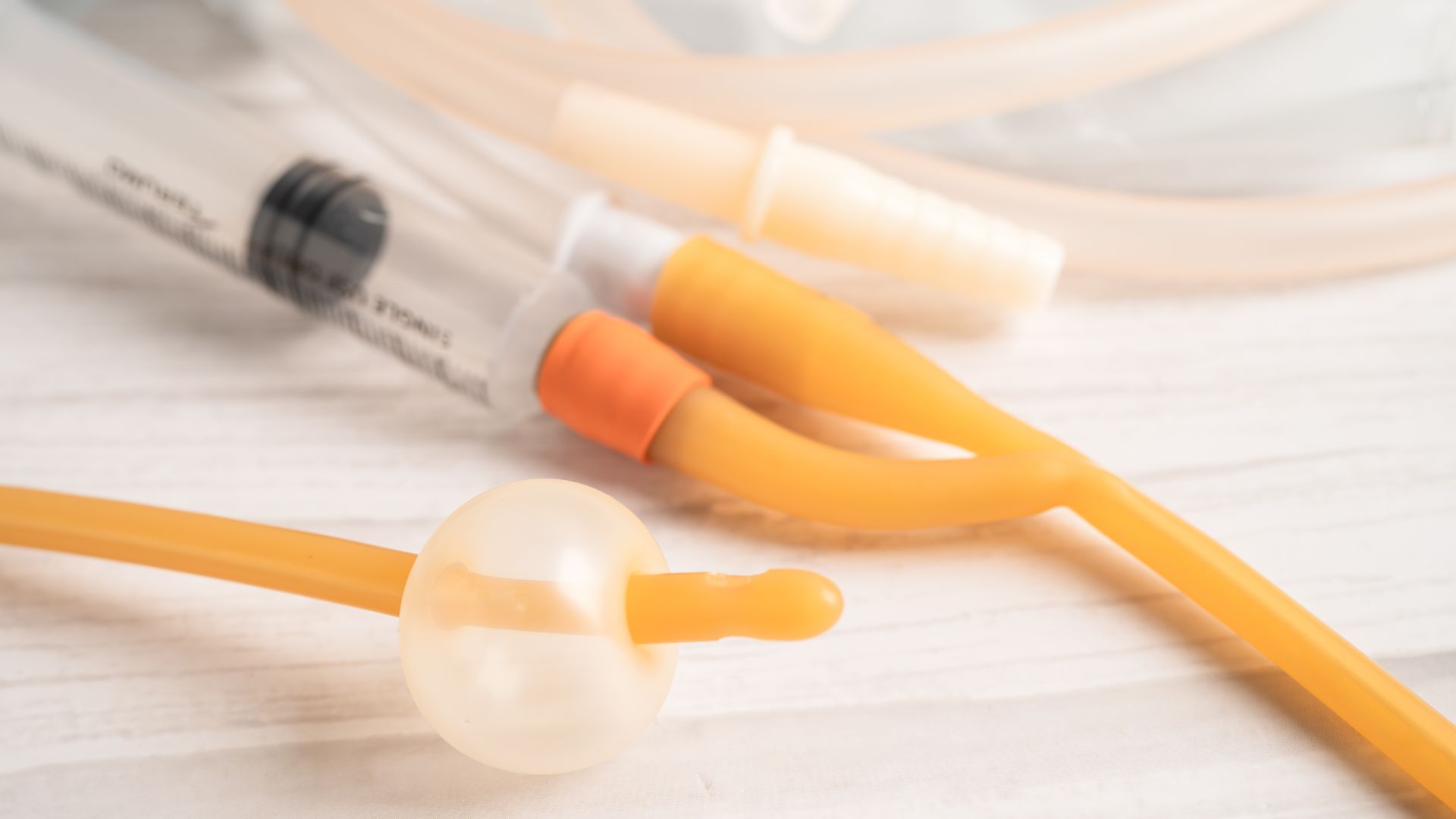
After some surgeries or after a brain stroke, sometimes a patient is not able to control his/her urinary bladder. This leads to social problems and poor hygiene. So a urinary catheter, called Foley’s catheter is left inside the bladder with a urine bag attached. This information will help you care for your own or your patient’s urinary (Foley) catheter.

Depending on the situation, either the patient himself/herself can manage the Foley’s catheter and bag, or a care provider, either a nurse or hospital trained staff should do it.

You may feel that you should do it for your father or mother, but remember, caring for your close and loved relative involves privacy issues and aversion to the work you are doing. Do it only if you feel confident that you can do it. Nurses and hospital staff are trained to do it. Is it’s not difficult, you only have to follow precise steps.

Table of Contents

About your urinary (Foley) catheter

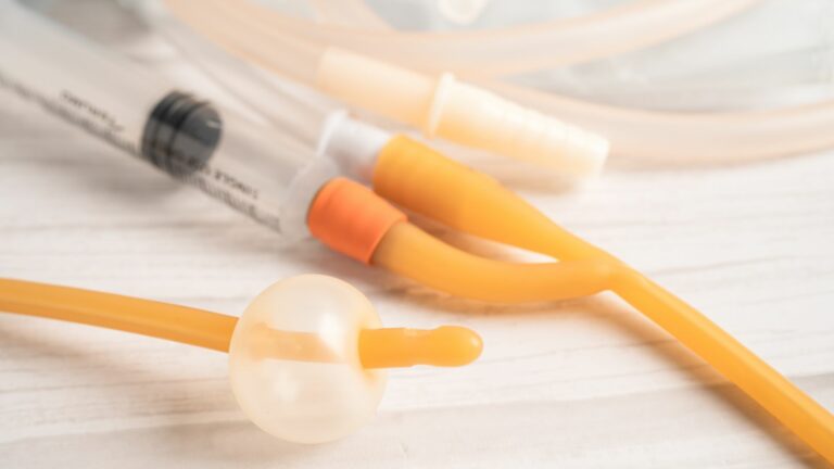
Foley catheter is a thin, flexible tube placed through your urethra (the small tube that carries urine from your bladder to outside your body) and into a urine bag. Your Foley catheter drains your urine. It’s held inside your bladder by a balloon filled with water.

How your Foley catheter works

Your urine goes from your bladder and through a drainage tube into a drainage collection bag. The drainage bag stays on your leg with a special tape.

How to take care of your Foley catheter

To take care of your Foley catheter, you will need to:

  • Clean your catheter every day.
  • Change your drainage bags. You will change your drainage bag 2 times a day.
  • Replace your drainage bags with new bags once a week. You should also change your drainage bag if it gets clogged or blocked.
  • Wash your drainage bags every day.
  • Drink 1 to 2 glasses of liquids every 2 hours while you’re awake to keep you hydrated.

You may see some blood or urine around where the catheter enters your body. This may happen when you’re walking or passing stool. This is normal if there’s urine draining into the drainage bag. If you do not have urine draining into the drainage bag, you must meet your doctor.

Penis or Vaginal care

Use a water-based lubricant to keep your penis or vagina opening from getting sore. Apply the gel where the catheter enters your body.

Keep your penis or vagina opening clean by taking a shower every day. This will help prevent infections when your Foley catheter is in place.

How to shower with your Foley catheter

  • You can shower while you have your catheter in place.
  • Do not take a tub bath until your catheter is removed. Taking a bath while you have your catheter in place puts you at risk for infections.
  • You may find it easier to shower in the morning before you change your bag to your leg bag.

How to clean your Foley catheter

You can clean your catheter while you’re in the shower. Follow these instructions.

  • Mild soap, such as Dove.
  • Wash your hands with soap and water or an alcohol-based hand sanitizer.
  • If you’re using an alcohol-based hand sanitizer, cover your hands with it. Rub them together until they’re dry.
  • Using mild soap and water, clean your penis or vagina.
  • If you have a penis, pull back your foreskin (the skin around the tip of your penis), if needed. Clean the area, including your penis.
  • If you have a vagina, separate your labia (the smaller folds of skin around your vaginal opening). Clean the area from front to back.
  • Clean the area where the catheter enters your body. This is called your urethra.
  • Clean the catheter from where it enters your body and then down, away from your body. Hold the catheter at the point it enters your body so that you do not put tension on it.
  • Rinse the area well and dry it gently.

When to change your drainage bags

You should change your drainage bag 2 times a day. Change it:

  • In the morning after you shower, change the bag.
  • At night before you go to bed, change the bag.
  • Replace your drainage bags with new bags once a week. You should also change your drainage bag if it gets clogged or blocked.

How to change your drainage bag

You will need:

  • A clean cloth (not one you’re using for bathing) or a 4 x 4 piece of gauze.
  • 2 alcohol pads.
  • Wash your hands with soap and water or an alcohol-based hand sanitizer thoroughly.
  • Empty the urine from the drainage bag into the toilet. Make sure the spout of the drainage bag never touches the side of the toilet or any emptying container. If it does, wipe it with an alcohol pad for 15 seconds.
  • Place the clean cloth or gauze under the connector to catch any leakage.
  • Pinch the catheter with your fingers and disconnect the used bag.
  • Wipe the end of the catheter with an alcohol pad.
  • Wipe the connector on the new bag with the second alcohol pad.
  • Connect the clean bag to the catheter and release your finger pinch. Make sure the catheter is tightly connected to the bag to keep it from opening or leaking.
  • Check all connections. Straighten any kinks or twists in the tubing.

How to take care of your drainage bags

  • Empty the leg bag into the toilet every 2 to 4 hours, as needed. You can do this through the spout at the bottom of the bag. Do not let the bag get completely full.
  • Do not lie down for longer than 2 hours while you’re wearing the leg bag. This can keep your urine from draining the way it should.

Caring for your drainage bag

  • Always keep the drainage bag below the level of your bladder.
  • When you go to sleep, hang your bag off the bed. You can do this by using a small trash can. Place a clean plastic bag inside the trash can. Hang your bag inside of the trash can.

Cleaning your drainage bags

Clean your bag every day. Follow these instructions.

You will need:

  • White vinegar.
  • Cool water.
  • Clean your hands with soap and water or an alcohol-based hand sanitizer.
  • Rinse the bag with cool water. Do not use hot water because it can damage the plastic.
  • To help get rid of the smell, fill the bag halfway with a mixture of 1part white vinegar and 3 parts water. Shake the bag and let it sit for 15 minutes.
  • Rinse the bag with cool water. Hang it up to dry.

How to prevent Foley catheter infections

Follow these guidelines to prevent getting infections while you have your catheter in place:

  • Keep the drainage bag below the level of your bladder.
  • Always keep your drainage bag off the floor.
  • Keep the catheter secured to your thigh to keep it from moving.
  • Do not lie on your catheter or block the flow of urine in the tubing.
  • Take a shower every day to keep the catheter clean.
  • Wash your hands before and after touching the catheter or bag.

Your catheter should be replaced about once a month, if it stops working, or if you have an infection.

Lumbar Disc Prolapse

Lumbar Disc Prolapse

Lumbar Disc Prolapse

By Dr.Ravindra Patil

Understanding, Managing, and Relieving Back Pain

Lumbar disc prolapse is a relatively common condition, affecting a significant portion of the population. Factors such as aging, genetics, and lifestyle contribute to its prevalence.

Table of Contents

Introduction

Lumbar disc prolapse, commonly known as a herniated disc, is a condition that can cause excruciating back pain and affect one’s overall spinal health. Understanding this condition, its causes, and treatment options is crucial for individuals seeking relief from the debilitating symptoms associated with disc herniation.

In mild cases of lumbar disc prolapse, individuals may experience minimal symptoms, often manifesting as localized back pain or discomfort. The pain might be intermittent and exacerbated by certain movements or positions. Mild numbness, tingling sensations, or a dull ache in the buttocks or legs may also occur. However, individuals with minimal symptoms usually maintain normal function and can perform daily activities without significant limitations. Conservative measures such as rest, over-the-counter pain medications, and lifestyle adjustments may be sufficient for managing these mild symptoms. It’s essential to note that even in cases of minimal symptoms, seeking medical attention is advisable for an accurate diagnosis and appropriate guidance on preventive measures to maintain spinal health and prevent the progression of disc prolapse symptoms.

Worst care scenarios of Lumbar disc prolapse is discussed at the end of this blog.

Understanding Lumbar Disc Prolapse

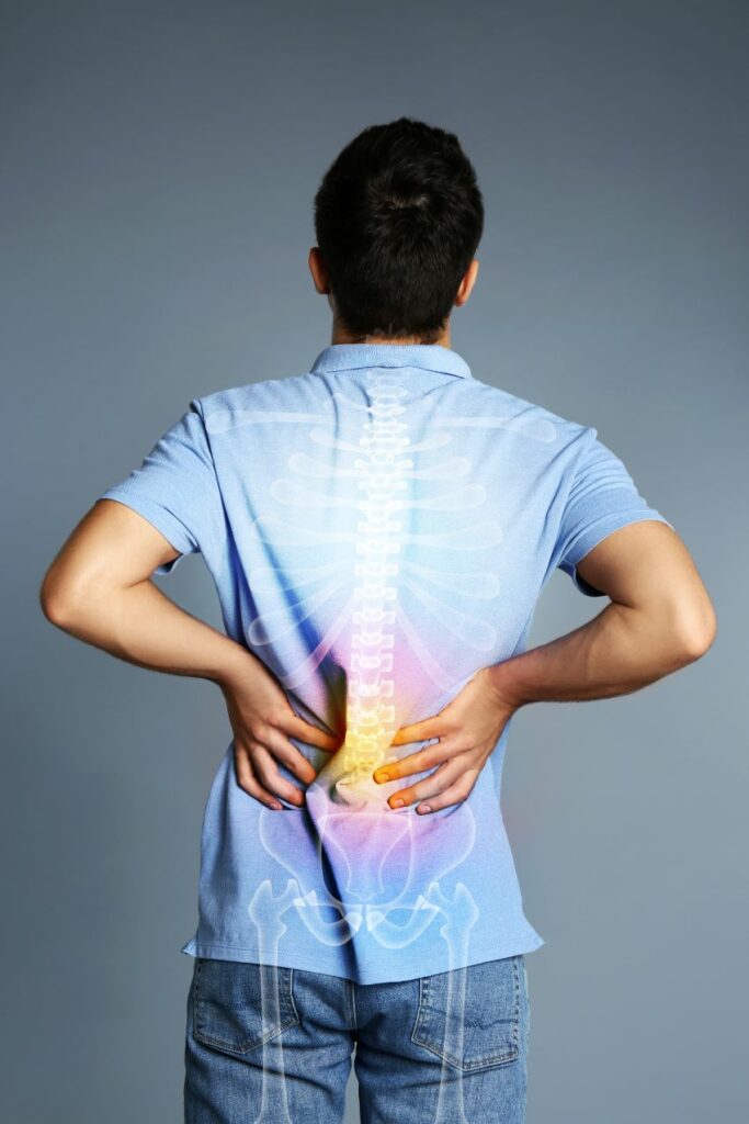
The lumbar spine consists of five vertebrae in the lower back, each separated by intervertebral discs responsible for cushioning and flexibility. In short, there are five lumbar vertebrae and four intervertebral discs. Lumbar disc prolapse occurs when the outer layer of a disc weakens or tears, causing the inner gel-like substance to protrude. This condition is often referred to as a herniated or slipped disc, and it can compress nearby nerves, leading to pain, numbness, and weakness in the affected area.

Common Causes of Disc Herniation

Several factors contribute to the development of lumbar disc prolapse. Aging is a primary factor, as discs naturally degenerate over time, losing water content and elasticity. Repetitive stress on the spine due to activities like heavy lifting or improper lifting techniques can also increase the risk of disc herniation. Other factors include obesity, genetic predisposition, and a sedentary lifestyle

Symptoms and Effects on Spinal Health

One of the hallmark symptoms of lumbar disc prolapse is persistent back pain, often radiating to the buttocks, thighs, and legs. Numbness, tingling sensations, and muscle weakness may also accompany the pain. In severe cases, disc herniation can lead to a condition known as Cauda Equina syndrome, characterized by bowel and bladder dysfunction, requiring immediate medical attention. It is discussed below.

Treatment Options for Disc Prolapse

Managing lumbar disc prolapse involves a combination of conservative and, in some cases, surgical interventions. Conservative treatments aim to alleviate pain and improve spinal health without resorting to surgery. These may include:

  1. Pain Medications: Non-steroidal anti-inflammatory drugs (NSAIDs) and muscle relaxants can help manage pain and inflammation.
  2. Physical Therapy: Targeted exercises can strengthen the muscles supporting the spine, improve flexibility, and promote overall spinal health.
  3. Hot and Cold Therapy: Applying heat or cold packs can help reduce inflammation and alleviate pain.
  4. Lifestyle Modifications: Adopting a healthy lifestyle, including maintaining a proper posture, regular exercise, and weight management, can contribute to preventing further disc herniation.

For cases where conservative treatments prove ineffective, surgical options may be considered. Procedures like discectomy or micro-discectomy aim to remove the protruding disc material and relieve pressure on the nerves.

Micro-Discectomy

Micro-Discectomy, also known as micro-decompression or micro-diskectomy, is a surgical procedure commonly used to treat lumbar disc prolapse or herniated discs. It is a minimally invasive surgery designed to alleviate the pressure on spinal nerves caused by the protrusion of disc material.

During a Micro-Discectomy, a small incision is made over the affected area of the spine. Unlike traditional open surgery, this minimally invasive approach involves the use of a microscope or magnifying lenses, allowing the surgeon to visualize the affected disc and surrounding structures with precision. The use of specialized instruments enables the removal of the herniated disc material, relieving compression on the nerves.

One of the primary advantages of Micro-Discectomy is its minimal disruption to surrounding tissues. By utilizing smaller incisions and specialized tools, the procedure reduces trauma to muscles and other structures in the spine. This not only results in less postoperative pain but also promotes a quicker recovery compared to traditional open surgery.

Patients undergoing Micro-Discectomy typically experience a significant reduction in symptoms such as back pain, leg pain, and numbness shortly after the procedure. The success rate of Micro-Discectomy in relieving symptoms associated with lumbar disc prolapse is generally high.

As with any surgical procedure, there are potential risks and complications, and it is crucial for individuals considering Micro-Discectomy to discuss these with their healthcare provider. However, for many patients, Micro-Discectomy offers an effective and minimally invasive solution to address the debilitating effects of lumbar disc prolapse, allowing them to regain mobility and improve their overall quality of life.

Managing Back Pain with Disc Compression Exercises

In addition to formal physical therapy, individuals with lumbar disc prolapse can benefit from disc compression exercises designed to alleviate pressure on the affected discs and promote healing. These exercises should be performed under the guidance of a healthcare professional. Some effective disc compression exercises include:

  1. Pelvic Tilts: Lie on your back with knees bent, gently tighten your abdominal muscles, and lift your pelvis off the floor, holding for a few seconds before lowering it back down.
  2. Cat-Cow Stretch: Start on your hands and knees, arch your back up like a cat, and then dip your back down, lifting your head and tailbone toward the ceiling.
  3. Child’s Pose: Kneel on the floor, sit back on your heels, and stretch your arms forward on the floor, lowering your chest towards the ground.
  4. Knee-to-Chest Stretch: Lie on your back, bring one knee toward your chest, and hold it with both hands. Repeat with the other knee.

Worst-Case Scenario of Lumbar Disc Prolapse

  • In the worst-case scenario of lumbar disc prolapse, severe and prolonged compression of spinal nerves can lead to debilitating complications. This may result in a condition known as cauda equina syndrome, characterized by a loss of bowel and bladder control, saddle anesthesia (loss of sensation in the buttocks and inner thighs), and progressive weakness in the legs.

    Cauda equina syndrome is a medical emergency requiring immediate intervention, typically through surgical decompression to alleviate pressure on the affected nerves. Failure to address cauda equina syndrome promptly may lead to permanent neurological damage. While such extreme complications are rare, it underscores the importance of seeking prompt medical attention when experiencing severe symptoms associated with lumbar disc prolapse to prevent irreversible consequences.

Conclusion

Lumbar disc prolapse can significantly impact one’s quality of life, but with a comprehensive understanding of the condition and proactive measures, individuals can manage back pain and promote spinal health. Whether through conservative treatments, surgical interventions, or targeted exercises, the key is to seek professional guidance to develop a personalized approach to address the unique challenges posed by lumbar disc prolapse. Remember, early intervention and a commitment to a healthy lifestyle play crucial roles in mitigating the effects of disc herniation and maintaining long-term spinal well-being.


Maxillary Sinusitis

Maxillary Sinusitis

Maxillary Sinusitis

By Dr.Ravindra Patil

It means an infectious disease of the maxillary sinus. As all the readers are not likely to be doctors or biologists, it is necessary to explain what is a sinus and then what is the maxillary sinus.

Table of Contents

Sinuses

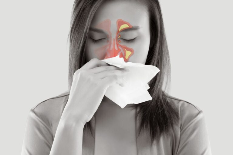
Sinuses are air-filled spaces in the forehead, cheekbones, and behind the bridge of the nose. They allow mucus to drain and air to circulate through the nasal passages. There are seven sinuses in the bones of our skull. Of those, three are located on each side of our head. The sphenoid sinus is located in the centre, and is single. The seven sinuses are:

  • Frontal sinuses: Located in the forehead, above the nose and behind the eyebrows.
  • Ethmoid sinuses: Located in the ethmoid bone, on the bridge of the nose, above the nasal cavity and between the eyes.
  • Sphenoid sinus: Located within the body of the sphenoid bone, just anterior and inferior to the sella turcica. It is a single, midline sinus.
  • Maxillary sinuses: Located on either side of the nostrils (cheekbone areas). They are the largest of the air sinuses.

What do the sinuses do?

The functions of sinuses include:

  • Produce mucus
  • Support the immune system of the nasal cavity
  • Lighten the weight of the skull

If the opening from a sinus cavity becomes plugged, the flow of mucus is blocked and pressure builds up, causing pain and inflammation.

The sinuses collectively are called the paranasal sinuses because they are on the sides of the nose. The sinuses are part of your nose and respiratory system. They connect to your nasal passages in a complex network of air flow and drainage passages. As you breathe in air through your nose and mouth, it moves through the sinus passages. The sinuses also produce mucus that coats and lubricates your nasal passages and the sinuses themselves.

Both air and mucus flow through your sinuses and drain into your nose, through tiny openings called ostia (or singular, ostium).

Little hairs called cilia help the mucus move through the sinus cavities. The mucus from the sinuses drains into your nasal passages and then down the back of your throat to be swallowed. The draining mucus helps keep your nose moist and it filters out dust and bacteria.

The sinuses also:

  • Give your voice resonance as the air vibrates
  • Help protect your face in case of trauma
  • Insulate against rapid temperature changes in the nose
  • Provide an immunological defence
  • The sinuses lighten the weight of the skull because they are hollow bones

Maxillary Sinusitis

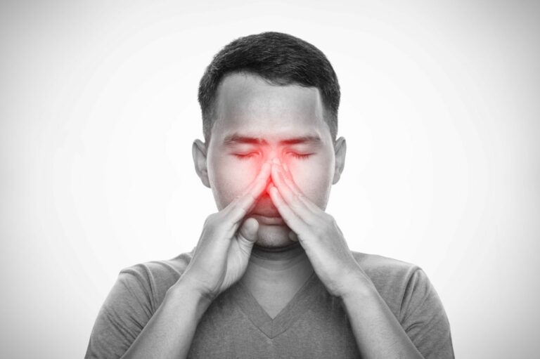
Inflammation of the sinuses is called Sinusitis. Inflammation of the Maxillary Sinus/s is called Maxillary Sinusitis. Infections of the maxillary sinuses are most common. This article is called ‘Maxillary Sinusitis’ because maxillary sinusitis is the most common sinus infection.

Sinusitis is a sinus infection that can be caused by:

  • Respiratory infections, such as a cold or flu
  • Allergies, like hay fever

Symptoms of sinusitis

Symptoms of a sinus infection are similar to those of a cold:

  • Depending on which sinuses are infected, you may feel pain or pressure in your forehead, cheeks, ears, or teeth.
  • You may have thick, sticky mucus coming from your nose.
  • Your mucus may be cloudy, or have a greenish-yellow color.
  • Mucus may drip down the back of your throat (postnasal drip), giving you a sore throat and cough.
  • Your nose may be stuffed, restricting your breathing.
  • Your face may feel tender, especially around the eyes.

Other symptoms

  • hoarseness
  • fever
  • fatigue
  • bad breath
  • decreased sense of smell and taste

The most common sinus problem

Any infection of your upper respiratory tract can easily spread to the sinuses. The resulting inflammation and pain are called sinusitis. It’s also known as rhinosinusitis. Many people refer to sinusitis as simply ‘sinus’ pain.Your sinuses can become infected by bacteria, a virus, or both. At any given time, millions of people all over the world are suffering from sinusitis.

Acute & Chronic sinus infections

A sinus infection can last from 10 days to as long as 8 weeks. This is called an acute sinus infection.

Sometimes a sinus infection can become chronic, getting better and then worse again, off and on for months.

Chronic sinusitis is medically defined as sinusitis that occurs more than four times a year. If your sinus infection lasts longer than eight weeks, it’s also usually considered chronic.

Chronic sinusitis is one of the most common chronic conditions.

The symptoms of acute and chronic sinusitis are similar. Fever is less likely, except in severe cases.

Factors involved with chronic sinusitis are…

  • Viral or bacterial infections
  • Fungal infections
  • Hay fever or allergies to dust mites, moulds, and so on
  • Frequent exposure to cigarette smoke or other airborne pollutants
  • Nasal polyps, a deviated septum, or a damaged nasal structure
  • Medical conditions such as asthma, hiv, or cystic fibrosis
  • Aspirin sensitivity
  • Respiratory tract infections

You’re at higher risk for sinusitis if you…

  • Have allergies
  • Smoke
  • Have a weakened immune system
  • Have a mechanical problem in the nose, such as a deviated septum
  • Have a dental infection
  • Spend time in a day care or kindergarten where germs are likely to be present

People often develop sinusitis after they have a common cold.  If the lining of your sinuses becomes https://blogpoet.com.ua blocked in a cold, the mucus gets thick and sticky. Your sinuses may not drain properly and bacteria can then build up in the mucus.

Treatment of Sinusitis

  • Getting plenty of rest
  • Drinking plenty of fluids
  • Taking painkillers, such as paracetamol or ibuprofen

Serious sinus infections can spread to the membranes and fluid around the brain and spinal cord. This infection is called meningitis. Other serious infections can spread to the bones, called osteomyelitis, or to skin, called cellulitis.

What should you do if you have Acute Sinusitis

  • Try moist heat or steam. You can make a steam inhaler by putting hot water in a bowl and inhaling the steam. To do this:
  • Keep at least 8 inches away from the water so that you don’t burn yourself.
  • Inhale for about 2 minutes.
  • Create an enclosed space for the moist, warm air to collect by draping a towel over your head as you lean over the bowl.
  • You can also buy a vaporizer, which creates steam from heat.

Other tips to manage symptoms

  • Take over-the-counter (OTC) pain medication to ease headaches.
  • Try OTC drugs such as guaifenesin (Mucinex) that thins your mucus, which can help to pass the mucus and lessen congestion.
  • Try acupuncture; there’s some evidence that it’s useful for sinus-related symptoms.

Chronic sinusitis care tips

Find and treat the condition that’s causing it. If another condition is associated with your chronic sinusitis, get treated for it and stick to your medications or treatment plan. Try treatments or management plans if it’s from allergies. See an allergy specialist to identify and have ongoing treatment for allergies.

Keeping sinuses healthy

You can take some steps to help keep your sinuses healthy and avoid sinusitis:

  • Keep your hands clean.
  • Maintain moisture in your nose and sinuses.
  • Drink plenty of fluids to keep your mucus thin.
  • Use a humidifier in the winter season, when the air tends to be drier.
  • Use an OTC nasal saline spray to irrigate your nose.
  • Keep the dust level down by dusting and vacuuming often, especially in the bedroom.

The good news is that if you’re otherwise healthy and your sinusitis is caused by a virus, it’s likely to resolve on its own within 7 to 10 days.

सायनुसायटिस कायमचा बरा कसा करावा

सायनुसायटिस कायमचा बरा कसा करावा

सायनुसायटिस कायमचा बरा कसा करावा

By Dr.Ravindra Patil

सायनसचा रोग कायमचा बरा करणे शक्य आहे का?

आम्ही प्रथम या प्रश्नाचे उत्तर देण्याचा प्रयत्न करू. बर्‍याच लोकांना ऍलर्जी असते ज्यामुळे बर्‍याचदा सर्दी होते. सर्दी हा विषाणूजन्य संसर्ग आहे. ते सायनसच्या आतपर्यंत वाढू शकतो. सर्दीचे वारंवार होणारे हल्ले सायनुसायटिस क्रॉनिक बनवू शकतात. क्रॉनिक म्हणजे दीर्घकाळ चालणारा रोग. अशा परिस्थितीत इम्युनोथेरपीद्वारे ऍलर्जीवर उपचार करणे आवश्यक आहे. शरीरातील ऍलर्जीक प्रतिक्रिया कमी करणारी इंजेक्शन्स सर्दी आणि सायनुसायटिसचे हल्ले कमी करू शकतात.

बहुतेक सायनस संक्रमणे प्रतिजैविकां (अ‍ॅन्टीबायोटीक्स) शिवाय बरे होतात.

शस्त्रक्रिया सर्वात उत्तम उपचार आहे असे मानले जाते. तथापि, तो शेवटचा उपाय आहे हे लक्षात घेणे.

सायनुसायटिसची बहुतेक प्रकरणे बरी होऊ शकतात परंतु जर ते पुन्हा पुन्हा उद्भवले तर शस्त्रक्रिया करावी लागते. बुरशीजन्य सायनुसायटिस बरा करणे कधीकधी कठीण असते.

Table of Contents

सायनुसायटिस म्हणजे काय?

सायनुसायटिस किंवा सायनस संसर्ग हा असा एक रोग आहे ज्यामध्ये पॅरानेसल सायनस फुगतात. पातळ श्लेष्माचे उत्पादन अनुनासिक मार्ग ओलसर करण्यास मदत करते. तथापि, जेव्हा श्लेष्माचे जास्त उत्पादन होते तेव्हा सायनसच्या आत सूज येऊ शकते. बॅक्टेरिया, ऍलर्जी, बुरशी आणि विषाणू (व्हायरस) यासारख्या विविध कारणांमुळे सायनुसायटिस होऊ शकते.

सायनुसायटिसचे अनेक प्रकार आहेत, प्रत्येकाची स्वतःची लक्षणे आहेत. काही लोकांना क्रॉनिक सायनुसायटिस, तीव्र सायनुसायटिस, वारंवार सायनुसायटिस किंवा सबएक्यूट सायनुसायटिस होतो. जरी या स्थितीमुळे अप्रिय संवेदना आणि वेदना होतात, तरीही तो सहसा वैद्यकीय मदतीशिवाय बरा होतो. तथापि, लक्षणे अधिक तीव्र झाल्यास, आपल्याला डॉक्टरांना भेटण्याची आवश्यकता असू शकते.

सायनसच्या समस्या कशामुळे होतात?

सायनुसायटिस हा विषाणू, बॅक्टेरिया किंवा बुरशीमुळे होऊ शकतो ज्यामुळे सायनसना आतून सूज येते आणि त्यातील नाकात उघडणारी छीद्रे बंद होतात. सामान्य सर्दीची होण्याची अनेक संभाव्य कारणे आहेत, ज्यात सर्दी विषाणू देखील आहेत. नाकातील ऍलर्जी आणि बूरशीची ऍलर्जी अनेकांना होते.

सायनुसायटिसचा धोका कोणाला आहे?

सायनसचा संसर्ग कोणालाही होऊ शकतो. परागकण, श्लेष्मा किंवा हवेतील इतर पदार्थांची ऍलर्जी, दमा किंवा नाकाची असामान्य रचना असलेल्या लोकांना सायनुसायटिस होण्याची शक्यता असते. याव्यतिरिक्त, धूम्रपानामुळे सायनस संसर्ग होण्याची शक्यता वाढते.

सायनुसायटिसचे निदान

  • तुमचे डॉक्टर खालील तपासणी प्रणाली द्वारे क्रॉनिक सायनुसायटिसचे निदान करू शकतात:

    • तुमच्या लक्षणांबद्दल विचारणे
    • तुमच्या नाकात आणि चेहऱ्यावर दाबल्यास दूखते का ते पाहणे
    • फायबरऑप्टिक एंडोस्कोपद्वारे तुमच्या नाकाच्या आत तपासणी करणे
    • सी.टी. स्कॅन किंवा एम.आर.आय. सारख्या इमेजिंग चाचण्या वापरणे

    बॅक्टेरिया किंवा बुरशी तुमच्या मेंदू, डोळे किंवा जवळच्या हाडांमध्ये पसरल्यास उपचार न केलेल्या सायनस संसर्गामुळे जीवघेणा संसर्ग होऊ शकतो.

तीव्र सायनुसायटिसची लक्षणे

    • चेहऱ्यावर वेदना किंवा दाब
    • वाहणारे नाक
    • चोंदलेले नाक
    • वासाची जाणीव कमी होणे
    • खोकला किंवा छातीत कफ
    • ताप
    • श्वासाची दुर्घंधी
    • थकवा
    • दातदुखी

    जर तुम्हाला दोन किंवा अधिक लक्षणे असतील किंवा नाकातून जाड, हिरवा किंवा पिवळा स्त्राव असेल तर तुम्हाला तीव्र सायनुसायटिस असू शकतो.

क्रॉनिक सायनुसायटिसची लक्षणे

तुमची सायनुसायटिस 12 आठवडे किंवा त्याहून अधिक काळ राहिल्यास तुम्हाला ही क्रॉनिक सायनुसायटिस असु शकतो व ही लक्षणे दिसू शकतात:

  • तुमच्या चेहऱ्यावर रक्तसंचय किंवा सूज जाणवणे
  • चोंदलेले नाक
  • अनुनासिक पोकळी मध्ये पू
  • ताप
  • वाहणारे नाक किंवा अनुनासिक स्त्रावाचा रंग बदलणे

तुम्हाला डोकेदुखी, श्वासाची दुर्गंधी आणि दातदुखीचा त्रास होऊ शकतो. तुमच्या आजारपणामुळे तुम्हाला खूप थकवा जाणवू शकतो. या लक्षणांची अनेक संभाव्य कारणे आहेत.

सायनुसायटिसचा संसर्ग एका व्यक्तीकडून दुसऱ्या व्यक्तीकडे जाऊ शकतो का?

बॅक्टेरियल सायनुसायटिस हा संसर्गजन्य नाही, परंतु व्हायरसमुळे संसर्ग पसरू शकतो. दूषित हातांद्वारे इतर लोकांना संसर्ग होऊ नये म्हणून हात धुण्याच्या चांगल्या पद्धतींचे अनुसरण करा. जर तुम्हाला शिंक किंवा खोकला येत असेल तर, इतरांपासून तोंड दूर ठेवा किंवा तुमचा चेहरा रुमालाने झाका.

सायनुसायटिससाठी उपचार

सायनुसायटिसचे उपचार करण्याचे अनेक मार्ग आहेत. काही प्रकार साध्या औषधांनी बरे होऊ शकतात, तर काहींना शस्त्रक्रियेची आवश्यकता असू शकते. तुम्हाला सायनसच्या संसर्गापासून मुक्त होण्यासाठी तुम्ही विविध घरगुती उपाय वापरू शकता. सायनुसायटिससाठी औषधांसह अनेक उपचार उपलब्ध आहेत. येथे काही उपचार दिले आहेत.

1. कॉर्टिकोस्टिरॉईड्सच्या गोळ्या घेणे

कॉर्टिकोस्टिरॉईड्सच्या गोळ्यांचा वापर गंभीर लक्षणे उद्भवल्यावर लवकर आराम होण्यासाठी केला जातो. ज्या रुग्णांना नाकातील पॉलीप्स आहेत ते देखील गोळ्या घेऊ शकतात. पण जर या गोळ्या जास्त काळ वापरल्या गेल्या तर त्यांचे दुष्परिणाम होऊ शकतात. कॉर्टिकोस्टिरॉईड्स फक्त तेव्हाच लिहून दिली जातात जेव्हा इतर उपचार जसे की नाकातील कॉर्टिकोस्टिरॉईड्सचा फवार अयशस्वी होतो.

2. नाकातील कॉर्टिकोस्टिरॉईड्सचा फवारा

नाकातील कॉर्टिकोस्टिरॉईड्स हे फवारण्याचे द्रव असतात व ते सूज कमी करण्यास मदत करतात. डॉक्टर विविध कॉर्टिकोस्टिरॉईड्स लिहून देऊ शकतात, जसे की बुडेसोनाइड, फ्लुटिकासोन इ. जर वैद्यकीय तज्ञांना असे वाटत असेल की नाक स्वच्छ धुवून मदत होत नाही, तर ते उपचारांच्या इतर पद्धती सुचवू शकतात. डॉक्टर एक उपाय लिहून देऊ शकतात ज्यामध्ये खारट आणि औषधे आहेत.

3. अनुनासिक फवारे

हे फवारण्या प्रत्येक फार्मसीमध्ये उपलब्ध आहेत आणि नाकातील रक्तसंचय दूर करण्यासाठी वापरले जातात. ते ड्रेनेज कमी करण्यात मदत करू शकतात आणि सूज दूर करण्यात मदत करू शकतात.

4. प्रतिजैविक

जर तुमच्या संसर्गाचे कारण जीवाणू असेल तर प्रतिजैविक मदत करू शकतात. कधीकधी, डॉक्टर या औषधासह इतर औषधे लिहून देतात. त्यामुळेच अनेक निदान चाचण्या केल्या जातात जेणेकरून योग्य मूल्यमापन करता येईल.

5. इम्युनोथेरपी

जर ऍलर्जी सायनुसायटिसचे कारण असेल तर त्या साठी इम्युनोथेरपी एक संभाव्य उपचार आहे. ऍलर्जीच्या कमी करण्याची इन्जेक्शने घेतल्यास तुम्हाला तुमच्या लक्षणांमध्ये घट दिसू शकते कारण तुमचे शरीर त्या नंतर ऍलर्जिनवर तितकी तीव्र प्रतिक्रिया देत नाही.

सायनुसायटिस बरा करण्यासाठी घरगुती उपाय

1. विश्रांती

तुम्हाला आराम करणे आवश्यक आहे जेणेकरून तुमचे शरीर संक्रमणाशी लढू शकेल. जेव्हा तुम्ही झोपायला जाता तेव्हा डोके वर करण्यासाठी उशीचा वापर करा. निरोगी फुफ्फुसांसाठी श्वास घेणे महत्वाचे आहे.

2. वाफ

स्टीम इनहेलेशन हे तुमचे सायनस स्वच्छ करण्याचा एक उत्तम मार्ग आहे. तुमचा सायनसच्या साफसफाईचा अनुभव अधिक आनंददायी बनवण्यासाठी तुम्ही वाफेच्या भांड्यात काही आवश्यक तेले घालू शकता. दुसरा पर्याय म्हणजे गरम शॉवर घेणे. तुमचा चेहरा थंड होण्यासाठी तुम्ही उबदार टॉवेल वापरू शकता.

3. अनुनासिक सिंचन

खारट द्रावणासह अनुनासिक सिंचन हा अनुनासिक जागा स्वच्छ करण्याचा एक चांगला मार्ग आहे. रक्तसंचय यांसारख्या श्वसनाच्या समस्यांसाठी तुम्ही नेटी पॉट, सलाईन कॅनिस्टर किंवा ह्युमिडिफायर वापरू शकता. पाणी स्वच्छ आहे याची खात्री करा आणि उपकरणे निर्जंतुक आहेत का ते तपासा.

4. मसालेदार पदार्थ

आपल्या आहारात मसालेदार पदार्थ खाऊन आपली भूक कमी होते आणि समाधान वाढते. मसालेदार पदार्थ खाल्ल्याने तुमचा नाकाचा मार्ग साफ होण्यास मदत होऊ शकते तथापि, जर तुमच्या इतर रोगांमुळे तुम्हाला मसालेदार पदार्थ खाण्यास मनाई असेल तर तुम्ही मसालेदार पदार्थ खाऊ नयेत.

5. उबदार कॉम्प्रेस

चेहेर्यावरील सूज कमी करण्यासाठी उबदार कॉम्प्रेसचा वापर केला जाऊ शकतो. हे अस्वस्थता आणि वेदनापासून आराम देईल.

6. द्रवपदार्थ

भरपूर द्रव पिणे आवश्यक आहे. हायड्रेटेड राहण्यासाठी अधिक रस आणि पाणी प्या. हे श्लेष्माचा थर कमकुवत करण्यास मदत करेल. तथापि, आपण अल्कोहोलयुक्त पेये पिणे टाळावे.

मी माझे सायनस जलद कसे बरे करू शकतो?

  • खूप पाणी प्या
  • बॅक्टेरियाविरोधी गुणधर्म असलेले पदार्थ खा
  • ओलावा जोडा
  • सायनस तेलाने स्वच्छ करा
  • नेटी पॉट वापरा
  • उबदार कॉम्प्रेसने चेहर्यावरील वेदना कमी करा
  • ओव्हर-द-काउंटर (OTC) औषधे वापरा

घरी सायनुसायटिसचा उपचार कसा करावा?

  • उष्णता वापरा.
  • अनुनासिक खारट द्रावण वापरून पहा. त्यात औषध नसले तरी (खारट हे खारट पाणी असते), ते तुमच्या अनुनासिक परिच्छेद ओलसर ठेवण्यास मदत करू शकतात.
  • तुमचे सायनस बाहेर काढा.
  • भरपूर द्रव प्या.

डॉक्टरांना कधी भेटायचे

तीव्र सायनुसायटिस असलेल्या बहुतेक लोकांना डॉक्टरांना भेटण्याची आवश्यकता नसते, परंतु तुमच्याकडे खालीलपैकी काही असल्यास तुमच्या डॉक्टरांशी संपर्क साधा:

  • एक आठवड्यापेक्षा जास्त काळ टिकणारी लक्षणे.
  • बरे झाल्यासारखे दिसू लागल्यानंतर आणखी वाईट होणारी लक्षणे.
  • सतत राहणारा ताप.
  • पुनरावृत्ती किंवा तीव्र सायनुसायटिसचा इतिहास.

तुम्हाला गंभीर संसर्गाची लक्षणे आढळल्यास ताबडतोब आरोग्य सेवा प्रदात्याला भेटा:

  • डोळ्यांभोवती वेदना, सूज किंवा लालसरपणा
  • खूप ताप
  • गोंधळ
  • दुहेरी दृष्टी किंवा इतर दृष्टी बदल
  • सतत मान ताठ राहणे

घरी डोके दुखणे ताबडतोब कसे थांबवायचे

घरी डोके दुखणे ताबडतोब कसे थांबवायचे

By Dr.Ravindra Patil

घरी डोके दुखणे ताबडतोब कसे थांबवायचे​ आपल्या डोकेदुखीला दृढ करण्यासाठी "आत्मशक्ती," "सामर्थ्य," "उत्साह," "शक्तिशाली," आणि "प्रबल" हे सकारात्मक शब्द आहेत.
जवळजवळ प्रत्येकाला कधी ना कधी डोके दुखते. डोकेदुखी हा पाठदुखीनंतर मानवांमध्ये दुसऱ्या क्रमांकाची सर्वात जास्त वेळा होणारी वेदना मानली जाते. सुदैवाने, बहुतेक डोकेदुखी किरकोळ कारणांमुळे होते आणि ती औषधांनी नाही तर घरगुती उपचारांनी बरी होऊ शकते. तुमच्या आई आणि आजीला डोकेदुखीसाठी अनेक उपचार माहीत असतील. आणि आश्चर्याची गोष्ट म्हणजे, त्यापैकी बहुतेक वेळा ते उपचार प्रभावी होतात.
पण सर्व डोकेदुखी इतके सौम्य नसतात. काही डोकेदुखीच्या उपचारांसाठी तज्ज्ञ डॉक्टरांची गरज असते. आम्ही विशिष्ट प्रकारच्या डोकेदुखीची यादी करतो ज्यांना डॉक्टरांच्या उपचारांची आवश्यकता आहे. त्यानंतरच्या परिच्छेदांमध्ये सुचविलेल्या उपचारांमुळे इतर सर्व प्रकारच्या डोकेदुखीपासून आराम मिळण्याची शक्यता आहे. परंतु प्रथम आपण डोकेदुखी पाहू या ज्यासाठी आपल्याला डॉक्टरांचा सल्ला घ्यावाच लागेल.

Table of Contents

विशेष प्रकारच्या डोकेदुखी

या डोकेदुखीसाठी डॉक्टरांच्या सल्ल्याची आवश्यकता असते:

  • तणाव डोकेदुखी
  • क्लस्टर डोकेदुखी
  • मायग्रेन डोकेदुखी
  • हेमिक्रानिया सतत
  • थंडरक्लॅप डोकेदुखी
  • ऍलर्जी किंवा सायनस डोकेदुखी
  • हार्मोनल डोकेदुखी (मासिक पाळीतील मायग्रेन म्हणूनही ओळखले जाते)
  • कॅफिन डोकेदुखी
  • परिश्रम डोकेदुखी
  • उच्च रक्तदाब डोकेदुखी
  • रिबाऊंड डोकेदुखी, जी औषधाचा परिणाम संपल्यावर परत येणे
  • आघातानंतरची डोकेदुखी
  • पाठीचा कण्याच्या रोगामुळे होणारी डोकेदुखी

जर तुमची डोकेदुखी तीव्र असेल आणि ती पुन्हा पुन्हा येत असेल, तर ती वरीलपैकी एक असू शकते आणि तुम्ही डॉक्टरांचा सल्ला घ्यावा.

आता गंभीर नसलेल्या डोकेदुखीवर घरगुती उपाय पाहू

तुमच्या टाळूवर किंवा डोक्यावरील दाब कमी करा

हे खालीलपैकी कोणत्याही पद्धतीद्वारे केले जाऊ शकते. तुम्हाला अनुकूल असलेली आणि तुम्ही सहजपणे अंमलात आणू शकता अशी पद्धत निवडा.

हीटिंग पॅड वापरा

इलेक्ट्रिकल हीटिंग पॅड किंवा गरम पाण्याची रबरी पिशवी कपाळावर किंवा मंदिरांना लावल्याने अनेकदा तुमची डोकेदुखी दूर होऊ शकते. तापमान सेटिंग ‘सौम्य’ ठेवा, ‘गरम’ नाही.

कोल्ड कॉम्प्रेस लावा

विरोधाभास म्हणजे, डोके किंवा मानेवर कोल्ड कॉम्प्रेस देखील वेदना कमी करू शकते! तुम्ही थंड शॉवर किंवा कोमट पाण्याचा शॉवर देखील घेऊ शकता. किंवा स्नान करा.

आपल्या मानेच्या मागील बाजूस मालिश करा

तुम्ही ते स्वतः करू शकता. तर्जनीने मानेच्या मागील बाजूस दाबल्याने डोकेदुखीपासून आराम मिळू शकतो. हे उपयुक्त ठरते कारण जेव्हा तुम्ही तणाव अनुभवता तेव्हा या भागातील स्नायू घट्ट होतात. तुमच्या कपाळ, मान आणि डोक्याच्या बाजुला काही मिनिटे मालिश केल्याने तणावामुळे होणारी डोकेदुखी कमी होण्यास मदत होते. किंवा वेदनादायक भागावर हलका, फिरणारा दाब लावा.

एक्यूप्रेशर

हातावर मागच्या बाजूला काही दाब बिंदू दाबल्याने डोकेदुखी दूर होऊ शकते. हा बिंदू तुमच्या अंगठ्याच्या मुळाच्या आणि तर्जनीच्या मध्ये स्थित असतो. तथापि, प्रशिक्षित एक्यूप्रेशर व्यक्तीचा सल्ला घ्या.

पाणी खूप प्या

पाणी किंवा हर्बल चहा प्यायल्याने डोकेदुखी दूर होऊ शकते.

दिवे मंद करा

दिवे बंद केल्याने डोकेदुखी कमी होण्यास मदत होते.

थोडी विश्रांती घ्या

तुम्ही एक लहानशी डुलकी घेतल्यानंतर, तुम्ही ताजेतवाने जागे व्हाल!

इतर घरगुती उपचारांमध्ये हे समाविष्ट आहे …

काहीही पदार्थ किंवा जेवण न चावण्याचा प्रयत्न करा

अन्न चावून खाल्ल्या मुळे काहीवेळा विशिष्ट प्रकारची डोकेदुखी वाढते.

थोडे कॅफीन घ्या

चहा किंवा कॉफीचा जुना उपाय विसरता येणार नाही.

विश्रांतीचा सराव करा

शरीराचे स्ट्रेचींग करा, योग, ध्यान किंवा प्रगतीशील स्नायू शिथिलता आणून डोकेदुखी कमी होते. तसेच, शांत कसे राहायचे हे शिकणे वेदना कमी करण्यास मदत करू शकते. जर तुमच्या मानेमध्ये स्नायूंचा त्रास होत असेल तर तुम्ही तुमच्या डॉक्टरांशी फिजिकल थेरपीबद्दल बोलू शकता.

थोडे आले घ्या

नुकत्याच झालेल्या एका लहानशा अभ्यासात असे आढळून आले आहे की नियमित ओव्हर-द-काउंटर वेदनाशामक औषधांव्यतिरिक्त आल्याचे सेवन केल्याने मायग्रेन असलेल्या लोकांच्या वेदना कमी होतात. दुसर्‍याला आढळले की आले जवळजवळ प्रिस्क्रिप्शन मायग्रेन औषधांप्रमाणेच काम करते. अनेक भारतीय कुटुंबांमध्ये, नेहमीच्या चहामध्ये आले घालण्याची पध्दत असते.

हर्बल चहा वापरून पहा

डोकेदुखी दूर करण्यासाठी अनेक खास हर्बल चहा उपलब्ध आहेत. ते पिऊन बघा.

औषधांचा वापर संयमाने करा

डोकेदुखीसाठी भरपूर औषधे उपलब्ध आहेत परंतु त्यांचा वापर शक्य तो कमी प्रमाणात करा. कमीत कमी जोखमीसह जास्तीत जास्त फायदा मिळवण्यासाठी, लेबलवरील सुचनांचे तंतोतंत अनुसरण करा:

  • तुम्हाला हृदयाचा रोग असला किंवा तुमची मूत्रपिंड निकामी झाल्यास इबुप्रोफेन आणि इतर नॉनस्टेरॉइडल अँटी-इंफ्लेमेटरी ड्रग्स (NSAIDs) टाळा.
  • १८ वर्षांखालील मुलास ऍस्पिरिन देऊ नका.
  • दुखायला लागताच गोळी घ्या. तुम्ही गोळी घेण्यास उशीर लावलात तर तुम्हाल जास्त डोस घ्यावा लागेल.
  • जेव्हा तुम्हाला डोकेदुखी होत असतांनाच तुमच्या पोटात बरे वाटतत नसल्यास कुठले औषध घ्यायचे ते तुमच्या डॉक्टरांना विचारा.
  • रिबाउंड डोकेदुखी टाळण्यासाठी काय घ्यायचे ते तुमच्या डॉक्टरांना विचारा. रिबाउंड डोकेदुखी म्हणजे जी वेदना औषधे बंद केल्यावर काही दिवसांनी वेदना सुरू होते ती.
  • आणि डोकेदुखीच्या कोणत्या लक्षणांवर तुम्ही घरी उपचार करू नये याबद्दल तुमच्या डॉक्टरांशी बोला.

वेदनांचे प्रकार

तुमच्या वेदनांचे प्रकार जाणून घेतल्याने निदान करण्यात मदत होते. चार प्रकारचे वेदना आहेत जे वैयक्तिकरित्या उपस्थित असू शकतात किंवा एकाच वेळी अनेक प्रकारच्या वेदना असू शकतात. अनेक प्रकारच्या वेदनांसह, प्रत्येक प्रकारच्या गुंतागुंतीनुसार विविध उपचार पर्याय आहेत. वेदनांचे चार प्रमुख प्रकार आहेत…

नोसिसेपटीव्ह वेदना

सामान्यत: ऊतकांच्या दुखापतीचा परिणाम. सामान्य प्रकारची नोसिसेपटीव्ह वेदना म्हणजे संधिवात वेदना, पाठदुखी किंवा पोस्ट-सर्जिकल वेदना.

दाहक वेदना

शरीराच्या रोगप्रतिकारक प्रणालीच्या अनुचित प्रतिसादामुळे उद्भवणारी एक असामान्य जळजळ. या श्रेणीतील स्थितींमध्ये गाउट आणि संधिवात यांचा समावेश होतो.

न्यूरोपथिक वेदना:

मज्जातंतूंच्या सुजेमुळे होणारी वेदना. यामध्ये न्यूरोपॅथी, रेडिक्युलर वेदना आणि ट्रायजेमिनल न्यूराल्जिया यासारख्या परिस्थितींचा समावेश होतो.

कार्यात्मक (फंक्शनल) वेदना:

म्हणजे स्पष्ट कारणाशिवयच्या वेदना. अशा परिस्थितीची उदाहरणे म्हणजे फायब्रोमायल्जिया आणि इरिटेबल बोवेल सिंड्रोम.

तुम्ही तुमच्या वेदनांचे तंतोतंत वर्णन तुमच्या डॉक्टरांना केल्यास ते तुम्हाला तुमच्या स्थितीचे निदान आणि उपचार करण्यात मदत करू शकतील. तसेच, आपल्या डॉक्टरांना आपल्या वेदनांचे वर्णन करताना, हे लक्षात ठेवा:

कशामुळे वेदना वाढतात किंवा कमी होतात

सुरुवातीला तुम्ही काय करत होता? तुमच्या वेदना कशामुळे वाढतात असे तुम्हाला वाटते? तुमच्या मते काय केले की वेदना कमी होतात?

गुणवत्ता/प्रमाण

वेदना कशासारखे वाटते आणि किती वेळ राहते? ती तीक्ष्ण, निस्तेज, वार झाल्यासारखी, चिरडल्या सारखी, धडधडल्य सारखी की मळमळल्या सारखी असते?

कुठे होते/किती पसरते

वेदना कुठे आहे? वेदनांचे अचूक स्थान दर्शवा किंवा ते चेहर्यावरील आकृतीवर काढा. वेदना कुठेही पसरते का? असल्यास, कुठे, कशी आणि कोणत्या बाजूने? डोक्याच्या दोन्ही बाजूंना सारखे दुखते का? तुम्ही जितकी अधीक व अचूक माहिती देऊ शकता तितके डॉक्टरांना निदान करणे सोपे जाते.

तीव्रतेचे प्रमाण (स्केल)

1-10 च्या स्केलवर किती दुखापत होते?

टायमिंग

दिवसाची वेळ, क्रियाकलाप, हवामान, वर्षाची वेळ किंवा स्थितीनुसार वेदनांची तीव्रता किंवा वर्ण बदलतो का?

तुमच्या डॉक्टरांना भेटायचे ?

तुम्ही सर्व घरगुती उपाय करून पाहिले आहेत पण तुमची डोकेदुखी कमी झालेली नाही किंवा ती आणखीनच वाढत आहे. तेव्हा डॉक्टरांना भेटायची वेळ आली आहे. तसेच, खालील गोष्टींसाठी त्वरित वैद्यकीय मदत घ्या:

  • डोक्याला दुखापत झाल्यानंतर डोकेदुखी
  • चक्कर येणे, बोलण्यात समस्या, गोंधळ किंवा इतर न्यूरोलॉजिकल लक्षणांसह डोकेदुखी
  • तीव्र डोकेदुखी जी अचानक येते
  • अशी डोकेदुखी जी तुम्ही औषधे घेतल्यानंतरही वाढते

आपत्कालीन (इमरजन्सी) वैद्यकीय उपचार केव्हा आवश्यक असतात ?

काही प्रकरणांमध्ये, डोकेदुखीसाठी त्वरित वैद्यकीय मदतीची आवश्यकता असू शकते. तुम्हाला तुमच्या डोकेदुखीसोबत खालीलपैकी कोणतीही लक्षणे जाणवत असल्यास तत्काळ वैद्यकीय मदत घ्या:

  • मान कडक राहणे व दुखणे
  • पुरळ
  • तुम्हाला आयुष्यात झालेली सर्वात वाईट डोकेदुखी
  • सोबत उलट्या होणे
  • मनाचा गोंधळ होणे
  • अचानक तोतरे बोलणे
  • १००.४° फॅ. (३८° से.) किंवा त्याहून अधिक ताप
  • तुमच्या शरीराच्या कोणत्याही भागात अर्धांगवायू
  • द्रष्टी कमी होणे

सामान्यतः असे मानले जाते की डोकेदुखीसारख्या साध्या लक्षणासाठी समर्थ न्यूरो आणि सूपरस्पेशालिटी हॉस्पिटल, मिरज, (जिल्हा सांगली) सारख्या सुपर स्पेशालिटी हॉस्पिटलमध्ये न्यूरोसर्जनचा सल्ला घेणे आवश्यक नसते. आश्चर्याची गोष्ट म्हणजे काही डोकेदुखीसाठी त्या हॉस्पिटलच्या सेवेची आवश्यकता असू शकते आणि त्या हॉस्पिटलचे संस्थापक डॉ रवींद्र पाटील यांच्यासारख्या तज्ञ न्यूरोसर्जनचा सल्ला घेणे देखील आवश्यक असू शकते.

मानेचा (म्हणजे सर्व्हायकल) स्पॉन्डिलायसिस कायमचा कसा बरा करावा

मानेचा (म्हणजे सर्व्हायकल) स्पॉन्डिलायसिस कायमचा कसा बरा करावा

By Dr.Ravindra Patil

मानेचा (म्हणजे सर्व्हायकल) स्पॉन्डिलायसिस कायमचा कसा बरा करावा स्पॉन्डिलायसिस उपचार सर्व्हायकल स्पॉन्डिलायसिस म्हणजे सकारात्मक आणि ताणशील गोंधळलेली असलेली समस्या

सर्व्हायकल स्पॉन्डिलायसिस म्हणजे काय

सर्व्हायकल स्पॉन्डिलोसिस हा मानेच्या मणक्याचा एक सामान्य, वय-संबंधित विकार आहे जो मानेच्या मणक्यांत असतो. या स्थितीला मान संधिवात किंवा सर्व्हायकल ऑस्टियोआर्थराइटिस असेही म्हणतात. वयानुसार, मानेच्या चकत्या हळूहळू ढासळतात आणि परिणामी मानेची संरचना बिघडते. सर्व्हायकल चकतींमधील द्रवपदार्थ गमावला जातो ज्यामुळे मान कडक बनते, व ज्यामुळे सर्व्हायकल भागात ग्रीवेचा वेदना होतात. यामुळे मानेच्या मणक्यातील हाडे, चकत्या आणि सांधे यामध्ये बदल होतात.

फ्लुइड डिहायड्रेशन आणि डिस्क्स आणि कूर्चा आजूबाजूच्या भागात झीज झाल्यामुळे, ऑस्टियोफाइट्स नावाच्या हाडांची असामान्य वाढ किंवा हाडांचे “स्पर्स” उद्भवू शकतात ज्यामुळे जिथे पाठीच्या नसा स्पायनल कॉर्डमधून बाहेर पडतात तिथले रस्ते अरुंद होऊ शकतात, व ज्यामुळे सर्व्हायकल स्पाइनल स्टेनोसिस नावाची स्थिती उद्भवते.

Table of Contents

स्पॉन्डिलायसिस म्हणजे काय?

स्पॉन्डिलायसिस हा शब्द पाठीच्या कण्यातील कोणत्याही ऱ्हासाचे वर्णन करण्यासाठी वापरला जातो. स्पॉन्डिलायसिसच्या लक्षणांमध्ये म्हातारपणात मान आणि पाठीत तीव्र वेदना होतात. सर्व्हायकल स्पॉन्डिलोसिस मध्यमवयीन आणि वृद्ध लोकांमध्ये प्रचलित असतो.

स्पॉन्डिलायटिस वि स्पॉन्डिलायटिस – फरक काय ?

स्पॉन्डिलायटिस आणि स्पॉन्डिलायटिस या दोन्हींचा मणकायंच्या माळेवर दुष्परिणाम होतो परंतु दोघांमध्ये मूलभूत फरक आहे. स्पॉन्डिलायटिसचा अर्थ असा आहे की ही एक किंवा अधिक सर्व्हायकल मणक्यांच्यात इन्फ्लेमेशन होणे व ज्यामुळे मणक्यामध्ये वेदना होतात. तर सर्व्हायकल स्पॉन्डिलोसिस हा वाक्प्रचार मुख्यतः मणक्याच्या कोणत्याही प्रकारच्या ऱ्हासाची स्थिती संदर्भित करतो. त्यामुळे, दोन्ही रोगांची लक्षणे व परिणाम सारखे असले तरी त्यांचे मूळ आणि कारणे पूर्णपणे वेगळी आहेत.

सर्व्हायकल स्पॉन्डिलायसिसची लक्षणे

सर्व्हायकल स्पॉन्डिलोसिसची लक्षणे प्रत्येक व्यक्तीमध्ये वेगळी असु शकतात आणि सर्व्हायकल मणक्याचे दुखणे रूग्णाच्या वयावर अवलंबून असते. स्पॉन्डिलोसिसची काही सामान्य लक्षणे आहेत:

  • मानेमध्ये जास्त कडकपणामुळे मानदुखी होणे
  • सर्व्हायकल स्पॉन्डिलोसिस मुळे डोकेदुखी होते, जी मुख्य सर्व्हायकल स्पॉन्डिलोसिसच्या वेदनांच्या भागात उद्भवू शकते
  • खांदा आणि हात दुखणे
  • डोके व्यवस्थित वळवण्यात अयशस्वी होणे
  • मान वळवण्याचा प्रयत्न करताना पीसण्याचा आवाज किंवा संवेदना
  • सकाळी आणि रात्री झोपण्यापूर्वी मानेतील वेदना तीव्र असतात
  • जर सर्व्हायकल स्पॉन्डिलोसिसमुळे स्पॉन्डिलोटिक मायलोपॅथी झाली, तर खालील लक्षणे दिसू शकतात:
    • दंड, हात, पोटर्या आणि पाय या मध्ये असामान्य मुंग्या येणे, सुन्नपणा आणि अशक्तपणा होणे
    • चालतांना समतोल आणि समन्वयाचा अभाव
    • असामान्य रीफ्लेक्स क्रिया, अचानक स्नायुंमध्ये पेटके येणे

मूत्राशय आणि आतड्यांवरील नियंत्रण गमावणे

सर्व्हायकल स्पॉन्डिलायसिसची कारणे

आधी मणक्यांच्या माळेची झीज होते आणि झीजेमुळे डीजेनेरेटिव्ह स्पॉन्डिलोसिस होतो. इतर कारणे आहेत:

  • हाडांची असामान्य वाढ होऊन ऑस्टिओफाईट्स नावाचे हाडांचे बारीक तुकडे बनतात.
  • स्पाइनल डिस्क निर्जलित होते
  • हर्निएटेड डिस्क्स- मणक्यांमधील चकती हलते व तिची जागा सोडते
  • मानेला दुखापत होणे
  • अस्थिबंधन (लिगामेन्ट) कडक होणे
  • मानेवर जास्त ताण आणि अतिवापर

सर्व्हायकल स्पॉन्डिलायसिसचे जोखीम घटक

मानेच्या स्पॉन्डिलोसिसचा सर्वात मोठा जोखीम घटक म्हणजे वृद्धत्व. इतर जोखीम घटक आहेत:

  • अवघड शारीरिक क्रिया जसे जड वजने उचलणे
  • जे लोक बैठे काम करतात व ज्यांची बसण्याची मुद्रा अयोग्य असते
  • अनुवांशिक घटक आणि कौटुंबिक इतिहास
  • धूम्रपान
  • जास्त वजन असणे आणि निष्क्रिय असणे

गर्भाशय ग्रीवाचा स्पॉन्डिलोसिस कसा टाळावा

  • डोक्यावर किंवा मानेवर जड वस्तू ठेवणे टाळा. जाड उशीवर झोपू नका.
  • बैठे काम करीत असलेल्या लोकांनी बसतांना योग्य पवित्रा ठेवावा
  • अधून मधून आपले डोके मागे टेकवा.
  • पोहणे हा सर्व्हायकल वेदनेसाठी आरामशीर व्यायाम मानला जातो
  • सामान्य शारीरिक क्रियाकलाप करत रहा.
  • दीर्घकाळ निष्क्रिय राहू नका, व्यायाम करा

सर्व्हायकल स्पॉन्डिलायसिसचे निदान

  1. शारीरिक तपासणी
  • रुग्णाच्या रिफ्लेक्सेसची तपासणी आणि स्नायूंच्या कमकुवतपणाची आणि संवेदी मज्जातंतूंच्या कार्यामध्ये कमतरता असल्याची तपासणी केली जाते.
  • मानेच्या हालचालीची श्रेणी निश्चित करण्यासाठी चाचण्या केल्या जातात
  • एकूण चालण्याची हालचाल व समतोलपणा तपासला जातो
  1. इमेजिंग चाचण्या
  • क्ष-किरण
  • सीटी स्कॅन
  • MRI स्कॅन
  • मायलोग्राम
  1. मज्जातंतू कार्य चाचण्या:
  • इलेक्ट्रोमायोग्राम (इ.एम.जी.) मज्जातंतूंच्या विद्युत क्रियाकलापांचे मोजमाप करून शरीरातील मज्जातंतूंमधील संकेतांच्या नोंद करतो.
  • नर्व्ह कंडक्शन स्टडी – शरीरातील मज्जातंतूंच्या टोकांनी पाठवलेल्या सिग्नलचा वेग आणि तीव्रता तपासली जाते.

सर्व्हायकल स्पॉन्डिलायसिसचा उपचार

स्पॉन्डिलायसिस उपचार प्रामुख्याने सर्व्हायकल स्पॉन्डिलोसिस कायमचा कसा बरा करायचा, वेदना कायमच्या कमी कशा करायच्या आणि तुमच्या मणक्यांना मोठ्या प्रमाणात नुकसान होण्याचा धोका कमी कसा करायचा यावर लक्ष केंद्रित करतात.

फिझिओथेरपी

  • सर्व्हायकल स्पॉन्डिलोसिससाठी फिजिओथेरपी हा वेदना कमी करण्याचा एक उत्तम मार्ग आहे. एक प्रशिक्षित फिजिकल थेरपिस्ट सर्व्हायकल स्पॉन्डिलोसिससाठी सर्व व्यायाम शिकवू शकतो.
  • मानेचे ट्रॅक्शन: हे मानेच्या सांध्यामधील जागा वाढवण्यासाठी आणि मानेच्या डिस्क आणि मज्जातंतूंच्या मुळांवर निर्माण होणारा दबाव कमी करण्यासाठी वजन वापरून देतात.

औषधे

ग्रीवाच्या स्पॉन्डिलायसिसच्या उपचारांसाठी सामान्यतः ही औषधे आहेत:

  • स्नायू शिथिल करणारे मसल रिलॅक्संट औषधे
  • गंभीर मानदुखीसाठी अंमली पदार्थांचा (नार्कोटीक्स चा) वापर केला जातो
  • जप्तीविरोधी औषधे मज्जातंतूंच्या नुकसानीमुळे होणारे वेदना कमी करण्यास मदत करतात
  • स्टिरॉइड्स: ऊतींचे इन्फ्लेमेशन कमी करण्यासाठी आणि वेदना कमी करण्यासाठी वापरली जातात
  • नॉन-स्टेरॉइडल अँटी-इंफ्लेमेटरी ड्रग्स [यांना एन.एस.ए.आय.डी. असे म्हणतात]
  • एंटिडप्रेसेंट औषधे गंभीर मान आणि खांद्याच्या वेदना कमी करण्यास मदत करतात
  • काही होमिओपॅथिक औषधे देखील मदत करतात

ग्रीवाच्या स्पॉन्डिलायसिससाठी घरगुती उपचार

  • पुरेशी विश्रांती घ्या
  • योग्य पवित्रा ठेवा
  • ज्या लोकांना लहान वयात स्पॉन्डिलोसिस होतो ते सामान्यतः अभ्यासाच्या वाईट सवयींमुळे होतो असे मानतात
  • उष्ण आणि थंड उपचारांमुळे आराम मिळण्यास मदत होते आणि मानेच्या आणि खांद्याच्या दुखापतीच्या स्नायूंना आराम देण्यास देखील मदत होते. गरम शेक किंवा कोल्ड शेक घेणे सर्व्हायकल स्पॉन्डिलोसिससाठी एक चांगला घरगुती उपाय आहे.
  • कायरोप्रॅक्टिक मॅनिप्युलेशन – हे जे सर्व्हायकल स्पॉन्डिलोसिसमुळे अधिक तीव्र वेदनांच्या घटनांवर नियंत्रण ठेवण्यासाठी मदत करू शकते. तथापि, कायरोप्रॅक्टरकडे मान हाताळण्याचे आवश्यक कौशल्ये असणे आवश्यक आहे
  • सर्व्हायकल स्पॉन्डिलोसिससाठी मानेवर कडक कॉलर घातल्याने मानेच्या हालचाली मर्यादित होण्यास मदत होते आणि मान आणि खांद्यांना आधार मिळतो. तथापि, हे नेक कॉलर जास्त काळ घालू नये कारण यामुळे स्नायू कमकुवत होऊ शकतात.
  • अनेक लोक गर्भाशयाच्या ग्रीवेच्या स्पॉन्डिलोसिससाठी आयुर्वेदिक औषधे आणि पतंजली औषधांचा आजार बरा करण्यासाठी उपयोग करतात.

गर्भाशय ग्रीवाच्या स्पॉन्डिलायसिस शस्त्रक्रियेची गरज

सर्व्हायकल स्पॉन्डिलोसिस हा प्रगतीशील रोग जरी नसला तरीही रुग्णांना आयुष्यभर तीव्र वेदना सहन कराव्या लागतात कारण ही अनेक वर्ष चालणारा रोग आहे. सर्व्हायकल स्पॉन्डिलायसिस शस्त्रक्रियेचा सल्ला डॉक्टरांनी केवळ क्वचित प्रसंगी देतील. त्या वेळी जेव्हा इतर उपचार पद्धती वेदना कमी करण्यास निरूपयोगी ठरतात. ज्या लोकांना दंड, हात आणि पाय यांच्या कार्यक्षमतेत प्रगतीशील रीते घट होते अशा लोकांसाठी ज्या बिंदूवर दोष आहे तो दूर करण्यासाठी शस्त्रक्रिया आवश्यक असू शकते. हे असे आहे कारण स्पायनल कॉर्डवर कोणत्याही प्रकारचे कॉम्प्रेशन आल्यास कायमस्वरूपी कार्यक्षम अपंगत्व येऊ शकते.

शस्त्रक्रियेमध्ये, पाठीचा कणा आणि मज्जातंतूंच्या टोकांवर अतिरिक्त दबाव आणणारे क्षेत्र किंवा स्त्रोत काढून टाकणे हे ध्येय असते. शस्त्रक्रियेच्या काही विस्तृत प्रकारांमध्ये स्थिरीकरण करण्यासाठी इम्प्लांट किंवा हाडांच्या कलमांचा (बोन ग्राफ्ट) उपयोग केला जाऊ शकतो. डॉक्टर सर्व्हायकल स्पॉन्डिलोसिसवर उपचार म्हणून विकृत डिस्क कायमची पण जोडू देखील शकतात.

ऑपरेशन नंतरची काळजी व बरे होण्याचा रस्ता

६० वर्षे ओलांडलेल्या लोकांमध्ये सर्व्हायकल स्पॉन्डिलोसिस असामान्य नाही. या रोगाचे लवकर निदान केल्याने डॉक्टरांना वेदना कमी करण्यासाठी ऑपरेशन शिवायच्या उपचारांची मदत होऊ शकते. माने दुखणे कायमचे कमी करता येत नसले तरी, योग्य काळजी, घरगुती उपचार आणि जीवनशैलीच्या पद्धतींच्यात योग्य बदल करून रूग्ण मानेचा कडकपणा आणि सतत होणार्‍या बारीक दुखण्यावर मात करू शकतात.

सर्व्हायकल स्पॉन्डिलोसिस कायमचा कसा बरा करावा?

या प्रश्नाचे उत्तर देणे खूप कठीण आहे. वर नमूद केल्याप्रमाणे, सर्व्हायकल स्पॉन्डिलोसिसचा उपचार प्रामुख्याने फिजिओथेरपी आणि औषधांद्वारे वेदना कमी करण्यासाठी केला जातो. मानेच्या व्यायामाचा उद्देश वेदना कमी करणे हा असतो. या अवस्थेत शस्त्रक्रिया तेव्हाच केली जाते जेव्हा पाठीच्या कण्यावर हाडांच्या स्पर दाबतो, किंवा मज्जातंतूंवर दाबणारी हर्निएटेड डिस्क किंवा कॉम्प्रेस्ड डिस्कमुळे मज्जातंतू ‘पिंच’ होतात म्हणजे चिमटीत पकडले जातात किंवा स्पाइनल कॅनाल अरुंद होतो. स्पायनल स्टेनोसिसचे व सर्व्हायकल स्पॉन्डिलोसिससाठी मिरजेतील समर्थ न्यूरो आणि सुपर स्पेशालिटी हॉस्पिटल वैद्यकीय आणि फिजिओथेरपी उपचार देते. तसेच गरज भासल्यास, गर्भाशय ग्रीवाच्या स्पॉन्डिलोसिसची शस्त्रक्रिया दक्षीण महाराष्ट्रातील मिरज येथील समर्थ हॉस्पिटलमध्ये करवता येते.

Is Bowel Leakage a Sign of Cancer?

Is Bowel Leakage a Sign of Cancer?

By Dr.Ravindra Patil

Talking about diarrhoeal disease is considered shameful. But this article describes about “is frequent diarrhoea a sign of cancer”. We hope to clarify myths and make people more aware about what is otherwise and embarrassing topic.

Another question which worries people is: “Is bowel leakage a sign of cancer?” In this context, anal leakage and bowel leakage are the same.

Cancer has many symptoms and blood in stool is one of them. Hence ‘Colon Cancer Poop” can often be bloody.

Many conditions can cause bowel leakage, and most of them are treatable, but if you experience bowel leakage that doesn’t seem to be connected to a temporary stomach illness or something you ate, you should see a doctor, as it can be a sign of cancer and other serious illnesses.

Table of Contents

What is bowel leakage?

Bowel leakage, or faecal incontinence, is characterized by difficulty controlling the release of faecal matter. It is the involuntary release of solid or liquid faeces.

  • Urge incontinence, where you know you need to defecate but can’t make it to the toilet in time
  • Passive incontinence, where you pass stool and are unaware

Faecal incontinence is not uncommon but is it likely that it is underreported. Minor illnesses, such as diarrhoea from many causes can lead to incontinence that typically resolves within a few hours to a couple of weeks. However, faecal incontinence may be cause for concern if it’s long-lasting or happens frequently.

Bowel leakage can range in severity. Some people may experience occasional leakage while passing gas, while others may lose control completely.

Expert Care at Samarth Neuro & Super Speciality Hospital

At Samarth Neuro & Super Speciality Hospital in Miraj, we understand that discussing bowel leakage and cancer can be uncomfortable. However, our dedicated medical teams are here to provide expert care, ensuring your well-being and peace of mind. Our experienced professionals are trained to address your concerns, provide accurate information, and offer appropriate guidance.

Causes of lasting bowel leakage

Numerous factors can trigger long-term bowel leakage:

  • Conditions associated with structural abnormalities in the muscles, nerves, tissue, or other structures
  • Inflammatory bowel disease
  • Previous injury or surgery (obstetrical injury or haemorrhoidectomy, for example)
  • Rectal prolapse, spinal cord injuries, and multiple sclerosis
  • Conditions associated with functional abnormalities, including diabetes mellitus and irritable bowel syndrome
  • Factors affecting stool characteristics include infections, certain medications, irritants (such as laxatives), and faecal impaction
  • Other factors and conditions include advancing age, dementia, antidepressant use, caffeine, and food intolerances

While it’s not among the most common causes, research indicates there are connections between bowel leakage and certain types of cancer.

Can neurological problems cause bowel problems?

Neurogenic bowel is the loss of normal bowel function due to a nerve problem. It causes constipation and bowel accidents. Nerve damage may be due to an injury or a health condition such as multiple sclerosis. Also spine tumours and injuries may in some cases cause faecal incontinence.

Relationships between bowel leakage and cancer

Fortunately, most cases of bowel leakage aren’t caused by cancer. However, there’s evidence of relationships between the two.

Among people with bowel leakage, the risk of developing cancer was still low

Compared to those without bowel incontinence, those with the condition were more likely to be diagnosed with one of the types of cancer within one year

Patients with bowel leakage went on to develop other types of cancer, including larynx, lung, and kidney cancer

Signs of colorectal cancer

Colorectal cancer often starts without symptoms, so screening is vital, but people with colorectal cancer may experience:

  • Blood in or on the stool
  • Changes in bowel habits
  • Diarrhoea
  • Constipation
  • A feeling that the bowel is not fully emptying
  • Abdominal pain, aches, or cramps
  • Unexplained weight loss
  • Signs of other gastrointestinal cancers

Gastrointestinal cancers can affect the oesophagus, liver, pancreas, and many other parts of the gastrointestinal system. The symptoms will depend on which part of the body is affected but may include:

  • Difficulty swallowing
  • Heartburn or indigestion
  • Abdominal pain or swelling
  • Loss of appetite
  • Vomiting blood or blood in the stool
  • Weakness and fatigue
  • Unexplained weight loss
  • Yellowing of the eyes and skin

Risk factors for colorectal cancer

About 5% of people with colorectal cancer have gene mutations passed down through families. But most people who develop colorectal cancer will have no family history of the condition. Other factors  include:

  • Crohn’s disease, ulcerative colitis
  • Sedentary lifestyle
  • Not eating enough fruits, vegetables, and fibre
  • Eating too much fat or too many processed meats
  • Overweight
  • Too much alcohol
  • Smoking
  • Advanced age, 50 or over

Causes of bowel cancer

  • A diet low in fibre.
  • High red meat consumption, especially processed meats
  • Being overweight or obese
  • Alcohol consumption
  • Smoking tobacco
  • Inherited genetic risk and family history
  • Inflammatory bowel disease such as Crohn’s disease.
  • Polyps

Risk factors for other gastrointestinal cancers

Like the associated symptoms, the risk factors for gastrointestinal cancers depend on the location. Risk factors may include:

  • Being 55 or older
  • Being a man
  • Pylori infection (the leading cause of GERD)
  • Having pernicious anaemia (when the body lacks B12 and can’t make enough red blood cells)
  • Having undergone stomach surgery
  • Having a hepatitis B or hepatitis C infection
  • Having alcohol-related liver disease
  • Having hemochromatosis (a condition in which the body stores too much iron)
  • Smoking
  • Drinking too much alcohol
  • Having a family history of gastrointestinal cancers
  • Low socioeconomic status
  • High-temperature beverages & foods

Treatment options for bowel leakage

To effectively treat your bowel leakage, you’ll need to determine the cause. If your incontinence is occasional and doesn’t last long, the solution may be as simple as avoiding certain foods. However, bowel leakage linked to an underlying illness may be more difficult to treat and will require gaining control of the underlying cause.

Bowel training

This sounds like you’re going back to being a toddler, but bowel training or retraining is used to treat both faecal incontinence and severe constipation. Encouraging bowel movement after meals using the gastro-colic reflex and encouraging defecating in a squatting position can be tried.

Pelvic floor exercises can be valuable if leakage is linked to muscle weakness.

Over-the-counter medicines

For diarrhoea, doctors usually recommend loperamide, codeine sulfate, or amitriptyline. Loperamide It slows down bowel movements and improves anal sphincter tone.

For constipation, your doctor may recommend a laxative, a stool softener, or a concentrated fibre supplement such as psyllium.

Consult your doctor before taking medications to help with your bowel leakage, even over-the-counter ones. Some may worsen your symptoms, and some may mask symptoms of a serious underlying condition that demands medical attention.

Dietary changes

Digestive troubles, including incontinence, may be caused or aggravated by your diet. A nutritionist is of help in such cases. You should keep a food diary, as disruptive foods can vary from person to person. Keep track of what you eat and how it makes you feel.

For constipation or haemorrhoids, the doctor may recommend increasing your fibre and fluid consumption. If you have diarrhoea, you should be cautious with alcohol, caffeine, dairy, spicy foods, sugar alcohols and foods with high levels of fructose.

Wearing absorbent pads

Wear absorbent pads in the worst cases. It will drastically improve quality of life.

When to see a doctor? Is Bowel Leakage a Sign of Cancer?​

You should always see a doctor if you have symptoms of bowel incontinence, because in some cases, it can be a sign of something more serious, including bowel cancer. Or, it is more likely to be related to a less severe, treatable condition.

You may be hesitant to see your doctor about bowel leakage, but remember, doctors and medical professionals have heard and seen it all. Seeing a doctor is essential to rule out serious conditions that may get worse with time or lead to severe complications.

नकळत मल निघणे हे हे कर्करोगाचे लक्षण आहे का?

नकळत मल निघणे हे हे कर्करोगाचे लक्षण आहे का कर्करोगाचे लक्षण

नकळत मल निघणे हे हे कर्करोगाचे लक्षण आहे का?

By Dr.Ravindra Patil

कोणाला नकळत शी होत असली तर त्या आजाराबद्दल बोलणे लज्जास्पद मानले जाते. परंतु हा लेख “वारंवार जुलाब होणे हे कर्करोगाचे लक्षण आहे का” या बद्दल आहे. आम्‍ही काही चुकीच्या समजुतींचे स्‍पष्‍टीकरण करणार आहोत आणि आम्‍ही आशा करतो की लोकांमध्ये या लाजिरवाण्‍या विषयाबद्दल अधिक जागरूक उत्पन्न होईल.

आणखी एक प्रश्न जो लोकांना चिंतित करतो तो म्हणजे: “नकळत शी होणे हे कर्करोगाचे लक्षण आहे का?” या संदर्भात, गुदद्वारासंबंधीचा गळती आणि आतड्याची गळती सारखीच मानली जाते.

कॅन्सरची अनेक लक्षणे आहेत आणि मलामध्ये रक्त हे त्यापैकी एक लक्षण आहे. त्यामुळे ‘कोलन कॅन्सर पूप (म्हणजे शी)’ अनेकदा रक्तरंजित असू शकते.

बर्‍याच रोगांमुळे मळ गळू शकतो, आणि त्यापैकी बहुतेक रोग उपचार करण्यायोग्य आहेत, परंतु जर तुम्हाला मळाची अशी गळती जाणवत असेल की जी पोटाच्या तात्पुरत्या आजाराशी किंवा तुम्ही खाल्लेल्या गोष्टीशी जोडलेली दिसत नाही, तर मात्र तुम्ही डॉक्टरांना दाखवावे, कारण ते लक्षण असू शकते कर्करोग आणि इतर गंभीर आजारांचे.

Table of Contents

आतड्याची गळती म्हणजे काय?

आतड्याची गळती, किंवा विष्ठा असंयम, विष्ठा बाहेर पडणे नियंत्रित करण्यात अडचण द्वारे दर्शविले जाते. हे घन किंवा द्रव विष्ठेची अनैच्छिक नकळत निघणे आहे.

  • असंयम वाढणे – तुम्हाला शौच करणे आवश्यक आहे हे माहित आहे परंतु वेळेत शौचालयात जाऊ शकत नाही
  • निष्क्रिय असंयम, जिथे तुमचे मल पास निघते पण तुम्हाला माहिती नसते

विष्ठा असंयम असणं ही असामान्य गोष्ट नाही पण ती कमी नोंदवली गेली असण्याची शक्यता आहे. किरकोळ आजार, जसे की अनेक कारणांमुळे अतिसारामुळे असंयम होऊ शकतो जो सामान्यत: काही तासांपासून ते दोन आठवड्यांत दूर होते. तथापि, विष्ठा असंयम जर ते दीर्घकाळ टिकत असेल किंवा वारंवार होत असेल तर चिंतेचे कारण असू शकते.

आतड्याची गळतीच्या तीव्रतेत फरक असू शकतो. काही लोकांना गॅस पास करताना अधूनमधून शी होते, तर काही लोक पूर्णपणे नियंत्रण गमावू शकतात.

दीर्घकालीन मल गळतीची कारणे

अनेक घटक दीर्घकाला पर्यांत मलाच्या गळतीस कारणीभूत ठरू शकतात:

  • स्नायू, नसा, ऊतक किंवा इतर संरचनांमधील संरचनात्मक विकृतींशी संबंधित रोग
  • दाहक आंत्र रोग
  • जूनी दुखापत किंवा शस्त्रक्रिया (उदाहरणार्थ प्रसूती इजा किंवा हेमोरायडेक्टॉमीची शस्त्रक्रिया)
  • रेक्टल प्रोलॅप्स, पाठीच्या कण्याला दुखापत आणि मल्टिपल स्क्लेरोसिस
  • मधुमेह आणि इरिटेबल बोवेल सिंड्रोम सारख्या कार्यात्मक विकृती
  • मलाच्या वैशिष्ट्यांवर परिणाम करणाऱ्या घटकांमध्ये, संक्रमण, काही औषधे, रेचक औषधे आणि विष्ठा आतंड्यात अडकून बसणे यांचा समावेश होतो.
  • इतर घटक आणि परिस्थितींमध्ये वाढलेले वय, स्मृतिभ्रंश, अँटीडिप्रेसस वापरणे, कॅफीन आणि अन्न असहिष्णुता यांचा समावेश होतो

जरी ते सर्वात सामान्य कारणांपैकी नसले तरी, आंत्र गळती आणि विशिष्ट प्रकारचे कर्करोग यांच्यात संबंध असल्याचे संशोधन सूचित करते.

न्यूरोलॉजिकल समस्यांमुळे आतड्यांचा त्रास होऊ शकतो का?

न्युरोजेनिक आतडी म्हणजे मज्जातंतूंच्या रोगामुळे आतड्याचे सामान्य कार्य कमी होणे. त्यामुळे मलावरोधही होतो आणि न कळत शी होण्याचे प्रसंगही होतात. नर्वच्या दुखापतीमुळे किंवा मल्टिपल स्क्लेरोसिससारख्या आरोग्याच्या स्थितीमुळे मज्जातंतूंचे नुकसान होऊ शकते. तसेच मणक्यातील ट्यूमर आणि जखमांमुळे काही प्रकरणांमध्ये विष्ठा असंयम होऊ शकतो.

आतड्याची गळती आणि कर्करोग यांच्यातील संबंध

सुदैवाने, आतडी गळतीची बहुतेक प्रकरणे कर्करोगामुळे होत नाहीत. तथापि, दोघांमधील संबंधांचे पुरावे आहेत. संशोधन हे दाखवते की…

आतड्याची गळती असलेल्या लोकांमध्ये देखील कर्करोग होण्याचा धोका अजूनही कमी असतो.

आतड्यांसंबंधी असंयम नसलेल्यांच्या तुलनेत, ही स्थिती असलेल्यांना एका वर्षाच्या आत कर्करोगाच्या प्रकारांपैकी एकाचे निदान होण्याची शक्यता असते.

आतड्याची गळती असलेल्या रुग्णांना स्वरयंत्र, फुफ्फुस आणि मूत्रपिंडाच्या कर्करोगासह इतर प्रकारचे कर्करोग विकसित होऊ शकतात.

कोलोरेक्टल कर्करोगाची चिन्हे

कोलोरेक्टल कॅन्सर सहसा लक्षणांशिवाय सुरू होतो, त्यामुळे स्क्रीनिंग अत्यावश्यक आहे, परंतु कोलोरेक्टल कॅन्सर असलेल्या लोकांना खालील गोष्टी होऊ शकतात:

  • मलात किंवा मलावर रक्त येणे
  • शौच्याला जाण्याच्या सवयींमध्ये बदल
  • जुलाब होणे
  • मलावरोध
  • शौच्याला जाऊनही पोट पूर्णपणे रिकामे होत नसल्याची भावना
  • ओटीपोटात दुखणे किंवा पेटके येणे
  • कारण न कळता वजन कमी होणे
  • इतर गॅस्ट्रोइंटेस्टाइनल कर्करोगाची चिन्हे

गॅस्ट्रोइंटेस्टाइनल कॅन्सर अन्ननलिका, यकृत, स्वादुपिंड आणि गॅस्ट्रोइंटेस्टाइनल सिस्टमच्या इतर अनेक भागांवर परिणाम करू शकतात. लक्षणे कॅंसर शरीराच्या कोणत्या भागावर होतो यावर अवलंबून असतील परंतु त्यात हे लक्षणे समाविष्ट असू शकतात:

  • गिळण्यात अडचण
  • छातीत जळजळ किंवा अपचन
  • पोटदुखी किंवा सूज
  • भूक न लागणे
  • स्टूलमध्ये रक्त किंवा रक्ताच्या उलट्या होणे
  • अशक्तपणा आणि थकवा
  • अकारण वजन कमी होणे
  • डोळे आणि त्वचा पिवळी पडणे

कोलोरेक्टल कर्करोगाचे जोखीम घटक

कोलोरेक्टल कॅन्सर असलेल्या सुमारे 5% लोकांमध्ये जीन उत्परिवर्तन कुटुंबांद्वारे झाले आहे. परंतु कोलोरेक्टल कॅन्सर विकसित करणार्‍या बहुतेक लोकांना या स्थितीचा कौटुंबिक इतिहास नसतो. इतर घटकांमध्ये हे समाविष्ट आहे:

  • क्रोहन रोग, अल्सरेटिव्ह कोलायटिस
  • बैठी जीवनशैली
  • पुरेशी फळे, भाज्या आणि फायबर न खाल्ल्याने
  • खूप जास्त चरबी किंवा खूप प्रक्रिया केलेले मांस खाण्याने
  • जास्त वजन
  • खूप जास्त दारू पिणे
  • धूम्रपान
  • 50 किंवा त्याहून अधिक वय

आतड्याच्या कर्करोगाची कारणे

  • फायबर कमी असलेला आहार.
  • लाल मांसाचा जास्त वापर, विशेषतः प्रक्रिया केलेले मांस
  • जास्त वजन किंवा लठ्ठ असणे
  • मद्य सेवन
  • धूम्रपान तंबाखू
  • अनुवांशिक जोखीम आणि कौटुंबिक इतिहास
  • क्रोन रोगासारखा दाहक आंत्र रोग.
  • पॉलीप्स

इतर गॅस्ट्रोइंटेस्टाइनल कर्करोगासाठी जोखीम घटक

संबंधित लक्षणांप्रमाणेच, गॅस्ट्रोइंटेस्टाइनल कर्करोगाचे जोखीम घटक स्थानावर अवलंबून असतात. जोखीम घटकांमध्ये हे समाविष्ट असू शकते:

  • 55 किंवा त्याहून अधिक वयाचे असणे
  • स्त्रीयांपेक्षा पुरुषांना जोखीस अधीक असते
  • एच. पायलोरी जीवाणूंचा संसर्ग
  • घातक अशक्तपणा असणे (जेव्हा शरीरात B12 ची कमतरता असते आणि पुरेशा लाल रक्तपेशी तयार होत नाहीत)
  • पोटावर शस्त्रक्रिया झालेली असणे
  • हिपॅटायटीस बी किंवा हिपॅटायटीस सी चा संसर्ग असणे
  • अल्कोहोल मुळे झालेला संबंधित यकृत रोग असणे
  • हेमोक्रोमॅटोसिस असणे (अशी स्थिती ज्यामध्ये शरीर खूप जास्त लोह साठवते)
  • धूम्रपान
  • जास्त दारू पिणे
  • गॅस्ट्रोइंटेस्टाइनल कर्करोगाचा कौटुंबिक इतिहास असणे
  • हालाखीची सांपत्तीक परिस्थिती
  • अति प्रमाणात गरम पेये आणि खाद्यपदार्थ सेवन

आतड्याच्या गळतीसाठी उपचार पर्याय

तुमच्या आतड्याच्या गळतीवर प्रभावीपणे उपचार करण्यासाठी, ते होण्याचे कारण निश्चित करणे आवश्यक आहे. जर तुम्हाला अधूनमधून असंयम होत असेल आणि दीर्घकाळ टिकत नसेल, तर उपाय काही खाद्यपदार्थ टाळण्याइतका सोपा असू शकतो. तथापि, अंतर्निहित आजाराशी संबंधित आतड्यांसंबंधी गळती उपचार करणे अधिक कठीण असू शकते आणि मूळ कारणावर नियंत्रण मिळवणे आवश्यक आहे.

आतड्यांचे प्रशिक्षण

हा उपचार घेतांना तुम्हाला असे वाटेल की तुम्ही पुन्हा लहान मूल बनत आहात, परंतु मल असंयम आणि गंभीर बद्धकोष्ठता या दोन्हींवर उपचार करण्यासाठी आतड्यांसंबंधी पुन्हा प्रशिक्षण दिले जाते. गॅस्ट्रो-कॉलिक रिफ्लेक्सचा वापर करून जेवणानंतर आतड्याची हालचाल करण्यास प्रोत्साहन देणे आणि स्क्वॅटिंग स्थितीत शौचास प्रोत्साहित केले जाते.

जर गळती स्नायूंच्या कमकुवततेशी निगडीत असेल तर पेल्विक फ्लोर व्यायाम उपयोगी ठरू शकतात.

ओव्हर-द-काउंटर औषधे

नकळत निघणार्या मलावर किंवा जुलाबांसाठी डॉक्टर सामान्यतः लोपरामाइड, कोडीन सल्फेट किंवा अमिट्रिप्टिलाइन देतात. लोपरामाइड हे आतड्याची हालचाल कमी करते आणि गुदद्वाराचा स्फिंक्टर टोन सुधारते.

बद्धकोष्ठतेसाठी, तुमचे डॉक्टर रेचक, स्टूल सॉफ्टनर किंवा सायलियम सारखे फायबर सप्लिमेंट देऊ शकतात.

तुमच्या आतड्याच्या गळतीतून बरे होण्यासाठी औषधे घेण्यापूर्वी तुमच्या डॉक्टरांचा अवश्य सल्ला घ्या. अगदी ओव्हर-द-काउंटर औषधे घेण्यापुर्वी पण डॉक्टरांना विचारा कारण काही औषधे तुमची लक्षणे वाढवू शकतात आणि काही गंभीर अंतर्निहित स्थितीची लक्षणे लपवू शकतात ज्यासाठी वैद्यकीय तज्ञाच्या देखरेखीची आवश्यकता असते.

आहारातील बदल

तुमच्या आहारामुळे असंयमसह पाचन समस्या होऊ शकतात किंवा वाढू शकतात. अशा परिस्थितीत पोषणतज्ञ (डायेटिशियन) मदत करतात. तुम्ही फूड डायरी ठेवावी, कारण व्यत्यय आणणारे पदार्थ व्यक्तीपरत्वे बदलू शकतात. तुम्ही काय खाता आणि ते तुम्हाला कसे वाटते याची माहिती फूड डायरीत ठेवा.

मलावरोध किंवा मूळव्याधी साठी, डॉक्टर फायबर आणि द्रवपदार्थांचा वापर वाढवण्याचा सल्ला देऊ शकतात. जर तुम्हाला अतिसार होत असेल तर तुम्ही अल्कोहोल, कॅफीन, दुग्धजन्य पदार्थ, मसालेदार पदार्थ, साखरेचे अल्कोहोल आणि उच्च पातळीचे फ्रक्टोज असलेले पदार्थ यापासून दूर रहावे.

शोषक पॅड घालणे

सर्वात वाईट परिस्थितीत शोषक पॅड घाला. हे जीवनाची गुणवत्ता लक्षणीयरीत्या सुधारेल.

डॉक्टरांना कधी भेटायचे

तुमच्यामध्ये मल असंयम असण्याची लक्षणे आढळल्यास तुम्ही डॉक्टरांना लगेच भेटावे, कारण काही प्रकरणांमध्ये, हे आतड्याच्या कर्करोगासह काहीतरी अधिक गंभीर रोगाचे लक्षण असू शकते. किंवा ते कमी गंभीर पण उपचार करण्यायोग्य स्थितीशी संबंधित असण्याची अधिक शक्यता असते.

आतड्याच्या गळतीबद्दल तुमच्या डॉक्टरांना भेटण्यास तुम्हाला संकोच वाटेल, परंतु लक्षात ठेवा, डॉक्टर आणि वैद्यकीय व्यावसायिकांनी हे सर्व माहित असते. वेळेवर वैद्यकीय सल्ला घेतल्यात वाढू शकणार्‍या किंवा गंभीर गुंतागुंत करू शकणार्‍या गंभीर रोगांना टाळता येते व त्यासाठी डॉक्टरांना भेटणे आवश्यक असते.