अ‍ॅटलांटो अ‍ॅक्सियल डिस्लोकेशन

अ‍ॅटलांटो-अ‍ॅक्सियल-डिस्लोकेशन मानसिक स्वास्थ्य विचारात्मक स्वास्थ्य सकारात्मक स्वास्थ्य डॉ. रवींद्र आर. पाटील समर्थ न्यूरो आणि सुपरस्पेशालिटी हॉस्पिटल, मिरज.

अ‍ॅटलांटो अ‍ॅक्सियल डिस्लोकेशन

By Dr.Ravindra Patil

[या लेखात अनेक इंग्रजी शब्द वापरले आहेत कारण त्यांना पर्यायी मराठी शब्द नाहीत.]
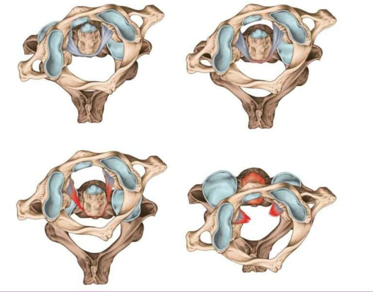
ही रोग परिस्थिती मानेचे पहिले दोन मणके हलले तर होते. सुदैवाने अ‍ॅटलांटो अ‍ॅक्सियल डिस्लोकेशन एक अत्यंत दुर्मिळ स्थिती आहे. पण जर कधी असे झाले तर त्याचे गंभीर परिणाम होऊ शकतात. रुग्णाला क्वाड्रिप्लेजिया, म्हणजेच मानेखालील सर्व शरीराचा अर्धांगवायू होऊ शकतो. परंतु अटलांटोअॅक्सियल डिस्लोकेशन म्हणजे काय हे समजून घेण्याआधी, आपण मानेच्या मणक्यांचे शरीरशास्त्र समजून घेतले पाहिजे, आणि विशेषत: स्पाइनल कॉलमचा पहिला मणका, ज्याला ‘अ‍ॅटलास’ म्हणतात, आणि दुसरा मणका, ज्याला ‘अ‍ॅक्सीस’ म्हणतात याची माहिती घ्यायला हवी. अॅटलास आणि अ‍ॅक्सीस हे दोन मणके एकमेकांशी जोडलेले असतात व या सांध्याला अटलांटो-अ‍ॅक्सीयल जॉइंट म्हणतात.F

मानेच्या मणक्याचे शरीरशास्त्र

Table of Contents

मानेचे मणके कवटी आणि छातीच्या मणक्यांच्या मधे असलेला मणकाच्या स्तंभाचा वरचा भाग आहे. यात सात मणके असतात आहेत, त्यापैकी दोन मणक्यांना विशेष नावे आहेत:

  • पहिला सर्व्हायकल मणका (C1) अॅटलास म्हणून ओळखले जाते.
  • दुसरा सर्व्हायकल मणका (C2) अॅक्सीस म्हणून ओळखला जातो.

सर्व्हायकल (म्हणजे मानेचे) मणके, एकूण सात, इतर मणक्यांपेक्षा वेगळे आहेत आणि थोडे अधिक नाजूक आहेत. आपण पहिल्या दोन सर्व्हायकल मणक्यांवर लक्ष केंद्रित करू. कारण या दोन मणक्यामधील सांधा हालला तर काय होते या विषयीच हा लेख आहे.

अ‍ॅटलास मणका

अ‍ॅटलास हा पहिला सर्व्हायकल म्हणजे मानेचा कशेरुक आहे आणि कवटी आणि अ‍ॅक्सीस मणक्यांच्या मध्ये असतो. हे दोन मणके इतर मानेच्या मणक्यांपेक्षा बरेच वेगळे असतात.

अ‍ॅक्सीस मणका

अ‍ॅक्सीस मणका अॅटलस आणि मानेच्या तिसऱ्या मणक्यांच्या मध्ये असतो. हा अटलांटो-अ‍ॅक्सीयल सांध्या द्वारे अ‍ॅटलास सोबत जोडलेला असतो. या सांध्यामुळे धडापासून डोके स्वतंत्रपणे फिरू शकते.

अ‍ॅक्सीस मणका दोन्ही बाजूला असलेल्या अटलांटो-अ‍ॅक्सीयल सांध्यात अॅटलसशी जोडलेला असतो.

मानेच्या मणक्याचे अस्थिबंधन (लिगामेन्ट)

मानेच्या मणक्यामध्ये सहा प्रमुख अस्थिबंधने असतात. अस्थिबंधने अतिशय मजबूत हाडे जोडणारी ऊतके असतात व ती सर्व मणक्यांना एकत्र ठेवतात.

मानेच्या मणक्याची दुखापत

उभेच्या उभे डोक्यावर पडल्यामुळे होणारी दुखापत

जर कोणी उंचावरून उथळ पाण्यात सूर मारला तर ओक्सीपीटल कॉन्डाइल्स आणि अ‍ॅक्सीस यांच्यातील अॅटलसच्या बाजूकडील वस्तुमान संकुचित होऊ शकतात. यामुळे एक किंवा दोन्ही आधीच्या/पुढील कमानी फ्रॅक्चर होऊन मणके वेगळे होऊ शकतात.

जर पडणे खूप वेगाने झाले तर, अॅटलसचे ट्रान्सव्हर्स लिगामेंट देखील फाटून जाऊ शकते.

हायपरएक्सटेन्शन (व्हिप्लॅश) इजा

ट्रॅफिक अपघातात मागील बाजूने जोराने टक्कर लागणे किंवा रग्बी फुटबॉलमध्ये टॅकल होणे या दोन्हीचा परिणाम म्हणजे डोके मागील बाजूस खांद्यावर जोरात फेकले जाऊ शकते. या मुळे व्हिप्लॅश इजा होऊ शकते. किरकोळ प्रकरणांमध्ये, मणक्याचे अ‍ॅन्टीरीयर लॉन्जीट्यूडीनल लिगामेन्टला दुखापत होते व त्यामुळे रुग्णाला तीव्र वेदना होत राहतात.

अटलांटोअॅक्सियल सबलक्सेशन

अटलांटोअॅक्सियल दुखापतींमध्ये सर्वात वाईट परिस्थिती म्हणजे अॅटलस आणि अॅक्सीस मणक्यांच्या सांध्यचे विघटन किंवा सबलक्सेशन होणे. सबलक्सेशन म्हणजे एकमेकांच्या संबंधात दोन मणक्यांची चुकीची जुळणी. सबलक्सेशन अनेक आरोग्य समस्यांचे कारण होते. हे बहुतेक वेळा अॅक्सीस मणक्याच्या पातळीवर त्या ठिकाणी घडते, जिथे त्याचे शरीर तिसऱ्या मानेच्या मणक्यांच्या संदर्भात पुढे सरकते. हे समजायला जरा क्लिष्ट आहे. अशा दुखापतीमुळे पाठीचा कणा खराब होऊ शकतो. क्वाड्रिप्लेजिया किंवा मृत्यू होऊ शकतो. सामान्यतः, मानेच्या मणक्याच्या खालच्या स्तरावर सबलक्सेशन उद्भवते.

हँगमॅनचे फ्रॅक्चर

हँगमॅनचे फ्रॅक्चर म्हणजे अॅक्सीस मणक्याच्या हाडांच्या स्तंभाचे फ्रॅक्चर होय. हे विशेषत: उच्च वेगाने हायपरएक्सटेन्शन होणे (डोके मागच्या बाजुला जोराने वळणे) आणि डोक्याची जागा हलल्यामुळे होते. अशी दुखापत प्राणघातक ठरण्याची शक्यता असते कारण पाठीचा कणा तुटतो, व त्यामुळे बेशुद्ध पडणे, श्वसनक्रिया बंद होणे आणि हृदयक्रिया बंद पडणे वगैरे होते.

डेन्स फ्रॅक्चर

हे अ‍ॅक्सीस मणकामध्ये उद्भवते. या प्रकारची फ्रॅक्चर अनेकदा अस्थिर असतात आणि त्यांच्यामुळे एव्हॅस्क्युलर नेक्रोसिसचा धोका असतो. मणक्याच्या कोणत्याही फ्रॅक्चर मधे होते त्याप्रमाणे, पाठीचा कण्याला गंभीर दुखापत होण्याचा धोका असतो.

अटलांटो-अ‍ॅक्सीयल सांध्याचे डिस्लोकेशन

अटलांटो-अ‍ॅक्सीयल सांध्याचे डिस्लोकेशन किंवा फ्रॅक्चरमुळे पाठीचा कणा गंभीर रीते दाबला जाऊ शकतो आणि त्यामुळे वरच्या पाठीच्या कण्याला आणि खालच्या मेडुला ओब्लॉन्गाटाला गंभीर न्यूरोलॉजिकल इजा होऊ शकते. रुग्णाला चारही अंगांचा अर्धांगवायू (क्वाड्रिप्लेजिया) होऊ शकतो. मानेच्या पाठीमागे आणि हातावर तीव्र मानदुखी होते. रुग्णाला सतत तीव्र वेदना होतात.

अटलांटो-अ‍ॅक्सीयल सांध्याचे डिस्लोकेशन (AADs) चे विस्थापनाच्या दिशा आणि समतलतेनुसार चार प्रकारांमध्ये वर्गीकरण केले जाऊ शकते जसे की पूर्ववर्ती-पोस्टरियर, रोटरी, मध्यवर्ती आणि मिश्रित विस्थापन. तथापि, शस्त्रक्रियेच्या दृष्टिकोनातून हे दोन श्रेणींमध्ये विभागले गेले आहेत जसे की रीड्युसीबल करण्यायोग्य [RAADs किंवा Reducible Atlanto Axial Dislocations] आणि इरीड्युसीबल [IAADs किंवा Irreducible Atlanto Axial Dislocations].

अटलांटो-अ‍ॅक्सीयल सांध्याचे डिस्लोकेशन किंवा फ्रॅक्चरमुळे पाठीचा कणा गंभीर संक्षेप होऊ शकतो ज्यामुळे वरच्या पाठीच्या कण्याला आणि खालच्या मेडुला ओब्लोंगाटाला गंभीर न्यूरोलॉजिकल इजा होऊ शकते. रुग्णाला चारही अंगांचा अर्धांगवायू (क्वाड्रिप्लेजिया) होऊ शकतो. मानेच्या पाठीमागे आणि हातावर तीव्र मानदुखी पसरलेली असते. रुग्णाला सतत तीव्र वेदना होतात.

अटलांटो-अ‍ॅक्सीयल सांध्याचे डिस्लोकेशन (AADs) चे विस्थापनाच्या दिशा आणि समतलतेनुसार चार प्रकारांमध्ये वर्गीकरण केले जाऊ शकते जसे की पूर्ववर्ती-पोस्टरियर, रोटरी, मध्यवर्ती आणि मिश्रित विस्थापन. तथापि, शस्त्रक्रियेच्या दृष्टिकोनातून हे दोन श्रेणींमध्ये विभागले गेले आहेत जसे की कमी करण्यायोग्य [RAADs किंवा Reducible Atlanto Axial Dislocations] आणि irreducible [IAADs किंवा Irreducible Atlanto Axial Dislocations] असे असते.

सर्जिकल उपचार

RAAD साठी, पोस्टरियर फ्यूजन केले जाते, म्हणजे मागच्या बाजुने मणके जोडले जातात. स्क्रू सांधे आणि वर्टिब्रल बॉडीमध्ये बसवले जातात आणि वायरिंग केले जाते. बहुतेकदा, IAAD डायनॅमिक एक्स-रे तपासणीमध्ये दिसते व ते स्नायूंच्या स्पाझम अथवा वांगामुळे देखील असू शकते.

वरील अतिशय गुंतागुंतीच्या शस्त्रक्रिया प्रक्रियेचे अतिशय सोप्या पद्धतीने स्पष्टीकरण आहे. AAD [अटलांटो-अक्षीय विस्थापन] वर उपचार करण्यासाठी विविध पद्धती आहेत. तथापि, हे वैद्यकीय अभ्यास व ज्ञान नसलेल्या लोकांना समजणे खूप क्लिष्ट होईल, म्हणून त्याचे तपशीलवार वर्णन केलेले नाही.

शस्त्रक्रियां करतांना अचूकता आवश्यक असते. त्यामुळेच मिरजेतील समर्थ न्यूरो आणि मल्टीस्पेशालिटी हॉस्पिटल यांसारख्या गुंतागुंतीच्या शस्त्रक्रिया करू शकतात. कारण समर्थ हॉस्पिटलमध्ये सर्जिकल नेव्हिगेशन सिस्टीम आहे जी अशा गुंतागुंतीच्या शल्याक्रिया योग्य रीतीने करू शकतात.

अटलांटो-अ‍ॅक्सीयल सांध्याचे सबलक्सेशन किंवा डिस्लोकेशनची जटिलता व अवघडता अतिशयोक्त रीते साकार केली आहे असे वाचकांना वाटत असले तरी, हे लक्षात ठेवले पाहिजे की जर शस्त्रक्रियेत थोडीशी जरी चूक झाली तर रुग्णाला क्वाड्रिप्लेजिया किंवा मृत्यू देखील होऊ शकतो. योग्य व यशस्वी शस्त्रक्रियेसाठी फक्त एकच संधी मिळते. अन्यथा रुग्णाला अयशस्वी शस्त्रक्रियेचा त्रास होऊ शकतो.

त्यामुळे सर्जिकल नेव्हिगेशनचा वापर, ज्यामध्ये सुरुवातीच्या चीर्याची नेमकी ठिकाणे आणि स्क्रू आणि वायर्स नेमक्या कोणत्या ठिकाणी बसवल्या पाहिजेत हे कळते, जेणेकरुन पाठीचा कणा आणि मणक्यांच्या दरम्यान बाहेर पडणाऱ्या पाठीच्या मज्जातंतूंना सर्जरी दरम्यान इजा होत नाही.

वैद्यकीय उपचार

अटलांटो-अ‍ॅक्सीयल अस्थिरता (ए.ए.आय.), सबलक्सेशन किंवा डिस्लोकेशनसाठी कोणतीही औषधे नाहीत. बहुतेक प्रकरणांमध्ये पेशंट येतो तेव्हा अस्थिरतेच्या तीव्रतेमुळे, कॉर्टिकोस्टिरॉईड्स औषधांचा थोडासा फायदा होतो. कॉर्टिकोस्टिरॉईड्समुळे न्यूरोलॉजिकल समस्या बर्या होत नाहीत. खरं तर कॉर्टिकोस्टिरॉईड्समुळे साइड इफेक्ट्स होऊ शकतात. तीव्र क्लेशकारक परिस्थितीमध्ये, कॉर्टिकोस्टिरॉईड्स द्यावेत की नाही हे विवादास्पद आहे. अमेरिकन अॅकॅडमी ऑफ न्यूरोलॉजिकल सर्जन्स (A.A.N.S.) द्वारे प्रदान केलेल्या मार्गदर्शक तत्त्वांमध्ये तीव्र आघात झालेल्या रुग्णामध्ये कॉर्टिकोस्टिरॉईड्स किंवा गॅंग्लीओसाइड्सच्या वापराविरूद्ध अनेक पुरावे आहेत.
पाठीचा कणा दाबला गेल्याची लक्षणे आढळल्याशिवाय, AAI ला उपचारांची आवश्यकता नसते. लक्षणे दिसू लागल्यानंतर, सर्जिकल स्थिरीकरण होईपर्यंत मानेच्या मणक्याचे मानेचा पट्टा बांधून स्थिरीकरण केले जाते.

रोटेटरी अटलांटोअॅक्सियल डिस्लोकेशनची लक्षणे काय आहेत?

मुख्य लक्षण म्हणजे टॉर्टिकॉलिस. टॉर्टिकॉलिस म्हणजे मान कायची वाकडी असणे. हे वेदनारहित आहे आणि बहुतेकदा, कोणतीही न्यूरोलॉजिकल लक्षणे नसतात. रोटेटरी अटलांटोअॅक्सियल डिस्लोकेशन कमी करण्यायोग्य (रीड्युसीबल) किंवा अपरिवर्तनीय (इरीड्युसीबल) प्रकारांमध्ये विभागले जाऊ शकते. घटता येण्याजोग्या प्रकाराचे रोटरी अटलांटोअॅक्सियल डिस्लोकेशन असे वर्गीकृत केले जाते जेथे डायनॅमिक प्रतिमांवर किंवा गर्भाशयाच्या ग्रीवेच्या कर्षणाच्या संस्थेनंतर अव्यवस्था कमी होते. टॉर्टिकॉलिस बहुधा वेदनारहित असते आणि बहुतेकदा, टॉर्टिकॉलिस असूनही कोणतीही न्यूरोलॉजिकल लक्षणे नसतात. रोटेटरी अटलांटोअॅक्सियल डिस्लोकेशन कमी करण्यायोग्य (रीड्युसीबल) किंवा अपरिवर्तनीय (इरीड्युसीबल) प्रकारांमध्ये विभागले जाऊ शकते. इरीड्युसीबल (कमी होण्याजोग्या) प्रकाराचे रोटरी अटलांटोअॅक्सियल डिस्लोकेशन तेव्हा वर्गीकृत केले जाते जेव्हा डायनॅमिक प्रतिमांवर डिस्लोकेशन कमी होते.

ऑक्सीपूटो-अटलांटल डिस्लोकेशन साठी उपचार काय आहे?

ऑक्सीपूटो-अटलांटल डिस्लोकेशन शी संबंधित प्रकार २ च्या जखमांसाठी आणि/किंवा निदान 14 दिवसांपेक्षा जास्त विलंब झाल्यास, पोस्टीरियर अटलांटोएक्सियल किंवा ऑक्सीपूटो-अटलांटो अॅक्सीयल आर्थ्रोडेसीस [म्हणजे सांध्याच्या दोन हाडांना कायमचे जोडून टाकणे] करवून घेण्याचा सलाल दिला जातो. इतर जखमांवर पोस्टरियर आर्थ्रोडेसिसने उपचार करण्यात येतात.

रोटरी अटलांटोअॅक्सियल अस्थिरतेचा उपचार कसा करावा?

अ‍ॅक्सीस, अ‍ॅटलसच्या मागील कमान, आणि अ‍ॅटलस आणि अॅक्सीसच्या पैलूंच्या स्पायनस प्रोसेसला धरून ठेवणे आणि योग्य प्रकारे हलवणे यामुळे रोटरी डिस्लोकेशन कमी करता येते.

थोडक्यात सांगायचे म्हणजे…

नमूद केल्याप्रमाणे, अटलांटोअॅक्सियल डिस्लोकेशन किंवा सबलक्सेशन फार दुर्मिळ आहे. बहुतेक लोकांसाठी वर दिलेली जटिल रोग स्थिती समजून घेणे सोपे नाही. समर्थ न्यूरो आणि मल्टीस्पेशालिटी हॉस्पिटलमध्ये अटलांटोअॅक्सियल डिस्लोकेशनवर उपचार करण्यासाठी पायाभूत सुविधा आणि सर्जिकल टीम आहेत.
How to Cure Cervical Spondylosis Permanently

मानेचा (म्हणजे सर्व्हायकल) स्पॉन्डिलायसिस कायमचा कसा बरा करावा स्पॉन्डिलायसिस उपचार सर्व्हायकल स्पॉन्डिलायसिस म्हणजे सकारात्मक आणि ताणशील गोंधळलेली असलेली समस्या

How to Cure Cervical Spondylosis Permanently

By Dr.Ravindra Patil

What is Cervical Spondylosis

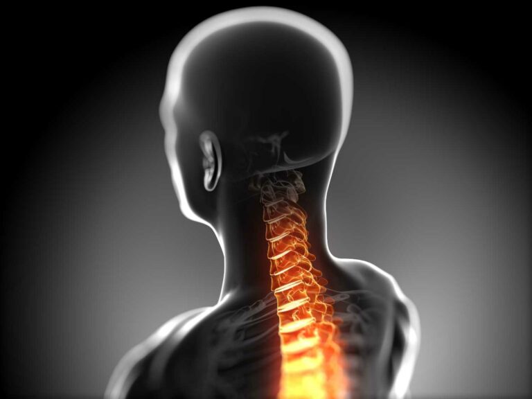
Cervical spondylosis is a common, age-related disorder of the cervical spine that is located in the neck region. This condition is also known as neck arthritis or cervical osteoarthritis. With age, the cervical discs gradually face wear and tear resulting in the breakdown of the structure. The fluid present between the cervical discs is lost which makes the spine stiffer, leading to cervical pain. This results in changes in the bones, discs, and joints at the spine of the neck.

As a result of the fluid dehydration and degeneration of the discs and cartilage in the surrounding area, abnormal growth or spurs of bones called osteophytes can occur leading to the narrowing of the passage from where spinal nerves exit, causing a related condition called cervical spinal stenosis.

Table of Contents

What is Spondylosis?

Spondylosis is an umbrella term that is used to describe any degeneration of the spinal cord. Spondylosis symptoms involve acute pain in the neck and back in old-age. Cervical spondylosis is prevalent among middle-aged and elderly people.

Spondylitis vs Spondylosis

Spondylitis and spondylosis both affect the spinal cord but there is a basic difference between the two. The meaning of spondylitis is that it is the inflammation of one or more typical cervical vertebrae leading to pain in the spine. Cervical spondylosis mostly refers to any kind of degeneration of the spine. So, even though the effects of both the diseases might be similar, their origin and cause are completely different.

Symptoms of Cervical Spondylosis: What is the difference?

Cervical spondylosis symptoms can vary from person to person and the extent of cervical spine pain depends on the age of the affected person. Some of the common spondylosis symptoms are:

  • Excessive stiffness at the neck leads to pain in neck
  • Cervical spondylosis headache that may originate at the main cervical spondylosis pain areas
  • Pain in the shoulder and arms
  • Failure to properly turn the head around
  • Grinding noise or sensation when trying to turn the neck
  • Cervical pain is severe in the morning and at night before bedtime
  • If cervical spondylosis leads to spondylotic myelopathy, the following symptoms may occur:
    • Abnormal tingling, numbness, and weakness in the arms, hands, feet, and legs
    • Lack of balance and coordination
    • Abnormal reflexes, random muscle spasms
    • Loss of control over urinary bladder and bowels

Causes of Cervical Spondylosis

Wear and tear of the vertebrae in the spinal cord causes degenerative spondylosis. Other causes are:

  • Bone spurs or abnormal outgrowths of bones called osteophytes
  • Dehydrated Spinal Discs
  • Herniated Discs:
  • Injury of the neck
  • Stiffness of the Ligament
  • Excessive Stress and Overuse

Risk Factors of Cervical Spondylosis

The greatest risk factor of cervical spondylosis is aging. Other risk factors are:

  • Heavy physical actions like heavy lifting
  • Poor posture in people who work at desk jobs
  • Genetic factors and family history
  • Smoking
  • Being overweight and inactive

How to prevent cervical spondylosis?

  • Avoid loading heavy objects on the head or neck. Do not sleep on a high-pillow.
  • For people with desk jobs, maintain a proper posture
  • Lean back your head once in a while.
  • Swimming is considered to be a relaxed exercise for cervical pain
  • Perform general physical activities.
  • Do not be inactive for a long period of time, take exercise

Diagnosis of Cervical Spondylosis

1. Physical Examination

  • Testing the reflexes of the patient and checking for the presence of muscle weakness and deficits in the sensory nerve function is done.
  • Tests are done to determine the range of neck movement
  • Overall walking movement is checked

2. Imaging Tests

  • X-Rays
  • CT Scan
  • MRI Scan
  • Myelogram

3. Nerve Function Tests

  • Electromyogram (EMG) records the passage of nerve signals in the body by measuring the electrical activity of the nerves.
  • Nerve Conduction Study – the speed and strength of the signals sent by the nerve endings in the body are checked.

Treatment of Cervical Spondylosis

Spondylosis treatments mainly focus on how to cure cervical spondylosis permanently, give pain relief and lower the risk of extensive damage to your vertebrae.

Physiotherapy

  • Physiotherapy for cervical spondylosis is a great way for pain relief. A trained physical therapist can teach all the exercises for cervical spondylosis.
  • Neck traction: it uses weights to help increase the space between the cervical joints and reduce the pressure forming on the cervical discs and nerve roots.

Medications

Commonly administered medications for the treatment of Cervical Spondylosis are:

  • Muscle relaxants
  • Narcotics are used for severe neck pain
  • Anti-seizure medications help to reduce pain caused due to damage of nerves
  • Steroids: Used to reduce the occurrence of tissue inflammation and reduce pain
  • Non-Steroidal Anti Inflammatory Drugs [NSAIDS]
  • Antidepressants help in relieving severe neck and shoulder pain
  • Certain homeopathic medicines also help

Home Remedies for Cervical Spondylosis

  • Take enough rest
  • Maintain a proper posture
  • People who acquire spondylosis at an early age normally do so because of bad study habits
  • Heat and cold treatment help provide relief and also works in relaxing the sore muscles of the neck and shoulders. Using a heating pack or a cold pack can be a good home remedy for cervical spondylosis
  • Chiropractic manipulation that can help in order to control events of more severe pain due to cervical spondylosis. However the chiropractor must have the requisite skills of manipulating the neck
  • Wearing a neck collar for cervical spondylosis helps limit movement and provides support for the neck and shoulders. However, these neck collars should not be worn for a longer period of time as it can cause muscle weakness.
  • Several people opt for ayurvedic medicine for cervical spondylosis and Patanjali medicine for cervical spondylosis to cure their ailment

The Need for Cervical Spondylosis Surgery

Cervical spondylosis is not progressive even though patients might have to live with severe pain for long periods of time all their life as it is a chronic condition. Cervical Spondylosis Surgery is only advised by doctors in rare cases when other forms of treatment do not help to ease the pain. For people who suffer from a progressive loss of function in the arms, hands, and legs, surgery may be required to fix the defect at the source point. This is because any form of compression at the cord may result in permanent functional disability.

In cases of the surgery, the main goal is to remove the areas or sources that have been applying extra pressure to the spinal cord and nerve endings. Some extensive forms of surgery may also involve the insertion of implants or bone grafts to add stabilization. Doctors also perform a fusion of the degenerated discs as a cure for cervical spondylosis.

Road to Recovery and Aftercare

Cervical spondylosis is not uncommon among people who have crossed their 60s. Early diagnosis of this disease can help doctors recommend a conservative line of treatment to help ease the pain. Though this condition is not reversible, with proper care, home remedies, and lifestyle practices, patients overcome the stiffness and discomfort.

How to cure cervical spondylosis permanently?

This is a very difficult question to answer as has been mentioned, the treatment of cervical spondylosis is primarily aimed at pain relief cervical exercises by physiotherapy and medicines. Surgery in this condition is done only when there is a physical problem like a bone spur pressing on the spinal cord, or a herniated disc pressing on the nerves or a compressed disk ‘pinches’ a nerve or the spinal canal is narrowed, due to spinal stenosis. Samarth Neuro and Super-speciality Hospital in Miraj offers medical and physiotherapy treatment for cervical spondylosis. If required, surgery for cervical spondylosis can also be done at Samarth Hospital in Miraj, Maharashtra.

Cranio-Vertebral Junction Anomalies

Cranio-Vertebral Junction Anomalies

Cranio-Vertebral Junction Anomalies

By Dr.Ravindra Patil

Anomaly

An anomaly is something that deviates from what is standard, normal, or expected. Cranio-vertebral junction anomalies mean any situation where there is some deviation from what is the ‘normal’ cranio-vertebral junction.

CVJ

The cranio-vertebral junction (CVJ) is present between the base of the skull and the upper region of the cervical spine. Any deviations or deformities in this region are known as cranio-vertebral junction anomalies. They can occur as congenital anomalies, developmental or secondary to other disease processes. Cranio-vertebral anomalies are classified into different types based on their causative factors.

The craniovertebral junction (CVJ) has unique anatomical structures that separate it from the cervical spine. In addition to housing vital nerves and blood vessels, the majority of head movements like flexion, extension, and looking sideways is accomplished at the CVJ.

The CVJ is a complex combination of osseous and ligamentous supports allow for stability despite a large degree of motion. To understand the anomalies of the CVJ, an understanding of anatomy and biomechanics in short is essential to understand the various anomalies and diseases that may affect this region.

The craniovertebral junction (CVJ) is the joint between the skull, atlas [first cervical vertebra], and axis [second cervical vertebra]. It is a complex area that houses vital nerves and blood vessels. This a joint has the most mobility as compared to any other joint of the vertebral column or the spine. The CVJ represents the transition between the brain and cervical spine. The majority of the spine’s rotation, flexion, and extension occur between the occiput, the atlas, and axis.

Table of Contents

Causes and risk factors of Cranio-vertebral Junction Anomalies

There are various factors associated with CVJ anomalies. Some of them include:

  • Skeletal structure abnormalities
  • Congenital systemic disorders such as Achondroplasia, Down syndrome, etc.
  • Infections
  • Metastatic Tumours effecting bones
  • Slow growing tumours in the CVJ
  • Rheumatoid Arthritis
  • Traumatic injuries by an accident, a fall etc.

Signs and symptoms of Cranio-vertebral Junction Anomalies

The signs and symptoms of CVJ anomalies vary based on the cause, severity and specific structures affected. However, some common signs and symptoms may include:

  • Neck pain
  • Headache in the back region of the head
  • Worsening head and neck pain with movement.
  • Decreased range of motion in the CVJ
  • Increased pain intensity while coughing or bending

Symptoms associated with spinal cord compression in the CVJ may include:

  • Weakness in arms, legs or both.
  • Numbness and tingling sensation on bending the neck.
  • Decreased sensations of heat, cold and pain in the legs/hands/feet.
  • Confusion and dizziness

Symptoms associated with brain stem dysfunction may include:

  • Sleep apnoea
  • Impaired balance and coordination (Ataxia)
  • Involuntary eye movements (Nystagmus)
  • Facial paralysis

Symptoms associated with lower cranial nerve dysfunction may include:

  • Difficulty in swallowing
  • Muscle weakness
  • Impaired hearing
  • Paralysis of soft palate and tongue atrophy
  • Diagnosis of Cranio-vertebral Junction Anomalies

CVJ anomalies are diagnosed based on medical history, physical examination and diagnostic imaging scans. Lateral view X-rays, CT scan and MRI are used to visualise the structural abnormalities in the CVJ. CT scan is the most common diagnostic tool used for CVJ anomalies. However, if further investigations are required, MRI and CT myelography are considered.

Treatment of Cranio-vertebral Junction Anomalies

Surgery is the treatment of choice for CVJ anomalies. The type of surgery and approach is decided based on some factors which include: traumatic causes, non-traumatic causes, reducible or irreducible lesions. Considering these factors, the basic surgical procedures involved are reduction, immobilization, decompression and fixation. Treatment may include any one of the procedures or a combination which is determined based on the nature, severity and risk factors of the disease.

Treatments

  • Full endoscopic cervical canal stenosis decompression
  • Spinal fixation
  • Craniovertebral junction surgeries
  •  

CVJ Fixation

Due to the complex anatomical nature as well as the significant mobility of the CVJ, fixation of this region remains at times a challenging decision and a difficult execution. A wide variety of fixation methods exist and may include a combination of the following: screws, rods, wires/cables, bone grafts, hooks, or plates. Furthermore, arthrodesis [surgical immobilization of a joint by fusion of the bones] is also a challenge as there is little space and bone surface for sufficient bone grafting.

Fixation of the CVJ to the skull can be accomplished via small burr holes and wire or a combination of screws and plates, which allows for fixation to the cervical spine through connection of rods or additional plates in the spine. Bur holes with wire are not currently used as often, as the screw/rod/plate constructs have shown to be bio-mechanically superior in terms of screw pullout strength and stiffness.

The screws used in the CVJ are generally a larger diameter than those used in the cervical spine. They have a smaller pitch and blunt tips to prevent piercing the dura mater [a thick tissue layer covering the brain and the spinal cord].

Fixation in the cervical spine can be a combination of a number of methods as well, usually with the intention of connecting to the occiput fixation. The screws used in the cervical spine are generally polyaxial and placed into the lateral masses of the vertebrae.

Generally, the skull and the first cervical vertebra [atlas] segment is not fixated alone due to the large moment arm created by the cranium on C-1. However, there are situations in which the occiput and atlas can be fixated using the above listed devices, like screws, rods, wires and so on.  Sometimes a bone graft is placed in between the posterior arch of atlas and the base of the skull. Successful fusion rates of such surgeries have been reported up to 89%, but the patient’s head is not immediately stable after surgery. Such patients are required to remain in a halo neck fixation for 12 weeks.

Another option involves the use of fixing screws across joints. Similar to the above construct, this is usually reserved for isolated Skull-Atlas instability and is not easily incorporated into multisegment constructs. This technique increases stiffness in rotation but is poor in flexion-extension. Thus, this technique is used in conjunction with supplemental fixation to allow for sufficient stability.

Skull to Atlantoaxial Segment fixation

The occiput to atlantoaxial region is classically difficult to fixate as both the occipitoatlantal and atlantoaxial segments are highly mobile in flexion and extension, and additionally the atlantoaxial segment is very mobile in axial rotation. Any number of combinations of the previously mentioned techniques can be used. Occipitocervical fixation constructs consist of points of fixation along the skull, atlas vertebra and axis vertebra, with connection to some type of longitudinal element. These longitudinal elements span the segments in the CVJ and allow them to be rigidly fixated.

The above description may seem to be very complex to the lay reader. However, all efforts have been made to removed complicated medical words and use as simple words as possible.

To develop a strong CVJ construct, the longitudinal elements should be able to have multiple points of fixation along the junction, interface with all of the fixation points, have the ability to interface with the thickest regions of bone in the suboccipital region, and have the ability to be crosslinked.  Various types of longitudinal elements are available and may include rods, structural bone grafts, reconstruction plates, and hybrid devices combining plates and rods in preformed shapes. As with all spinal instrumentation, it is critical to choose the type of longitudinal element that best suits the individual patient’s needs based on the goal of surgery and the patient’s anatomy.

Samarth Neuro and Multispeciality hospital in Miraj, Maharashtra, has the infrastructure and team of doctors headed by neurosurgeon Dr Ravindra Patil to accomplish such complex surgeries. Besides as the hospital is located in a tier 2 city, the overall cost of such surgeries is much less in Samarth Hospital of Miraj as compared to surgeries done in large metropolitan cities.

Spina Bifida Causes, Symptoms and Treatment

Spina Bifida - Samarth neuro and super speciality hospital

Spina Bifida Causes, Symptoms and Treatment

By Dr.Ravindra Patil

What is Spina Bifida?

Spina bifida is a birth defect in which an area of the spinal column doesn’t form properly, leaving a section of the spinal cord and spinal nerves exposed through an opening in the back. Spinabifida occurs in 1 per 2,000 live births.

Table of Contents

Myelomeningocele

The most common and serious form of spinabifida is called myelomeningocele, in which a part of the spinal cord and surrounding nerves push through the open bones (vertebrae) in the spine and protrude from the foetus’s back. In myelomeningocele, spinal cord and nerves protrude from the back in a sac that is exposed to the amniotic fluid. Continuous bathing of the fragile developing spinal cord in amniotic fluid over the course of gestation is thought to result in progressive neurologic injury.

Other types of spina bifida

Other types of spinabifida include myeloschisis, lipomeningocele and myelocystocele. Open neural tube defects such as myelomeningocele and myeloschisis are treatable by foetal repair. Closed neural tube defects such as lipomeningocele and myelocystocele are not treatable by foetal repair.

Causes of spina bifida

Myelomeningocele has genetic and micronutrient causes. Mothers who have had a baby with spinabifida have up to a 4 percent risk of recurrence in subsequent pregnancies.

Spina bifida symptoms

The myelomeningocele lesion can occur at any level on the developing spine, but most are found in the lumbo-sacral region. Depending on the lesion’s location, myelomeningocele may cause:

  • Bladder and bowel problems (incontinence)
  • Sexual dysfunction
  • Weakness and loss of sensation below the defect
  • Inability to move the lower legs (paralysis) and other cognitive impairments
  • Orthopaedic malformations such as club feet or problems of the knees or hips
  • Generally, the higher the defect is located on the spine, the more severe the complications.

In many cases, the brain develops an Arnold-Chiari II malformation, in which the hindbrain herniates or descends into the upper portion of the spinal canal in the neck. This herniation of the hindbrain blocks the circulation of cerebrospinal fluid, causing hydrocephalus (accumulation of fluid in the brain), which can injure the developing brain. Ventricular shunting (placement of a thin tube into the ventricles of the brain) is used to drain fluid and relieve hydrocephalus.

Hindbrain Herniation and Ventricular Shunting

Ventricular shunting is done to treat hydrocephalus. It involves placement of a thin tube into the ventricles of the brain to drain fluid and relieve hydrocephalus. The other end of the tube is usually put in the peritoneum [abdomen] of the patient for continuous drain.

Evaluation and diagnosis of spina bifida

As with other birth defects, most myelomeningoceles are discovered by routine ultrasound evaluation between 16 and 18 weeks into the pregnancy. Sonographic features suggestive of myelomeningocele include a cystic mass anywhere on the spine. Abnormally high alpha-fetoprotein (AFP) will support the diagnosis.

Other evaluation methods for suspected spinabifida are:

  • High-resolution level II ultrasound — to confirm the diagnosis and determine the location of the lesion, and to assess for any other birth defects such as club feet
  • Ultrafast foetal MRI — to confirm presence of the Chiari II malformation and to screen for evidence of any other neurologic abnormalities
  • Foetal echocardiogram — to determine any problems with the heart
  • Amniocentesis — to confirm the presence of elevated amniotic fluid alpha-fetoprotein (AFAFP) levels and acetylcholinesterase (AChE), which indicate open neural tube defects
  • Maternal serum alpha-fetoprotein (MSAFP) test — to confirm elevated AFP levels in the mother’s blood

After evaluation, a multidisciplinary team works together to review test results, confirm the diagnosis, explain spina bifida treatment options and potential outcomes, and answer all the patient’s questions.

Treatment

The two main spina bifida treatment options are foetal surgery during pregnancy or surgery on the baby right after birth.

Deciding whether prenatal or postnatal spinabifida repair is appropriate is influenced by several considerations, including gestational age, the level of the myelomeningocele lesion on the spine, presence of the Chiari II malformation, and a number of important maternal health factors.

Spina bifida surgery after birth

If postnatal spina bifida surgery is recommended or selected, your pregnancy is monitored and a caesarean delivery is planned at 37 weeks.

Traditional spina Bifida treatment takes the form of surgical repair 24 to 48 hours after birth. Your child will undergo general anaesthesia. A paediatric neurosurgeon removes the MMC sac, if one is present, and closes the surrounding tissue and skin over the defect to protect the spinal cord. After surgery, your baby will receive care in the Neonatal Intensive Care Unit (NICU).

Spina bifida surgery before birth

Because spinal cord damage is progressive during gestation, prenatal repair of myelomeningocele may prevent further damage. Scientific studies have demonstrated that prenatal repair can offer significantly better results than traditional postnatal repair.

  • It reduced the need to divert fluid from the brain using ventricular shunting
  • Reduced the incidence or severity of neurological effects such as impaired motor and sensory function of the legs
  • Improved mobility and improved the chances that a child will be able to walk independently
  • Reversed the hindbrain herniation component of the Arnold-Chiari II malformation

Foetal surgery for spinabifida is a complex and challenging procedure, requiring the most expert, comprehensive care for both mother and foetus. The surgical team’s level of experience and all aspects of care surrounding the operation are of paramount importance.

If your unborn baby has been diagnosed with myelomeningocele, the surgery is performed between 23 weeks and 25 weeks of gestation.

Spina bifida prognosis

Rarely, children can die due to complications associated with Chiari II malformation or hydrocephalus. Many patients require shunts (85 percent), and 45 percent of shunts develop complications within one year.

Follow-up care

Children operated for spina Bifida require comprehensive long-term follow-up, from the time of repair through adolescence. It is team work of a paediatrician, nurse, social worker, physical therapist, neurosurgeon, orthopaedic specialists, urologist and genetic counsellor.

Depending on the location of the lesion and outcome after surgery, follow-up care may also include:

  • Clean intermittent catheterization
  • Bowel management
  • Lower extremity bracing
  • Physical therapy evaluation and guidance to outside physical therapists and early intervention
  • Pressure sore management
  • Referral to appropriate psychosocial and financial resources
  • Consultation with other subspecialties including ophthalmology, nephrology, nutrition and feeding specialists, and plastic surgery

The Spina Bifida team works closely with each child’s primary care paediatrician and school, and helps families access appropriate community services. Ongoing follow-up is critical when it comes to improving outcomes and advancing treatment for patients with myelomeningocele.

Spina Bifida can lead to a variety of social and emotional challenges and lifelong quality-of-life issues. Hence proper diagnosis during pregnancy is essential.

Spina bifida occulta

Spina bifida occulta is the mildest type of spinabifida. It is sometimes called “hidden” spinabifida. In this birth defect, there is a small gap in the spine, but no sac comes out of the back. The spinal cord and the nerves usually are normal.

At Samarth Neuro & Super Speciality Hospital in Miraj, we are dedicated to providing expert care, ensuring the well-being and quality of life for individuals facing this condition.

Lumbar Spondylosis

Lumbar Spondylosis -Samarth

Lumbar Spondylosis

By Dr.Ravindra Patil

It is a painful condition of area of the lower back above the buttocks. But first let us understand each term clearly. Let us see ‘Lumbar’ means and what ‘Spondylosis’ means.

Table of Contents

What is ‘Lumbar’ Spine?

It refers to that part of the spinal column which has five vertebrae and is located at the lower back but above the buttocks. Painful conditions occur in the lumbar spine because it carries the weight of the upper body and is prone to many stresses and injuries during routine work and severe hard exertions.

Understanding spondylosis

Spondylosis refers to the development of age-related arthritis that affects the spine. More specifically, it involves a number of different degenerative issues that affect the disks and joints of the spine. It may occur in any part of the spine, from the neck to the back to the lower back. In this article we focus on the lower back or the ‘Lumbar’ region. Hence we call it Lumbar Spondylosis.

Why does spondylosis occur? Over time, disks between two vertebrae [called intervertebral disks] may become dehydrated and become compressed, meaning their height is reduced as a result of dehydration. Thus, they provide less cushioning and can actually “crack,” which can lead to herniation. Herniation means a part of our body protrudes out of a normal or an abnormal opening.

Similarly, the cartilage in the spine’s joints can wear down, resulting in the formation of bone spurs.  

These arthritic changes can not only cause pain in the back but can also lead to a narrowing of the spinal canal (also known as stenosis), which may compress nerves. Nerve compression causes symptoms like pain and numbness, which slowly increases with the passage of time. Later there may be loss of vital functions like bladder and bowel control. Individuals with spinal stenosis may have neurologic symptoms in their extremities, such as numbness, tingling, or weakness radiating into their arms or legs. Loss of bladder or bowel control may lead to further complications. 

What causes spondylosis?

The most common cause of spondylosis is the cumulative joint stress that occurs as people age. It predisposes them to osteoarthritis, a common form of arthritis typically associated with progressive “wear and tear” on joint cartilage. 

Spondylosis may also arise as a result of previous trauma to the spine. For instance, patients who:

  • Have had car accidents, falls, or other spinal injuries
  • Play competitive sports
  • Perform strenuous physical activity at work
  • Have had a previous neck or back surgery

The lumbar region is most affected by spondylosis because of its exposure to mechanical stress.

What are the risk factors for spondylosis?

The risk factors for spondylosis include the following:

  • Age 60 or older
  • Osteoarthritis affecting other joints
  • Physically demanding jobs that may require heavy lifting or bending
  • High-level athletics
  • Neck or back injury
  • Previous spine surgery
  • Obesity
  • Smoking
  • Physical inactivity

What are the symptoms of lumbar spondylosis?

Many with spondylosis do not have pain or neurologic symptoms. Some people may experience symptoms. They are:

  • Neck or back pain that worsens when coughing or sneezing
  • Decreased flexibility in and range of motion of the neck or back
  • “Clicking” sounds from the spine
  • Pain radiating from the lower back into the legs
  • Numbness, tingling, or weakness in the arms or legs
  • Unsteady gait
  • Muscle spasms
  • Headaches
  • Bladder or bowel dysfunction in severe cases

How is spondylosis diagnosed?

A person can be diagnosed with lumbar spondylosis by a medical history, physical exam, and reviews of diagnostic tests. When providing your medical history, it is important to share any history of spinal injuries or previous surgeries. Physical activity information must also be shared.

Your doctor will observe your walking ability and assess the range of motion of your spine to determine if you have any pain or stiffness. Palpation of the neck and back can also help identify the location of any tenderness associated with spondylosis. Your neurologic function will also be evaluated, including your sensation, strength, and reflexes. Special tests may also be performed to check for any signs of nerve impingement (from a disk herniation, for example). 

Finally, imaging studies like X-rays, computed tomography (CT scan), and magnetic resonance imaging (MRI) may also be used to identify any arthritic changes in the spine. Lab tests may also be useful for ruling out more concerning diseases, such as an infection or cancer.

Differential Diagnosis

When a patient is suffering from low back pain, there are a lot of other possible diseases that have to be considered and ruled out:

  • Rheumatoid arthritis
  • Excessive exercise pain
  • Ankylosing Spondylitis
  • Coccyx Pain
  • Spinal Masses
  • Infection
  • Discitis
  • Lumbar Compression Fracture
  • Mechanical Low Back Pain
  • Overuse Injury

Lumbar Spondylosis’s degeneration can influence spinal alignment, potentially exacerbating pre-existing Spinal Deformities, emphasizing the need for comprehensive diagnosis and treatment.

How is spondylosis treated?

Different treatments are available to treat spondylosis, depending on the type and severity of symptoms. 

Treatments for milder spondylosis include:

  • Activity modification, such as avoiding activities that cause pain
  • Medications, such as non-steroidal anti-inflammatory drugs (NSAIDs) like Ibuprofen and Diclofenac or Paracetamol
  • Hot/cold compresses, massage, and traction
  • Physical therapy, such as strengthening and flexibility exercises and ergonomic education
  • Spinal injections, such as an epidural around nerves and facet joints

In more severe cases, surgery might be the best course of action. It may involve the following procedures:

  • Decompression (laminectomy, discectomy), which involves the removal of bone spurs, disk herniations, and arthritis to relieve compression of the nerves and address neurologic symptoms
  • Fusion, which is the formation of bone across adjacent vertebral bodies to stabilize the spine and relieve pain

Other Surgical treatment for Lumbar Spondylosis

  • This surgery is used to relieve pain associated with a compressed nerve in the spine.
  • Disk replacement.
  • Interlaminar implant.
  • Physical Therapy Management
  • Educating the patient: may include reviews of lumbar anatomy, explanations of the concept of posture, ergonomics and giving appropriate back exercises.

Other non-surgical treatment modalities include

  • Lumbar Back Support: Can be beneficial for patients suffering from chronic LBP. It occurs to limit spine motion, stabilize, correct deformity and reduce mechanical forces. There is no consensus if it may function as a placebo or really improve pain and functional ability.
  • Special type of taping is done on the back to prevent moment in certain directions, but again it is thought to give more of a placebo effect of pain relief.

Clinical Bottom Line

Lumbar spondylosis can be described as a degeneration of the lumbar vertebrae.

  • It is a form of low back pain and is an important clinical, social, economic and public health problem affecting the worldwide population.
  • It is a disorder with many possible causes and many definitions.
  • Clinical diagnosis of lumbar spondylosis is done by MRI, CT, SPECT and X-ray and a general examination of the spine.
  • Mild cases are managed with medicines.
  • Severe cases need surgery.
  • The physical therapy has an important role, and may include lumbar traction, manipulation of the spine, massage therapy, TENS, back school and Lumbar back supports.
  • The choice of therapy depends on the individual wishes and the surgeons advice.

Lumbar Spondylosis’s degeneration can influence spinal alignment, potentially exacerbating pre-existing Spinal Deformities. This complexity was underscored when a patient, who had tumours in his spine twice, faced a critical situation. Here, surgical navigation emerged as a crucial factor in saving him. This advanced technique seamlessly complemented the comprehensive approach discussed earlier, highlighting the intricate nature of spinal health management.

Outlook for people with spondylosis?

Fortunately, most individuals with spondylosis will not experience symptoms and may never even realize that they have arthritis in their spines. However, for those who experience pain or neurologic changes due to nerve compression, safe and effective treatments are available.

गॅस्ट्रिक व्हर्टिगो: पोटदुखी आणि व्हर्टिगो एकत्र येऊ शकतात का?

Vertigo - Samarth

गॅस्ट्रिक व्हर्टिगो: पोटदुखी आणि व्हर्टिगो एकत्र येऊ शकतात का?

By Dr.Ravindra Patil

आश्चर्याची गोष्ट म्हणजे उत्तर ‘हो’ आहे.

‘गॅस’ (म्हणजे पोटात फुगलेली भावना) मुळे चक्कर येऊ शकते का? होय, हे शक्य आहे. पोटाच्या स्थितीमुळे चक्कर येऊ शकते. म्हणूनच याला कधीकधी ‘जठरासंबंधी व्हर्टिगोची लक्षणे’ असेही संबोधले जाते. जठराची समस्या कधीकधी चक्कर येण्याशी संबंधित असते. गॅस्ट्रिक समस्येची लक्षणे आणि चक्कर येण्याशी त्यांचा संबंध पाहू या.

ओटीपोटात दुखणे हे संक्रमण, अन्न विषबाधा, अल्सर किंवा काही कर्करोगांसारख्या विविध परिस्थितींमुळे होऊ शकते.

ओटीपोटात दुखणे, किंवा पोटदुखी आणि चक्कर येणे अनेकदा हाताशी असते. या लक्षणांचे कारण शोधण्यासाठी, हे जाणून घेणे महत्त्वाचे आहे की कोणते लक्षण पहिले आले – पोटाची समस्या का चक्कर ?

तुमच्या पोटाच्या आजूबाजूच्या वेदना स्थानिकीकृत किंवा सर्वत्र जाणवू शकतात, ज्यामुळे शरीराच्या इतर भागांवर परिणाम होतो. दुय्यम लक्षण म्हणून अनेक वेळा पोटदुखीनंतर चक्कर येते.

चक्कर येणे आणि व्हर्टिगो हे सहसा सारखेच मानले जाते, तथापि ते भिन्न आहेत. चक्कर येणे ही भावनांची एक श्रेणी आहे ज्यामुळे तुम्हाला असंतुलितता किंवा अस्थिरपणा वाटतो. व्हर्टिगो आल्यावर तुम्हाला असे वाटते की खोली किंवा जग तुमच्याभोवती फिरत आहे. चक्कर येणे अनेकदा किरकोळ कारणांमुळे होते आणि ते क्षणिक असते. व्हर्टिगो ही एक अशी स्थिती आहे ज्यासाठी अनेकदा औषधे आणि इतर उपचारांची आवश्यकता असते. चक्कर येणे आणि व्हर्टिगो होणे ही अनेक रोगांची लक्षणे असू शकतात. पण चक्कर येणे हा आजार मानला जात नाही. चक्कर येणे हे फक्त एक लक्षण आहे. काही प्रकरणांमध्ये, व्हर्टिगो हा आजार समजला जातो.

Table of Contents

लक्षणे

आपण ओटीपोटात दुखणे आणि त्याच्या विविध प्रकारांपासून सुरुवात करूया. ते असू शकते:

  • तीक्ष्ण वेदना
  • कंटाळवाण्या वेदना
  • चावल्यासारखी वेदना
  • सतत वेदना
  • वेदना येतात जातात
  • जळजळणार्या वेदना
  • क्रॅम्प (आटी) सारखी वेदना
  • एपिसोडिक किंवा नियमित पणे होणारी वेदना
  • एकसारख्या वेदना

कोणत्याही प्रकारच्या तीव्र वेदनांमुळे तुम्हाला डोके हलके होऊ शकते किंवा चक्कर येऊ शकते. पोटदुखी आणि चक्कर येणे अनेकदा उपचाराशिवाय निघून जाते. थोडी विश्रांती घेतल्यावर तुम्हाला बरे वाटू शकते. एकतर बसा किंवा झोपा आणि तुम्हाला फरक जाणवतो का ते पहा.

परंतु जर तुमच्या ओटीपोटात दुखणे आणि चक्कर येणे ही इतर लक्षणांसह, जसे की दृष्टी बदलणे आणि रक्तस्त्राव होणे, हे अंतर्निहित गंभीर वैद्यकीय स्थितीचे लक्षण असू शकते.

जर तुमची लक्षणे दुखापतीमुळे उद्भवली असतील, तुमच्या दैनंदिन कामांमध्ये व्यत्यय येत असतील किंवा उत्तरोत्तर वाईट होत असतील तर तुमच्या डॉक्टरांशी भेट घ्या.

क्वचित प्रसंगी, छातीत दुखणे पोटदुखीचे अनुकरण करू शकते. छातीत दुखणे सुरू असले तरीही वेदना तुमच्या पोटाच्या वरच्या भागात जाते.

जर या गोष्टी झाल्या तर ताबडतोब हॉस्पिटलच्या आपत्कालीन कक्षाला भेट द्या:

    • हृदयाचा एक असामान्य ठोका
    • डोके हलके होणे
    • छातीत दुखणे
    • धाप लागणे
    • तुमच्या खांदा, मान, हात, पाठ, दात किंवा जबड्यात वेदना किंवा दाब
    • घामयुक्त आणि चिकट त्वचा
    • मळमळ आणि उलटी

    ही हृदयविकाराच्या झटक्याची लक्षणे आहेत आणि त्यांना त्वरित वैद्यकीय मदतीची आवश्यकता आहे.

ओटीपोटात वेदना आणि चक्कर येण्याची संभाव्य कारणे

आश्चर्याची गोष्ट म्हणजे, अशा अनेक परिस्थिती आहेत ज्यामुळे पोटदुखी किंवा अस्वस्थता आणि चक्कर येते. त्यापैकी काही कारणे इतके महत्त्वाचे वाटत नाहीत, परंतु लक्षात ठेवा, हा डेटा अनेक वर्षांच्या अभ्यास आणि संकलनात गोळा केला जातो. यापैकी काही परिस्थिती वैद्यकीय आपत्कालीन आहेत आणि त्यांना त्वरित काळजी घेणे आवश्यक आहे.

  • अपेंडिसाइटिस
  • स्थानभ्रष्ट गर्भधारणा (एक्टोपीक प्रेगनन्सी)
  • स्वादुपिंडाचा रोग (पॅन्क्रियाटायटीस)
  • अन्न विषबाधा
  • गॅस्ट्रोइंटेस्टाइनल रक्तस्त्राव
  • खते आणि वनस्पती अन्न विषबाधा
  • विषारी मेगाकोलन
  • आतड्यांसंबंधी किंवा जठरासंबंधी छिद्र
  • पोटातील महाधमनीचे एन्युरिझम
  • पेरिटोनायटिस
  • जठराचा कर्करोग
  • एडिसोनियन संकट (तीव्र एड्रीनल संकट)
  • अल्कोहोलिक केटोअॅसिडोसिस
  • चिंता विकार
  • ऍगोराफोबिया
  • मूत्रखडे
  • हायपोग्लाइसेमिया (कमी रक्त शर्करा)
  • इलियस (आतडे आकुंचन पावण्यास असमर्थता यामुळे अन्नपदार्थ आतड्यांत जमून राहतात)
  • रासायनिक बर्न्स
  • पोटाचा फ्लू
  • ओटीपोटात मायग्रेन
  • ड्रग ऍलर्जी
  • अपचन
  • प्रीमेन्स्ट्रुअल सिंड्रोम (PMS)
  • वेदनादायक मासिक पाळी
  • पेरिफेरल व्हास्क्युलर रोग
  • आयसोप्रोपाईल अल्कोहोल विषबाधा
  • एंडोमेट्रिओसिस
  • मोशन सिकनेस
  • अतिरेकी व्यायाम करणे
  • निर्जलीकरण

खाल्ल्यानंतर ओटीपोटात दुखणे आणि चक्कर येणे कशामुळे होऊ शकते?

पोस्टप्रॅन्डियल हायपोटेन्शन

म्हणजे जेवणानंतर कमी रक्तदाब. खाल्ल्यानंतर तुम्हाला ओटीपोटात दुखणे आणि चक्कर आल्यास, तुमचे रक्तदाब स्थिर न झाल्यामुळे असे होऊ शकते. जेवणानंतर अचानक रक्तदाब कमी होणे याला पोस्टप्रान्डियल हायपोटेन्शन म्हणतात.

साधारणपणे, जेव्हा तुम्ही खाता तेव्हा तुमच्या पोटात आणि लहान आतड्यात रक्त प्रवाह वाढतो. तुमच्या शरीराच्या उर्वरित भागात रक्त प्रवाह आणि दाब राखण्यासाठी तुमचे हृदय देखील वेगाने धडधडते. पोस्टप्रॅन्डियल हायपोटेन्शनमध्ये, तुमचा रक्तप्रवाह पाचन तंत्राशिवाय सर्वत्र कमी होतो. पोस्टप्रॅन्डियल हायपोटेन्शनखालील असंतुलनास कारणीभूत ठरू शकते:

  • चक्कर येणे
  • पोटदुखी
  • छातीत दुखणे
  • मळमळ
  • धूसर दृष्टी

वृद्ध प्रौढ आणि खराब झालेले मज्जातंतू रिसेप्टर्स किंवा रक्तदाब सेन्सर असलेल्या लोकांमध्ये ही स्थिती अधिक सामान्य आहे. हे खराब झालेले रिसेप्टर्स आणि सेन्सर आपल्या शरीरातील इतर भाग पचनाच्या वेळी कशी प्रतिक्रिया देतात यावर परिणाम करतात.

गॅस्ट्रिक अल्सर

अल्सर म्हणजे त्वचा किंवा पोटाच्या आतील आवरण विस्कळीत होणे व ज्यामुळे खालील ऊतींचे भाग उघडे राहतात. जठरातील अल्सर हा तुमच्या पोटाच्या आतील आवरणात फाटलेला हिस्सा आहे. पोटदुखी अनेकदा खाल्ल्यानंतर काही तासांतच होते. सामान्यतः गॅस्ट्रिक अल्सर झाल्यास इतर लक्षणे खालील प्रमाणे असतात:

  • सौम्य मळमळ
  • भरल्यासारखे वाटणे
  • पोटाच्या वरच्या भागात दुखणे
  • मल किंवा लघवीमध्ये रक्त
  • छातीत दुखणे

रक्तस्रावासारखी गंभीर गुंतागुंत होईपर्यंत पोटातील बहुतेक अल्सर कोणाच्याही लक्षात येत नाहीत. यामुळे पोटदुखी आणि रक्त कमी झाल्यामुळे चक्कर येऊ शकते.

वैद्यकीय मदत कधी घ्यावी

सात ते 10 दिवस टिकणाऱ्या किंवा तुमच्या दैनंदिन कामांमध्ये व्यत्यय आणणाऱ्या कोणत्याही वेदनांसाठी नेहमी त्वरित वैद्यकीय मदत घ्या.

तसेच, जर तुम्हाला पोटदुखी आणि चक्कर याच बरोबर खालील लक्षणे असल्यास डॉक्टरांना भेटा:

  • दृष्टीमध्ये बदल
  • छाती दुखणे
  • खूप ताप
  • मान कडक होणे
  • तीव्र डोकेदुखी
  • शुद्ध हरपणे
  • तुमच्या खांद्यावर किंवा मानेत वेदना
  • तीव्र पेल्विक वेदना
  • धाप लागणे
  • अनियंत्रित उलट्या किंवा अतिसार
  • योनिमार्गात वेदना आणि रक्तस्त्राव
  • अशक्तपणा
  • तुमच्या लघवी किंवा मलामध्ये रक्त

याव्यतिरिक्त, तुम्हाला खालीलपैकी कोणतीही लक्षणे 24 तासांपेक्षा जास्त काळ जाणवल्यास तुमच्या डॉक्टरांचा सल्ला घ्या:

  • ऍसिड सारखे जळजळीत पाणी उलटी सारखे अन्ननळीतून वर येणे
  • तुमच्या लघवीत रक्त
  • डोकेदुखी
  • छातीत जळजळ
  • खाज सुटणे, फोड येणे
  • वेदनादायक लघवी
  • कारण न कळणारा थकवा
  • वरील लक्षणे बिघडत जाणे

ही माहिती केवळ आपत्कालीन लक्षणांचा सारांश आहे. तुम्हाला ओटीपोटात दुखणे आणि चक्कर येणे ही किरकोळ स्थिती आहे की गंभीर स्थिती आहे हे ठरवण्यासाठी हुषार राहणे आवश्यक आहे.

ओटीपोटात दुखणे आणि चक्कर येणे याचे निदान कसे केले जाते?

तुमचे डॉक्टर शारीरिक तपासणी करतील आणि निदान करण्यात मदत करण्यासाठी तुमच्या वैद्यकीय इतिहासाबद्दल विचारतील. तुम्ही तुमची लक्षणे तपशीलवार समजावून सांगितल्यास, ते तुमच्या डॉक्टरांना कारण निश्चित करण्यात मदत करेल.

उदाहरणार्थ, वरच्या ओटीपोटात दुखणे हे पेप्टिक अल्सर, स्वादुपिंडाचा दाह किंवा पित्ताशयाच्या आजाराचे लक्षण असू शकते. खालच्या उजव्या ओटीपोटात दुखणे हे किडनी स्टोन, अॅपेन्डिसाइटिस किंवा ओव्हरीयन सिस्टचे लक्षण असू शकते.

तुमच्या चक्कर येण्याच्या तीव्रतेकडे लक्ष द्या. हे लक्षात घेणे महत्त्वाचे आहे की हलके-डोकेपणा असे वाटते की आपण बेशुद्ध होणार आहात असे वाटते, तर ही संवेदना आहे की आपले वातावरण हलत आहे.

तुम्हाला व्हर्टिगो असल्यास तुमच्या संवेदी प्रणालीमध्ये समस्या असण्याची शक्यता जास्त असते. हा सहसा आतील कानाचा विकार असतो.

ओटीपोटात दुखणे आणि चक्कर येणे यावर उपचार कसे केले जातात?

काही प्रकरणांमध्ये, ओटीपोटात दुखणे आणि चक्कर येणे उपचारांशिवाय दूर होते. हे अन्न विषबाधा, पोटाचा फ्लू आणि मोशन सिकनेसमध्या वगैरे सामान्य आहे. दुर्दैवाने, असे नेहमीच होत नसते.

पोटदुखी आणि चक्कर येण्याचे उपचार प्राथमिक लक्षण आणि मूळ कारणावर अवलंबून असतात. उदाहरणार्थ, गॅस्ट्रिक अल्सरसाठी औषध किंवा शस्त्रक्रिया आवश्यक असू शकते. तुमचे डॉक्टर या स्थितीवर उपचार करण्यासाठी विशिष्ट उपचार करण्याचा सल्ला देऊ शकतात.

पोटदुखीसोबत उलट्या आणि जुलाब होत असल्यास भरपूर पाणी किंवा इतर द्रव पिण्याचा प्रयत्न करा. लक्षणे सुधारण्याची प्रतीक्षा करत असताना झोपणे किंवा बसणे मदत करू शकते. पोटदुखी आणि चक्कर येणे कमी करण्यासाठी तुम्ही औषधे देखील घेऊ शकता.

मी ओटीपोटात दुखणे आणि चक्कर येणे कसे टाळू शकतो?

तंबाखू, अल्कोहोल आणि कॅफिन हे ओटीपोटात दुखणे आणि चक्कर येण्याशी जोडलेले आहेत. जास्त प्रमाणात सेवन टाळल्याने ही लक्षणे कमी होण्यास मदत होऊ शकते.

तीव्र व्यायामादरम्यान पाणी प्यायल्याने पोटातील पेटके (क्रॅंप्स) आणि निर्जलीकरण कमी होण्यास मदत होते. तुम्ही व्यायाम करत असताना व तुमचे शरीर गरम असतांना दर 15 मिनिटांनी किमान 4 औंस पाणी प्यावे.

तसेच, अतिव्यायाम करून उलट्या होणे, भान गमावणे किंवा स्वतःला इजा होणार नाही याची काळजी घ्या.

काही त्रास वाटल्यास मिरजच्या समर्थ न्युरो एन्ड मल्टी स्पेशालिटी हॉस्पिटल सारख्या हॉस्पिटल मध्ये जाऊन वैद्यकिय सल्ला जरूर घ्या.

Vertigo and Dizziness

Vertigo and Dizziness​

Vertigo and Dizziness

By Dr.Ravindra Patil

Dizziness

Overview

Dizziness is a term used to describe a range of sensations, such as feeling faint, woozy, weak or unsteady. Dizziness that creates the false sense that you or your surroundings are spinning or moving is called vertigo. Dizziness is one of the more common reasons adults visit their doctors. Frequent dizzy spells or constant dizziness can significantly affect your life. But dizziness rarely signals a life-threatening condition.

Treatment of dizziness depends on the cause and your symptoms. It’s usually effective, but the problem may recur.

Table of Contents

Symptoms

People experiencing dizziness may describe it as any of a number of sensations, such as:

  • A false sense of motion or spinning (vertigo)
  • Light-headedness or feeling faint
  • Unsteadiness or a loss of balance
  • A feeling of floating, wooziness or heavy-headedness

These feelings may be triggered or worsened by walking, standing up or moving your head. Your dizziness may be accompanied by nausea or be so sudden or severe that you need to sit or lie down. The episode may last seconds or days and may recur.

Causes of Dizziness

Dizziness has many possible causes, including inner ear disturbance, motion sickness and medication effects, poor circulation, infection or injury.
Inner ear problems that cause dizziness (vertigo): Your sense of balance depends on the combined input from the various parts of your sensory system. These include the following:

  • Eyes, which help you determine where your body is in space and how it’s moving
  • Sensory nerves, which send messages to your brain about body movements and positions
  • Inner ear, which houses sensors that help detect gravity and back-and-forth motion
  • A viral infection of the vestibular nerve
  • Meniere’s disease: excessive build-up of fluid in your inner ear.
  • People who experience migraines may have episodes of dizziness

Circulation problems that cause dizziness

You may feel dizzy, faint or off balance if your heart isn’t pumping enough blood to your brain. Causes include:

  • Drop in blood pressure
  • Poor blood circulation

Neurological conditions that cause dizziness

  • Parkinson’s disease
  • Multiple Sclerosis
  • Dizziness can be a side effect of certain medications such as anti-seizure drugs, antidepressants, sedatives and tranquilizers and blood pressure lowering medications
  • Anxiety disorders like panic attacks and a fear of leaving home or being in large, open spaces

Other causes of Dizziness

  • Low iron levels (anaemia)
  • Low blood sugar (hypoglycaemia)
  • Carbon monoxide poisoning
  • Overheating and dehydration

Risk factors

Factors that may increase your risk of getting dizzy include:

  • Older adults are more likely to have dizziness
  • A past episode of dizziness

Complications

Dizziness can increase your risk of falling and injuring yourself. Experiencing dizziness while driving a car or operating heavy machinery can increase the likelihood of an accident.

मणक्याचे विकृती

Spinal Deformity

आजारी रुग्णांसाठी बॅक केअर

By Dr.Ravindra Patil

स्पाइनल विकृती म्हणजे काय?

जेव्हा तुमचा पाठीचा कणा “निरोगी” वक्रतेपासून 10 अंशांपेक्षा जास्त विचलित होतो तेव्हा पाठीच्या कण्यातील विकृती उद्भवते. पण, याचा नेमका अर्थ काय?

लोकप्रिय समजुतीच्या विरुद्ध, तुमचा पाठीचा कणा 100% सरळ नसतो. पण तरीही, तुमचा पाठीचा कणाही सरळ आहे! ही दोन विरुद्धार्थी विधाने कशी स्पष्ट करायची?

वस्तुस्थिती अशी आहे की आपल्या मणक्यामध्ये सौम्य वक्राकारपणा असतो. तुमचा कमरेजवळचा पाठीचा कणा, किंवा पाठीचा खालचा भाग, किंचित मागील बाजूस झुकतो आणि तुमचा वक्षस्थळाचा मणका, किंवा पाठीचा वरचा भाग, सूक्ष्मपणे पुढे वाकतो. तुमच्या पाठीच्या खालच्या बाजूच्या वक्र आकाराला लॉर्डोसिस म्हणतात आणि तुमच्या खांद्याच्या ब्लेडच्या दरम्यान जाणारा बाह्य वक्र कायफोसीस म्हणून ओळखला जातो. एखाद्या व्यक्तीच्या मणक्याकडे बाजूने पाहताना हे वक्र आकार दिसतात.

पण, समोरून पाहताना तुमचा पाठीचा कणा सरळ खांबासारखा दिसला पाहिजे. (म्हणून, डॉक्टर बहुतेक वेळा मणक्यांच्या स्तंभाला “वर्टेब्रल कॉलम” म्हणून संबोधतात. स्तंभ ही अशी गोष्ट आहे जी पूर्णपणे सरळ आणि उभी असते.)

मणक्याचे वक्र इतके समायोजित असतात की तुमच्या मणक्याचे वक्र आणि सरळ ताणणे सममिती शक्य करतात. यामुळे तुमचे डोके बरोब्बर तुमच्या ओटीपोटाच्या वर असते, कारण तुमच्या पाठीच्या खालच्या भागाचा लॉर्डोसिस आणि तुमच्या वरच्या मणक्याचा कायफोसीस एकमेकांना संतुलित करतात.

Table of Contents

मणक्याचे असामान्य वक्रता

यातील एक वक्र, लॉर्डोसिस आणि दुसरा वक्र कायफोसीस, दुसर्‍यापेक्षा जास्त किंवा कमी झाल्यास समस्या उद्भवू शकतात. आम्ही याला सजायटल असंतुलन म्हणून संबोधतो, कारण यात डोके आणि ओटीपोट एका रेषेच नसतात. सजायटल पातळी, म्हणजे प्लेन, ही एक काल्पनिक पातळी आहे जी मानवी शरीराला उजव्या आणि डाव्या भागात समान रीतीने विभाजित करते.

अशा प्रकारे, नितंबांना खूप मागे झोकणे म्हणजे लॉर्डोसिस आणि पाठीच्या वरच्या बाजूला खूप पुढे वाकणे याला कायफोसीस म्हणतात. हे पाठीच्या वक्रता विकृतीचे दोन प्रमुख प्रकार आहेत.

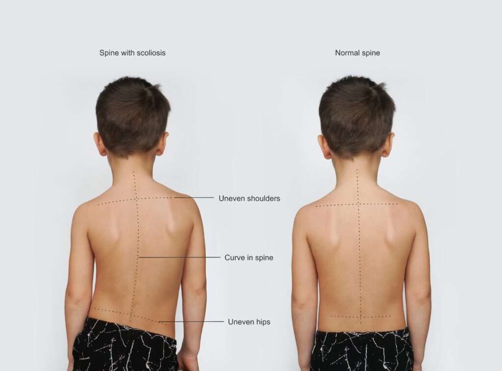
त्याचप्रमाणे, शरीराच्या मध्यरेषेपासून पाठीचा कणा बाजूला देखील झुकू शकतो, म्हणजे, डॉक्टर या समस्येला कोरोनल असंतुलन किंवा स्कोलियोसिस म्हणतात. कोरोनल पातळी अथवा प्लेन ही एक काल्पनिक पातळी आहे जी मानवी शरीराला पुढील आणि मागील भागामध्ये समान रीतीने विभाजित करते. “कोरोनल” पातळीतील असमानता (जेव्हा आपण समोरून मानवी शरीर पाहतो तेव्हा आपण कोरोनल पातळी (प्लेन) पाहू शकतो) मणक्यांच्या स्तंभात असममितता निर्माण करते. यामुळे नितंब आणि खांदे किंवा बरगड्यांचा असमान होतात व छातीत किंवा पाठीत एकतर्फी फुगवटा असू शकतो.

पाठीचा कण्याची विकृती

आधी म्हटल्याप्रमाणे, मणक्याच्या सजायटल वक्रतेतील कोणतेही विचलन किंवा मणक्याच्या कोरोनल प्लेनमधील कोणतेही विचलन मणक्यांची विकृती किंवा पाठीच्या कण्याची विकृती म्हणून ओळकली जाते. मणक्याच्या विकृतीचे अनेक प्रकार आहेत.

पाठीच्या विकृतीची लक्षणे काय आहेत

पाठीच्या विकृतीची गंभीरता सौम्य ते मध्यम किंवा गंभीर असू शकते. तथापि, प्रत्येक मणक्याच्या विकृतीमध्ये, वेदना नेहमीच होतात.

पण, तुम्हाला मणक्याच्या कोणत्याही भागात वेदना किंवा विकृती असल्यास तुम्हाला सतत दुखणं सहन करत राहण्याची आफत नाही. मिरजेसारख्या छोट्या शहरातील समर्थ न्यूरो आणि मल्टी स्पेशालिटी हॉस्पिटलचे डॉ रवींद्र पाटील यांच्यासारखे कुशळ न्युरोसर्जन तुम्हाला तुमच्या वेदनादायक लक्षणांपासून आराम मिळवून देऊ शकतात आणि काही केसेस मध्ये तुमची पाठीची विकृती देखील सुधारू शकतात.

पाठीच्या विकृतीचे प्रकार

नमूद केल्याप्रमाणे, पाठीच्या विकृतीमुळे मणक्याची योग्य वक्रता किंवा मणक्याची योग्य सरळता बदलू शकते. याव्यतिरिक्त, काही प्रकरणांमध्ये ऑस्टियोपोरोसिसमुळे कशेरुका कोसळू शकते आणि यामुळे मणक्याचा भाग लहान होऊ शकतो.

स्कोलियोसिस विकृती

तंदुरुस्त व्यक्तिला जर पुढून पाहिले की तर ती अगदी सरळ दिसते. पुढून पाहिले की कुठल्याही एका बाजूला मणक्यांच्या स्तंभाचा वक्र आकार म्हणजे स्कोलियोसीस. जर तुमचा पाठीचा कणा उजवीकडे वळला असेल तर तुम्हाला डेक्स्ट्रोस्कोलिhओसिस आहे. जर तुमचा पाठीचा कणा डावीकडे वळला असेल तर तुम्हाला लेव्होस्कोलिओसिस आहे. तथापि, बर्‍याचदा, कशेरूदंडाच्या एका बाजूला असलेला बाक असलेल्या प्रौढ आणि बालरोग रूग्णांच्या मणक्यामध्ये एकापेक्षा जास्त वळणे देखील असु शकतात, म्हणजे मणक्याला उजव्या आणि डाव्या दोन्ही बाजूंनी वळण येते.

कायफोसीस

काफोसीस म्हणजे कुबड. हे तेव्हा होते जेव्हा पाठीच्या वरच्या बाजूस कुबड येते. खांदे पुढे वाकतात व डोके बाहेर जाईल आणि धड खाली वाकलेले किंवा खाली पडलेले दिसेल.

लॉर्डोसिस

हे बहुतेकदा किफोसिसचा दुष्परिणाम म्हणून उद्भवते. हायपर-लॉर्डोसिस किंवा स्वे-बॅकमुळे पाठीचा खालचा भाग मागच्या बाजूला वळतो आणि उदर पुढच्या बाजूला वळते.

फ्लॅटबॅक सिंड्रोम

जेव्हा मणक्यांचा स्तंभ त्याची नैसर्गिक वक्रता गमावतो तेव्हा ही स्थिती उत्पन्न होते. हे सौम्य वाटू शकते. शेवटी, पाठीचा कणा “सरळ” झाला आहे. परंतु, फ्लॅटबॅक सिंड्रोममुळे रूग्ण चालतांना पुढे पडू शकतात. त्यामुळे चालताना गंभीर समस्या निर्माण होतात.

अँकिलोझिंग स्पॉन्डिलायटीस

संधिवातचा हा प्रकार पाठीच्या कशेरुकाला एकत्र जोडण्यास कारणीभूत ठरू शकतो. जेव्हा हे घडते, तेव्हा तुमचा पाठीचा कणा त्याची लवचीकता गमावतो आणि तुमची मुद्रा एकाच स्थितीत लॉक होते.

स्पॉन्डिलोलिस्थेसिस

ही परिस्थिती घसरलेली कशेरुका म्हणूनही ओळखली जाते. ही स्थिती मणक्यातील जन्मजात दोषामुळे होऊ शकते. सर्वात गंभीर प्रकरणांमध्ये, एक घसरलेला मणका पाठीच्या स्तंभातून पूर्णपणे सरकतो. ही स्थिती स्पॉन्डिलोप्टोसिस म्हणूनही ओळखली जाते.

पाठीच्या कण्यातील विकृती कशामुळे होतात?

डॉक्टर पाठीच्या विकृतीची त्यांच्या कारणांनुसार वेगवेगळ्या गटांमध्ये वर्गीकरण करतात. जरी बर्‍याच समस्यांमुळे पाठीचा कणा विकृती होऊ शकते, परंतु सर्वात जास्ती खालील कारणे असतात :

  • जन्मजात: मूल जन्माला येण्यापूर्वी पाठीच्या विकृतीचे काही प्रकार विकसित होतात. गर्भातील आनुवंशिकता आणि जन्माच्या वेळच्या दुर्घटनांमुळे असे होऊ शकते.
  • बालरोग: पाठीच्या विकृतीचे इतर प्रकार बालपणात अधिक स्पष्ट होतात. स्कोलियोसिस, उदाहरणार्थ, बहुतेकदा 10-12 वयोगटातील उद्भवते. विशेषत:, वाढीच्या वेगामुळे हे होऊ शकते.
  • पोस्‍चरल: पाठीचा कणा वाढण्‍याच्‍या प्रमुख कालावधीत खराब आसनामुळे पाठीचा वरचा भाग पुढे गोलाकार होऊ शकतो. या समस्येचे निराकरण करण्यासाठी, स्नायू-मजबुतीकरण आणि पवित्रा योग्या ठेवण्याचे धडे आवश्यक असू शकतात.
  • न्यूरोमस्क्यूलर: सेरेब्रल पाल्सीसारखे काही विकार स्नायूंच्या क्रियाकलापांमध्ये बदल करू शकतात. ताठर स्नायू मणक्याला खेचतात म्हणून, पाठ आणि मान त्यांचा नैसर्गिक आकार गमावू शकतात.
  • प्रौढ किंवा डीजनरेटिव्ह: मणक्यावरील झीजेमुळे तुमच्या पाठीतील डिस्क खराब होऊ शकतात. कमकुवत कशेरुका फ्रॅक्चर होऊ शकतात आणि त्यांचा आकार पाचरी सारखा होऊ शकतो. त्यामुळे कायफोसीस होतो आणि, मणक्याला आधार देणारे अस्थिबंधक कडक होऊ शकतात, तुमची पाठ संरेखनातून बाहेर काढून विकृत करू शकतात.
  • आघातामुळे : पाठीला अचानक मार लागल्यामुळे तुमच्या पाठीच्या वक्रतेत बदल होऊ शकतो. कशेरुकाला त्यांची जागा सोडू शकतात.
  • आयट्रोजेनिक: हा शब्द स्पाइनल समस्यांशी संबंधित आहे, जसे की फ्लॅटबॅक सिंड्रोम, जे अयशस्वी स्पाइनल फ्यूजनच्या परिणामी विकसित होतात.
  • इडिओपॅथिक: जेव्हा अज्ञात कारणास्तव पाठीचा कणा विकृत होतो तेव्हा त्या स्थितीला इडिओपॅथिक म्हणतात. बहुतेक स्कोलियोसिस प्रकरणे इडिओपॅथिक स्कोलियोसिस म्हणून वर्गीकृत करतात.

पाठीचा कणा विकृती ही गंभीर परिस्थिती आहे. ही विकृती स्वाभिमान नष्ट करू शकते आणि तुम्हाला तुमची दैनंदिन कामे पूर्ण करण्यात अडचणी आणू शकतात. त्याच बरोबर खूप वेदनाही होऊ शकतात.

सुदैवाने मणक्यांच्या विकृच्या उपचारासठी अनेक प्रकारचे औषधोपचार आणि सर्जिकल उपचार वेदना कमी करण्यासाठी आणि रेग्याची स्थिती पूर्ववत करण्यासाठी उपलब्ध आहेत. तुमच्या स्थितीला उपचारांची आवश्यकता असल्यास प्रथम शस्त्रक्रिया न करता उपचार तुम्ही करून पहाल. यामध्ये स्कोलियोसिस ब्रेसिंग किंवा फिजिकल थेरपीचा असू शकते.

जर तुमची स्थिती या पद्धतींनी सुधारली नाही, तर तुम्हाला पाठीच्या विकृतीची शस्त्रक्रिया करवून घ्यावी लागेल. पाठीच्या विकृतीसाठी ऑपरेटिव्ह उपचार तीन मुख्य मार्गांनी सुधारणा साध्य करतात. ते पाठीला सरळ करतात आणि मणक्याला चांगल्या स्थितीत धरतात (स्पाइनल फ्यूजन). ते हाडे (ऑस्टियोटॉमी) काढून टाकतात. किंवा, ते तुमच्या मणक्याला अधिक आदर्श पद्धतीने वाढण्यास प्रोत्साहित करण्यासाठी फ्यूजनलेस सिस्टम वापरतात.

अशा शस्त्रक्रिया करण्यासाठी तुम्हाला मोठ्या शहरांमध्ये जाण्याची गरज नाही. अशा प्रकारच्या शस्त्रक्रिया मिरजेतील समर्थ न्यूरो आणि सुपरस्पेशालिटी  हॉस्पिटलसारख्या विशेष केंद्रांमध्ये केल्या जाऊ शकतात, जेथे मुख्य न्यूरो आणि स्पाइन सर्जन डॉ रवींद्र पाटील यांनी अशा अनेक प्रकरणांवर यशस्वी उपचार केले आहेत.

पाठीची काळजी घेण्यास गुगलची खूप मदत होते. तुम्ही फक्त इंग्रजीत ‘बॅक केअर पीपीटी’ टाइप करा आणि तुम्हाला बॅक केअरच्या चरणांचे स्पष्टीकरण देणारी अनेक प्रेझेन्टेशन मिळतील.

आजारी रुग्णांसाठी बॅक केअर

Back care - Ravindra patil

आजारी रुग्णांसाठी बॅक केअर

By Dr.Ravindra Patil

पाठीची काळजी म्हणजे काय?

पाठीची काळजी समजून घेण्याआधी, प्रेशर सोर्स किंवा बेडसोर्स म्हणजे काय हे समजून घेतले पाहिजे. प्रेशर सोर्स टाळण्यासाठी बॅक केअर (पाठीची विशेष काळजी) घेतली जाते.

सोर अथवा अल्सर म्हणजे मानवी शरीराचे कातडे नष्ट होऊन त्वचेखालील ऊतक दिसते ती स्थिती.

प्रेशर सोर्सना प्रेशर अल्सर किंवा डेक्यूबिटस अल्सर किंवा बेडसोर्स असेही म्हणतात. त्वचेवर दीर्घकाळापर्यंत दाब पडल्यामुळे त्वचेला आणि त्वचेखालील ऊतींना झालेल्या जखमा आहेत. बेडसोर्स बहुतेकदा त्वचेवर विकसित होतात जे शरीराच्या हाडांच्या भागांना कव्हर करतात, जसे की टाच, घोटे, नितंब, शेपटीचे हाड आणि डोक्याच्या मागील बाजूस अल्सर होतात.

ज्या लोकांना बेडसोर्सचा धोका असतो अशा लोकांना वैद्यकीय परिस्थिती असते ज्यामुळे त्यांची स्थिती बदलण्याची क्षमता मर्यादित होते. ते पलंगावर किंवा खुर्चीवर बसलेले असतात. त्यांना अंथरुणावर किंवा खुर्चीत वेळ घालवायला भाग पाडले जाते.

बेडसोर्स तास किंवा दिवसात विकसित होऊ शकतात. बहुतेक फोड उपचाराने बरे होतात, परंतु काही पूर्णपणे बरे होत नाहीत. बेडसोर्स टाळण्यासाठी आणि त्यांना बरे करण्यात मदत करण्यासाठी आम्ही पावले उचलू शकतो.

Table of Contents

लक्षणे

बेडसोर्स किंवा प्रेशर अल्सरची चेतावणी चिन्हे आहेत:

•         त्वचेचा रंग किंवा पोत मध्ये असामान्य बदल

•         सूज येणे

•         पू सारखा स्त्राव

•         त्वचेचा एक भाग जो स्पर्शास इतर भागांपेक्षा थंड किंवा उबदार वाटतो

•         स्पर्श करताच दुखणारे क्षेत्र

बेडसोर्स त्यांची खोली, तीव्रता आणि इतर वैशिष्ट्यांवर आधारित अनेक टप्प्यांपैकी एका टप्प्यात असतात. त्वचा आणि ऊतींचे नुकसान, त्वचेचा रंग बदलण्यापासून ते स्नायू आणि हाडांच्या खोल दुखापतीपर्यंत असते.

प्रेशर अल्सरच्या सामान्य जागा

ज्या लोकांना सतत अंथरुणावर राहण्याची गरज असते त्यांना बेडसोअर्स होऊ शकतात व ते सहसा खालील जागी होतात :

  • डोक्याची मागील बाजू
  • खांद्याच्या मागे
  • नितंब, पाठीचा खालचा भाग किंवा शेपटीचे हाड
  • टाच, घोटे

व्हीलचेअरवर सतत बसावे लागते अशा लोकांना देखील प्रेशर पॉईंट्सवर प्रेशर सोर्स होऊ शकतात.

डॉक्टरांना कधी भेटायचे

जर तुम्हाला बेडसोअरची चेतावणी चिन्हे दिसली तर, त्या भागावरील दबाव कमी करण्यासाठी तुमच्या शरीराची स्थिती बदला. 24 ते 48 तासांत तुम्हाला सुधारणा दिसत नसल्यास, तुमच्या डॉक्टरांशी संपर्क साधा. जर तुम्हाला संसर्गाची लक्षणे दिसत असतील, जसे की ताप, बेडसोर मधून पू निघणे, घसा दुखणे व खराब वास येणे, त्वचेचा रंग बदलणे, बेडसोरच्या आसपास उबदारपणा जाणवणे किंवा फोडाभोवती सूज येणे.

बेडसोर्सची कारणे

बेडसोर्स त्वचेवर दाबामुळे होतात. त्या दाबा मुळे त्वचेला रक्त प्रवाह मर्यादित होतो. मर्यादित हालचालीमुळे त्वचेचे नुकसान होऊ शकते आणि बेडसोर्सचा विकास होऊ शकतो.

बेडसोर्सची कारणे खालील आहेत :

  • दबाव. तुमच्या शरीराच्या कोणत्याही भागावर सतत दबाव राहिल्याने ऊतींमधील रक्त प्रवाह कमी होतो. योग्य रक्तप्रवाहाशिवाय आवश्यक पोषक तत्वे त्वचेपर्यंत पोहोचत नाहीत आणि ती खराब होते.
  • मर्यादित हालचाल असलेल्या लोकांमध्ये स्नायू किंवा चरबीचे हाडांवर पुरेसे पॅड नसल्याने आणि मणक्याचे, शेपटी हाडाच्या किंवा खांद्याच्या मागे, नितंब, टाच आणि कोपर यासारख्या हाडांवर जर जोर आला आणि हालचाल कमी असली तर बेडसोर्स होतात.
  • घर्षण. जेव्हा त्वचा कपडे किंवा पलंगावर सतत घासली जाते तेव्हा घर्षण होते. हे घर्षण नाजूक त्वचेला, विशेषतः जर त्वचा ओलसर असेल तर, दुखापतीसाठी तयार करवू शकते.
  • कातरणे. जेव्हा दोन पृष्ठभाग विरुद्ध दिशेने ओढले जातात तेव्हा कातरणे होते. उदाहरणार्थ, जेव्हा पलंग डोक्यावर उंचावला जातो तेव्हा तुम्ही गुरूत्वाकर्षणाने अंथरुणावर खाली घसरत जाता. जसजसे शेपटीचे हाड खाली सरकते तसतसे हाडावरील त्वचा जागी कातलरी जाऊ शकते आणि बेडसोर होतात.

जोखीम घटक

जर तुम्हाला हालचाल करण्यात अडचण येत असेल आणि बसून किंवा अंथरुणावर सहज स्थिती बदलता येत नसेल तर तुम्हाला बेडसोर्स होण्याचा धोका जास्त असतो. जोखीम घटकांमध्ये खालील घटक समाविष्ट आहेत :

  • हलता न येणे. हे खराब आरोग्य, पाठीच्या कण्याला दुखापत आणि इतर कारणांमुळे असू शकते.
  • लघवी आणि मल यांवर असंयम. लघवी आणि मल यांच्या दीर्घ संपर्कामुळे त्वचा अधिक असुरक्षित होते.
  • संवेदना कमी होणे. पाठीचा कणा दुखापत, मज्जासंस्थेसंबंधीचा विकार आणि इतर परिस्थितीमुळे संवेदना नष्ट होऊ शकतात. वेदना किंवा अस्वस्थता जाणवण्यास असमर्थता होते आणि बेडसोरची चेतावणी चिन्हे कळत नाहीत आणि स्थिती बदलण्याची आवश्यकता भासत नाही व बेडसोर होतात.
  • कुपोषण आणि हायड्रेशन. निरोगी त्वचा राखण्यासाठी आणि ऊतींचे तुटणे टाळण्यासाठी लोकांना त्यांच्या दैनंदिन आहारात पुरेसे द्रव, कॅलरी, प्रथिने, जीवनसत्त्वे आणि खनिजे आवश्यक असतात.
  • रक्त प्रवाह कमी करणारे रोग – मधुमेह आणि रक्तवहिन्यासंबंधी रोग रक्तप्रवाहावर दुष्परिणाम करू शकतात आणि त्यामुळे बेडसोर्स होण्याचा धोका वाढू शकतो.

बेडसोर कसे टाळायचे

त्वचेवर ताण येऊ नये म्हणून तुम्ही स्वतः शक्य होईल तितक्या वेळा शरीराची स्थिती बदलत रहावे. तुमच्या त्वचेची चांगली काळजी घेणे. चांगले पोषण आणि द्रवपदार्थाचे सेवन करणे. धूम्रपान सोडणे, तणावाचे व्यवस्थापन करणे आणि दररोज व्यायाम करणे या इतर आवश्यक गोष्टी आहेत.

जागरूक रुग्णांनी घ्यायची पाठीची काळजी

बेड किंवा खुर्चीवर पुनर्स्थित होतांना अथवा करतांना खालील गोष्टी विचारात घ्या :

  • तुमचे शरीर वारंवार हलवा. तासातून एकदातरी पुनर्स्थित होण्यासाठी मदतीसाठी विचारा.
  • शक्य असल्यास, स्वतःला हातांवर उचला. तुमच्या शरीराच्या वरच्या भागावर पुरेशी ताकद असल्यास, खुर्चीच्या हातावर दाबून तुमचे शरीर सीटवरून वर करून व्हीलचेअरवर पुश-अप करा.
  • काही व्हीलचेअर्स तुम्हाला त्यांना झुकवण्याची सोय देतात. त्यामुळे दबाव कमी होतो.
  • दाब कमी करणारी छोटी उशी किंवा गादी वापरा.
  • दाब कमी करण्यासाठी उशी किंवा ‘एअर बेड’ नावाची विशेष गादी वापरा.
  • तुमच्या पलंगाची उंची समायोजित करा. जर तुमचा पलंग डोक्यावर उंच केला जाऊ शकत असला तरी तो 30 अंशांपेक्षा जास्त वाढवू नका. हे कातरणे टाळण्यास मदत करते.

नर्सिंगमध्ये बॅक केअर टप्पे

रूग्णालयांमध्ये सतत पलंगावर झोपून असणार्या रूग्णांना ‘एअर बेड’ दिला जातो. हा बेडसोर्स टाळण्यासाठी नर्सिंग केअर करण्यासाठी महत्वाची भूमिका बजावतो. नर्सिंगमध्ये पाठीच्या काळजीच्या चरणांचा सारांश खाली दिला आहे:

  • त्वचा स्वच्छ आणि कोरडी ठेवा. हलक्या क्लीन्सरने त्वचा धुवा आणि कोरडी करा. त्वचेचा ओलावा, लघवी आणि मल यांच्याशी होणारा संपर्क कमी करण्यासाठी ही साफसफाई नियमितपणे करा अथवा करवा.
  • त्वचेचे रक्षण करा. लघवी आणि मल यांच्यापासून त्वचेचे संरक्षण करण्यासाठी ओलावा ठेवणारे क्रीम वापरा.
  • गरज भासल्यास पलंग आणि कपडे वारंवार बदला. कपड्यांवरील बटणे आणि पलंगावरील सुरकुत्या त्वचेला त्रास देऊ शकतात, त्या टाळा.
  • त्वचेची दररोज तपासणी करा. प्रेशर सोअरच्या चेतावणी चिन्हांसाठी दररोज आपल्या त्वचेकडे बारकाईने पहा.
  • जेव्हा आपण आपल्या पाठीवर झोपतो तेव्हा आपण वळत राहतो, परंतु आजारी रुग्ण वळू शकत नाहीत. डॉक्टरांच्या सल्ल्यानुसार त्यांच्या शरीरची स्थिती बदलत रहावा.

पाठीच्या काळजीचे हे टप्पे महत्वाचे आहेत.

पाठीच्या काळजीचा उद्देश आहेः

  • रक्ताभिसरण उत्तेजित करा आणि सामान्य आराम द्या.
  • बेडसोअर प्रतिबंधित करा
  • रुग्णाला दिलासा द्या.

बॅक केअर मसाजचे सात टप्पे

  • रुग्णाला त्याच्या ओटीपोटावर किंवा कुशीवर झोपवा. रूग्णाची पाठ नर्सकडे ठेवा आणि त्याचे शरीर पलंगाच्या काठावर ठेवा.
  • रुग्णाचा गाऊन काढा किंवा वर सरकवा.
  • पाठीवर नीट चोळून औषधी लोशन लावा.
  • पाठीवर घासताना मजबूत लांब स्ट्रोक आणि मालीश करण्याच्या हालचाली वापरा.
  • किती दबाव आणायचा हे रुग्णाच्या स्थितीवर अवलंबून असते.
  • मान आणि खांद्यापासून सुरुवात करा नंतर संपूर्ण मालीश करत खालपर्यंत जा.
  • जोरदार स्ट्रोकसह दोन्ही हातांनी मसाज करा. शक्यतो हात वरच्या दिशेने न्या. लोशनमध्ये घासताना जेथे कातडीवर दबाव येतो त्या विशेष लक्ष द्या.

रुग्णाचा गाऊन नीट करून परत त्याला त्याच्या पाठीवर फिरवा आणि गाऊन खाली खेचा. नर्सिंग कर्मचार्यांनी या पाठीच्या मसाजचे टप्पे पायऱ्या नियमितपणे पाळले पाहिजेत.

पाठीला मालीश करतांना हाताच्या हालचाली कशा कराव्यात

इफ्ल्युरेज – ही हातांच्या तळव्याने केलेली पाठीच्या पृष्ठभागावर लांब स्वीपिंग हालचाल आहे. मानेसारख्या लहान पृष्ठभागांवर अंगठा आणि बोटे वापरली जातात. स्ट्रोक हळू, लयबद्ध आणि सौम्य असावेत आणि दाब स्थिर असावेत. रक्ताच्या शिरांमधील रक्त प्रवाहाच्या दिशेत चोळा.

नीडिंग – हाताच्या करंगळीच्या बाजूने केलेला मसाज. यात बोटे आणि अंगठा त्वचा आणि त्वचेखालील ऊतींना पकडतात आणि मसाज देणार्याच्या हाता प्रमाणे हलतात.

घर्षण – हाताच्या पूर्ण पृष्ठभागाने किंवा बोटांनी आणि अंगठ्यांसह मर्यादित भागांवर घर्षण केले जाते. ही हालचाल गोलाकार आहे आणि ऊतींच्या अशा भागावर दाब देऊन मालीश केली जाते ज्या भागांना पकडता येत नाही.

बॅक केअर ppt पॉवर पॉइन्ट प्रेझेन्टेशन

पाठीची काळजी घेण्यास गुगलची खूप मदत होते. तुम्ही फक्त इंग्रजीत ‘बॅक केअर पीपीटी’ टाइप करा आणि तुम्हाला बॅक केअरच्या चरणांचे स्पष्टीकरण देणारी अनेक प्रेझेन्टेशन मिळतील.

Back Care for Sick Patients

Back care - Ravindra patil

Back Care for Sick Patients

By Dr.Ravindra Patil

What is back care?

Before we understand back care, we must understand what are pressure sores or bedsores. Back care procedure is done to prevent pressure sores.

Pressure sores are also called pressure ulcers or decubitus ulcers or bedsores. They are injuries to the skin and underlying tissue resulting from prolonged pressure on the skin. Bedsores most often develop on skin that covers bony areas of the body, such as the heels, ankles, hips, tailbone and back of the head.

People who are at risk of bedsores have medical conditions that limit their ability to change positions. They are bed ridden or chair ridden. They are forced to spend their time in a bed or chair.

Bedsores can develop over hours or days. Most sores heal with treatment, but some never heal completely. We can take steps to help prevent bedsores and help them heal.

Table of Contents

Symptoms

Warning signs of bedsores or pressure ulcers are:

  • Unusual changes in skin colour or texture
  • Swelling
  • Pus-like discharge
  • An area of skin that feels cooler or warmer to the touch than other areas
  • Tender areas

Bedsores fall into one of several stages based on their depth, severity and other characteristics. The degree of skin and tissue damage ranges from changes in skin colour to a deep injury involving muscle and bone.

Common sites of pressure ulcers

For people who need to stay in bed, bedsores may happen on:

  • The back or sides of the head
  • The shoulder blades
  • The hip, lower back or tailbone
  • The heels, ankles and skin behind the knees

Wheelchair bound people also may suffer pressure sores at pressure points.

When to see a doctor

If you notice warning signs of a bedsore, change your position to relieve the pressure on the area. If you don’t see improvement in 24 to 48 hours, contact your doctor. Seek immediate medical care if you show signs of infection, such as a fever, drainage from a sore, a sore that smells bad, changes in skin colour, warmth or swelling around a sore.

Causes of bedsores

Bedsores are caused by pressure against the skin that limits blood flow to the skin. Limited movement can make skin vulnerable to damage and lead to development of bedsores.

Primary contributing factors for bedsores are:

  • Constant pressure on any part of your body can lessen the blood flow to tissues. Without a good blood flow, essential nutrients don’t reach the skin and it is damaged.
  • For people with limited mobility, bedsores occur in areas that aren’t well padded with muscle or fat and that lie over a bone, such as the spine, tailbone, shoulder blades, hips, heels and elbows.
  • Friction occurs when the skin rubs against clothing or bedding. It can make fragile skin more vulnerable to injury, especially if the skin is also moist.
  • Shear occurs when two surfaces move in the opposite direction. For example, when a bed is elevated at the head, you can slide down in bed. As the tailbone moves down, the skin over the bone might stay in place — essentially pulling in the opposite direction.

Risk factors

Your risk of developing bedsores is higher if you have difficulty moving and can’t change position easily while seated or in bed. Risk factors include:

  • This might be due to poor health, spinal cord injury and other causes.
  • Skin becomes more vulnerable with extended exposure to urine and stool.
  • Lack of sensory perception. Spinal cord injuries, neurological disorders and other conditions can result in a loss of sensation. An inability to feel pain or discomfort can result in not being aware of warning signs and the need to change position.
  • Poor nutrition and hydration. People need enough fluids, calories, protein, vitamins and minerals in their daily diets to maintain healthy skin and prevent the breakdown of tissues.
  • Medical conditions affecting blood flow. Health problems that can affect blood flow, such as diabetes and vascular disease, can increase the risk of tissue damage such as bedsores.

Prevention

You can help prevent bedsores by frequently repositioning yourself to avoid stress on the skin. Other strategies include taking good care of your skin, maintaining good nutrition and fluid intake, quitting smoking, managing stress, and exercising daily.

Back care steps which conscious patients must take

Consider the following recommendations related to repositioning in a bed or chair:

  • Shift your weight frequently. Ask for help with repositioning about once an hour.
  • Lift yourself, if possible. If you have enough upper body strength, do wheelchair push-ups by raising your body off the seat by pushing on the arms of the chair.
  • Some wheelchairs allow you to tilt them, which can relieve pressure.
  • Select small cushions or a mattress that relieves pressure.
  • Use cushions or a special mattress called an ‘air bed’ to relieve pressure.
  • Adjust the elevation of your bed. If your bed can be elevated at the head, raise it no more than 30 degrees. This helps prevent shearing.

Back care steps in nursing

In hospitals, an ‘air bed’ is given to patients who are immobile. Nursing care plays a vital role in preventing bedsores. Steps of back care in nursing are summarised below:

  • Keep skin clean and dry. Wash the skin with a gentle cleanser and pat dry. Do this cleansing routine regularly to reduce the skin contact with moisture, urine and stool.
  • Protect the skin. Use moisture barrier creams to protect the skin from urine and stool.
  • Change bedding and clothing frequently if needed. Watch for buttons on the clothing and wrinkles in the bedding that irritate the skin.
  • Inspect the skin daily. Look closely at your skin daily for warning signs of a pressure sore.
  • When we sleep on our back, we keep on turning, but sick patients cannot turn. Try and turn them under the doctor’s advice.

These five steps of back care are crucial.

Back care procedure

Aim of back care is to:

  • Stimulate the circulation and give general relief.
  • Prevent bedsore
  • Give comfort to the patient.

7 steps of back care massage

  • Help the patient to turn on his abdomen or on his side with his back toward the nurse and his body near the edge of the bed.
  • Raise the patient’s gown.
  • Apply medicated lotions to the back to reduce friction.
  • In rubbing the back use firm long strokes and kneading motions.
  • The amount of pressure to exert depends upon the patient’s condition.
  • Begin from the neck and shoulders then proceed over the entire back.
  • Massage with both hands working with a strong stroke. In upward than in downward motions. Give particular attention to pressure areas in rubbing in the lotions.

Turn patient on his back and pull down the gown. These back massage steps in nursing should be regularly followed.

Hand Movements Used

Effleurage (stroking—is a long sweeping movement with palm of hand conforming to the contour of the surface treated, over small surface (on the neck) the thumb and fingers are used. Strokes should be slow, rhythmical and gentle with pressure constant and in the direction of venous stream.

Kneading—performed with the ulnar side palm resting on the surface and the fingers and thumb grasping the skin and subcutaneous tissues which move with the hand of the operator.

Friction—is performed with the whole palmar surface of the hand or fingers and thumbs over limited areas. This movement is a circular and kneading with pressure against the underlying part of tissue which cannot be grasped.

Back care ppt

Google is of such great help, just type ‘back care ppt’ and you will get many presentations explaining steps of back care.